Human spine, characterization and modeling 1

12:00 - 13:30 Monday, 9th July, 2018

Wicklow Hall 2A

Track Musculoskeletal

Posters for this session are displayed on Monday 9th July in the Forum.

Chairs: Hans-Joachim Wilke and Fabio Galbusera

P1178 An experimentally validated computational study of changes in static and fatigue properties following balloon kyphoplasty

Dr. Philip Purcell1, Mr. Stephen Tiernan1, Dr. Fiona McEvoy1, Mr. Derek Sweeney2, Mr. Seamus Morris3
1IT Tallaght, Dublin, Ireland. 2CADFEM UK & Ireland, Dublin, Ireland. 3Mater Misericordiae University Hospital, Dublin, Ireland

Abstract

Utilisation of minimally invasive procedures such as balloon kyphoplasty for the treatment of vertebral compression fractures, has increased considerably during the past decade. While many studies have examined the kyphoplasty treatment using in-vitro biomechanical tests and continuum finite element models, they have limited capability to address clinical observations of vertebral re-collapse after the treatment. The following study proposes an experimentally validated computational analysis of the kyphoplasty treatment that can address these adverse events.

The experimental protocol utilised a series of static and fatigue tests on specimens randomly assigned to either an untreated control group or kyphoplasty treated group. A random specimen was selected from each test group for micro computed tomography scans, which were subsequently skeletonised using the opensource BoneJ software. Specimen specific beam finite element models of the skeletonised structure were then created using the ANSYS® Parametric Design Language. The computational study utilised specimen specific models with a custom strain dependant material damage law for bone that enabled simulation of both static and cyclic loading conditions for comparison with the experimental data.

Experimental and computational results indicated significant increases in static stiffness and strength after kyphoplasty, by factors of approximately 2 and 1.5 respectively. Specimens treated with kyphoplasty also exhibited rates of damage accumulation 41% lower than untreated samples under fatigue conditions. Similarly, cyclic loading simulations predicted higher levels of total strain for untreated specimens, with failure rapidly ensuing at levels above 1.4%. Computational simulations also illustrated how a 50% reduction in the number of microscale cement interlocks can diminish strength and stiffness below untreated levels, thereby increasing the risk of vertebral re-collapse. Consideration of these findings in the context of clinical evidence showing discontinuity at the bone-cement interface highlights the importance of this region to sustaining the stabilisation achieved during the initial treatment.  

Acknowledgements
Funded by the Irish Research Council EMBARK Postgraduate Scholarship RS/2011/399 awarded to Philip Purcell.


P1179 Simulation of the flexion motion of human cervical spine segments associated with contraction of the main agonist muscle

Prof. Satoshi Shimawaki, Mr. Takuya Suda, Prof. Masataka Nakabayashi
Utsunomiya University, Utsunomiya, Japan

Abstract

Introduction: Cervical spine motion was studied by analyzing the passive behavior of the cervical spine in reaction to external loads, as in the field of impact biomechanics, or by analyzing the active behavior of the cervical spine associated with muscle contractions, as in the field of medical biomechanics. In the latter situation, studies focused on investigating the effects of arthrodesis and muscle/ligament damage on cervical spine motion. In this study, we simulated a multibody model of the human upper trunk, which was constructed using human CT images and anatomical data, to analyze the flexion motion of the cervical spine segments associated with contraction of the main agonist muscle.

Methods: The model comprised bone models connected with soft-tissue models. The bone models were created based on CT images (slice interval, 1 mm) of a single subject (22-year-old Japanese male: height, 170 cm; weight, 70 kg) and included the skull (C0), cervical spine (C1~C7), thoracic spine (T1–T12), clavicle, scapula, costa, and sternum. Bones other than the skull, cervical spine, and thoracic spine (T1, T2) were deemed to be a single unit and were fixed in space. Soft tissues (articular cartilage, intervertebral disk, ligament, and muscle) were set using an anatomy book as reference. Five flexors (such as sternocleidomastoid and scalenus anterior) and eight ligaments (such as nuchal ligament and ligamenta flava) were approximated with a spring model connecting the attachment points. The cartilage and intervertebral disk were approximated by multiple spring models in such a manner that adjacent bones did not collide. Each muscle was subjected to a constant contractile force that was 50% of the maximum voluntary contraction of the muscle. The effect of gravity was ignored.

Results: The rotation angle and intervertebral distance were calculated for each cervical spine segment (C0-C1~C6-C7, C7-T1) at 36° flexion. The center of gravity of each vertebra was determined, and a unit vector was set from the center of gravity in the horizontal direction. The distance between the centers of gravity of two adjacent vertebrae was defined as the intervertebral distance, and flexion-associated changes in the intervertebral distance were determined. The angle between the unit vectors set for both vertebrae was determined, and flexion-associated changes in this angle were defined as rotation angles. At 36° flexion, the maximal change in the intervertebral distance was 2.0 mm that occurred at C6-C7. The minimum change in the intervertebral distance was -0.4 mm that occurred at C1-C2. The maximum rotation angle (contribution percentage) was 8.8° (24.3%) that occurred at C0-C1. The minimum rotation angle was 2.4° (6.8%) that occurred at C2-C3.


P1180 Biomechanical investigation of different posterior fixation surgeries for spinal burst fracture

Dr. Wengpin Chen1, Dr. Jen-Chung Liao2, Mr. Hao Wang1, Dr. Ching-Lung Tai3, Dr. Po-Liang Lai2
1Department of Mechanical Engineering, National Taipei University of Technology, Taipei, Taiwan. 2Department of Orthopaedic Surgery, Chang Gung Memorial Hospital, Taoyuan, Taiwan. 3Graduate Institute of Medical Mechatronics, Chang Gung University, Taoyuan, Taiwan

Abstract

Introduction
    Burst fracture approximately accounts for 20% of thoracolumbar fractures. Surgery is usually indicated for a patient suffering from severe deformity. One-above and one-below posterior short-segment instrumentation with fusion has been widely used for unstable thoracolumbar burst fracture.  Various anterior augmentation methods include transpedicular calcium sulfate or phosphate cement injection or two additional screws fixation at the injured vertebra had been proposed to prevent early implant failure and donor site complications. Each fixation method had been reported clinically, however, there was no study to discuss the effects of different fixation methods. In this research, a biomechanical comparison of the four surgical methods on the thoracolumbar spine based on finite elements (FE) analysis was proposed.

Methods
    Four fixation models were established to simulate the unstable thoracolumbar fracture with different fusion surgeries:  short fixation with crosslink (S-C), short fixation with intermediate screws (S-I), short fixation with crosslink and calcium sulfate (S-C-C), short fixation with intermediate screws and calcium sulfate (S-I-C). Four FE models under four different loading (flexion, extension, lateral bending, and axial rotation) conditions were analyzed. By using FE analysis, the range of motion (ROM), as well as the maximum value and distribution of the implant stress, and the facet joint stress were compared.

Results
    The results showed that when S-I-C fixation was used under the four types of loadings, the ROM’s are almost all the lowest, while the S-C fixation has the largest ROM’s. The von Mises stress results showed that under lateral bending and axial rotation loadings, maximum stress values were larger than those under flexion and extension loading. The von Mises stress distribution results showed that high stress is concentrated at the crosslink and rod junctions. The value of the maximal von Mises stress on the superior vertebral body for all loadin These findings were also consistent with clinical outcome.


Discussion
    According to the simulation results, S-I-C and S-I fixation methods possesses better biomechanical performance for the treatment of burst fracture. However, the facet joint load at the adjacent level is larger than those of the two other fixation methods. Since the current study focused on the treatment of burst fracture patients, The internal fixation devices will be removed when bone fusion is achieved. This is different from the case of fusion surgery for degenerative disc disease. The adjacent level degeneration problems need to be taken into consideration when fixation devices were not removed for those patients.

Conclusions
    It is recommended that surgeons can use intermediate bilateral pedicle screws together with or without calcium sulfate (S-I or S-I-C) to provide better stability and avoid stress concentration at the screw-crosslink junctions for the S-C and S-C-C surgeries.


P1181 Biomechanics of compensatory mechanisms of spine

Mr. Dmitriy Ivanov1, Dr. Leonid Kossovich1, Dr. Irina Kirillova1, Dr. Alexey Kudyashev2
1Saratov State University, Saratov, Russian Federation. 2Military-medical Academy, Saint-Petersburg, Russian Federation

Abstract

Transpedicular fixation is the most effective method of spine injuries and diseases treatment, when stabilization and correction of spine segments is necessary. However, transpedicular fixation is often accompanied by degenerative-dystrophic changes in functional units located above the fixation level. This leads to the formation of proximal junctional kyphosis (PJK) in 26–39 % of patients [1]. Researchers associate these changes with incorrectly formed sagittal vertebral-pelvic relationships [1].

This work presents biomechanical study aimed at identifying mechanical factors leading to the formation and progression of degenerative changes in functional units located above the fixation. An attempt was also made to substantiate the need for spine correction and fixation in accordance with calculated optimal sagittal balance parameters.

Surgical operation of decompressive inter-aminefascectomy at L3-L4, L4-L5, L5-S1 level, removal of herniated intervertebral discs, transpedicular fixation of L3-L4, L4-L5, L5-S1 segments was simulated. Biomechanical model was constructed on the basic of CT data and contained vertebrae, ribs, pelvic bones, upper thirds of femur, intervertebral discs, archicular joints and ligaments of the spine. Spatial arrangement of spine and pelvic complex elements was corrected according to Full Body X-Ray data and corresponded to sagittal profile in standing position.

Transpedicular fixation at L3-S1 level was considered for two models: with unbalanced sagittal profile and sagittal profile after correction. Lumbar lordosis was corrected to 58° with prevalence of lower lordosis arch (LA=38°). Position of pelvis was also changed (SS=34°; PT=5°). In ANSYS software, spine was loaded by stationary force (400 N) directed normally to the upper plate of C7 vertebra and mechanical stresses in vertebrae and intervertebral discs were analyzed for both models.

Displacements of corrected spine turned out to be significantly smaller in comparison with the model of unbalanced spine (displacements of C7 vertebra were 84 mm and 143 mm respectively). Similar picture was observed for effective stresses fields in vertebrae: 351 MPa and 475 MPa respectively. Maximum strains in intervertebral discs above the fixation level were 0.37 and 0.63 respectively, which indicates a significantly higher load on intervertebral disks located above fixed vertebrae for unbalanced spine model compared to the balanced one.

Numerical results provided biomechanical justification for clinical data [2], devoted to the problem of PJK formation in patients with transpedicular fixation.

References

1. Kim Y.J., Lenke L.G., Bridwell K.H., et al. Proximal junctional kyphosis in adolescent idiopathic scoliosis after 3 different types of posterior segmental spinal instrumentation and fusions: incidence and risk factor analysis of 410 cases. Spine J. 2007;32(24):2731-2738.

2. Matsumoto T., Okuda S., Maeno T., et al. Spinopelvic sagittal imbalance as a risk factor for adjacent-segment disease after single-segment posterior lumbar interbody fusion. J. Neurosurg Spine. 2017;26(4):435-440.


P1182 A Finite Element Study of Spinal Fusion and PJK

Mr. Hank Ballard, Dr. Jason Pittman, Dr. Alan Eberhardt
University of Alabama at Birmingham, Birmingham, AL, USA

Abstract

Proximal junction kyphosis (PJK) is a condition that may occur after a long spinal fusion, such as that typically seen in deformity correction surgery. PJK is defined as when the angle of kyphosis between the uppermost instrumented vertebra and two vertebrae above that is greater than 10°. PJK may progress, resulting in pain and discomfort characterized by degradation of the adjacent intervertebral disc or collapse of the vertebral body. It is conjectured that PJK arises from increased stress in the adjacent intervertebral disc due to the rigidity of the fused vertebrae.

Presently, the lower thoracic vertebrae (T7-T12) of a severe scoliotic patient were modeled through computed tomography (CT) scans. The CT scans were imported into Simpleware ScanIP software where vertebral segmentation was performed. A cortical shell was created based on the visual density and translated into 3D surfaces. The vertebrae were realigned using surface tools according to postoperative x-rays. Each intervertebral disc was separated into a nucleus pulposus and annulus fibrosus. An intact, non-instrumented model was created. Pedicle screws and rods were modeled as cylinders, connected as one continuous object and applied with the guidance of a practiced surgeon. Five mm (diameter) long screws were applied where the tip of the screw was 5 mm from the inner cortical face. Other screws (5 mm diameter short and 4 mm diameter long/short) were also investigated. The models were meshed using an automated meshing algorithm with linear tetrahedral elements with 1.5 mm (cancellous bone, instrumentation) and 1 mm (cortical shell, intervertebral disc) target edge lengths. The finite element (FE) models contained roughly 1 million elements. 

The models featured nonlinear hyperelastic (neo-Hookean) annuli fibrosus, elastic and nearly non-compressible nuclei pulposus, and linearly elastic ligaments (LINK180), bone, and instrumentation. Fixed boundary conditions were applied to the bottom vertebra and posterior faces of the instrumentation. FE analysis in ANSYS was used to calculate stresses and strains for the fusion model (T12-T8) and the intact model, subject to a compressive load (500 N) with a forward bending moment (10.5 kNmm), each applied with 10 load steps. Large deformations were considered and the automatic re-mesher was run (skewness threshold = 0.75). For the baseline model, the T7/T8 disc showed stress/strain levels at 2.92 MPa and 0.34 mm/mm, respectively. The T12-T8 fusion model (5 mm long) revealed higher levels of stress/strain (roughly 20% increase) in the T7/T8 disc when compared to the baseline model. Stress/strain levels were reduced modestly (1-3%) with reduced screw length and diameter. The baseline model predictions compared favorably to previously published models, suggesting its value as a platform to investigate spinal fusion on the likelihood for PJK.

P1183 Raster stereographic measurement of adolescent idiopathic scoliosis: a case study

Ms Lin Fu1, Professor Yaodong Gu1,2, Dr Dongdong Xia3, Mr Qichang Mei1,2, Dr Justin Fernandez1,2
1Research Academy of Grand Health Interdisciplinary, Ningbo University, Ningbo, China. 2Auckland Bioengineering Institute, University of Auckland, Auckland, New Zealand. 3Ningbo First Hospital, Ningbo, Ningbo, China

Abstract

Introduction

Adolescent Idiopathic Scoliosis (AIS) is a structural spinal deformity in the coronal plane that affects 1-3% of children in the United States [1]. The treatment of AIS depends on the severity and may include observation, bracing, surgery, physical therapy, chiropractic treatment and electrical stimulation. A scoliotic angle less than 10º [2], kyphotic angle within the range of 20-40º, and/or lydotic angle within the range of 20-60º [3] is classed as normal. Outside of this range observation and/or intervention may be required. The purpose of this study is to evaluate a 3D raster stereographic measurement technique for assessing before and after scoliosis surgery.

Methods

A 15-year-old girl who had not had any surgical intervention presented with AIS (which had been observed for 6 months). Raster stereographic analysis was conducted on her prior to surgery and following scoliotic correction surgery. The angles computed included the scoliotic angle (coronal plane), thoracic kyphotic angle and lumbar lordotic angle (both in the sagittal plane).


Results

Prior to surgery the patient exhibited a scoliotic angle of 45° (classed as severe), and kyphotic and lordotic angles of 34° (classed as normal). Following surgery the patient exhibited a reduced scoliotic angle of 13° (classed as mild), kyphotic angle of 37° and lordotic angle of 26°, both remaining in the normal range.

Uncaptioned visual Uncaptioned visual

Uncaptioned visual Uncaptioned visual

Figure 1: Coronal view of spinal orientation prior to surgery (left) and following surgical correction (right).


Discussion

Following surgery the scoliotic angle was reduced to 13°, now classed as mild scoliosis. While the kyphotic and lordotic angles also changed, though they remained in the normal range. This suggests surgeons should pay attention to the 3D modifications in the sagittal plane when intervening in the coronal plane to correct scoliosis. This is best revealed using raster stereography. However, raster stereography is not common in the clinic, but prior studies have shown that it is correlated to standard 2D radiography (>0.7) [4], which is known as the classic Cobb angle. This technique is non-invasive, portable and may play a role in accurately assessing surgery outcomes in the clinic.


References

  1. Soucacos P N et al., Orthopedics, 23(8):833, 2000.
  2. Kondrad E Bloch et al. ERS Handbook: self-assessment in respiratory medicine: page 460, 2015.
  3. Bernhardt M et al. Spine, 14(7): 717-721, 1989.
  4. Frerich J M et al., Open Orthopaedics Journal, 6(1):261-265, 2012.

P1184 Dynamic stability of the lumbar spine during unstable push-ups

Mr. Adrian Nizzero, Dr. Sylvain Grenier
Laurentian University, Sudbury, Canada

Abstract

Introduction
Spine stability is the ability to resist a perturbation. Stability training has gained popularity in both rehabilitative settings and athletic development for its ability to produce similar levels of muscle activity under a reduced load. Challenging the nervous system while limiting the load may decrease the potential for load-induced injury. Muscle activity in unstable exercises has been researched; the resultant loads and stability of the lumbar spine has yet to be examined. 
Methods
An LED triad is placed on the L1 and L5 vertebral processes. With the use of three HD action cameras and a computer software digitization program, we record the movement of these LED triads during unstable exercise and transform the data into 3-dimensional space. We calculate the cross product of the two vectors to generate a vector orthogonal to the vertebra and thus a local coordinate system, and determine how the LCS of the L1 and L5 vertebra move in relation to each other using Lyapunov analysis. More movement indicates a less stable lumbar spine. This process is performed for stable lunges and pushups, for a moderately unstable variation of the lunge and pushup, and an extremely unstable variation of lunge and pushup.
Results
This project is still in progress. Preliminary results indicate a decrease in lumbar spine stability as the exercise becomes progressively more unstable. The triads move in syncronicity during the most stable variation. During unstable variations, the L1 LCS moves much more in relation to the L5 LCS, indicating a decrease in lumbar spine stability.
Discussion
The results suggest that exercises which induce a great reduction of lumbar spine dynamic stability should be performed with caution as they may place the lumbar spine in a vulnerable environment. Stability training requires some degree of instability, however too much may prove catastrophic and induce injury. Recommendations include a slow progression of exercise instability, limiting the degrees of freedom of the movement until the client or athlete demonstrates sufficient ability to maintain spinal stability before progressing. Rapid progression may prove injurious, especially in a rehabilitative setting.

P1185 Assessment of the effects of bone-preserving decompression procedures on lumbar spine loads through musculoskeletal modeling approach

Mr. Tito Bassani, Mr. Fabio Galbusera
IRCCS Istituto Ortopedico Galeazzi, Milan, Italy

Abstract

Introduction: In the treatment of lumbar stenosis, new bone-preserving decompression techniques have been introduced to reduce the risk of iatrogenic instability, but have never been extensively investigated from a biomechanical point of view. The effects of these surgical techniques (i.e. laminotomy, laminectomy and fixation) can affect spine flexibility, distribution of the intervertebral loads and muscle activation. Investigating this issue accounting for in vivo measurements would result highly invasive. Conversely, musculoskeletal modeling allows to compute the internal biomechanical loads in response to imposed motion conditions. To this aim, the present work exploits motion data obtained in vitro and describing the flexibility of six L2-L5 human spine specimens in intact conditions and after the decompression techniques. Motion data were used to drive the kinematics of the AnyBody full-body musculoskeletal model (Figure1), in order to compute the corresponding loads at decompression level and paraspinal muscles activation.

Methods: Motion data allowed characterizing vertebral kinematics during flexion-extension, lateral bending and axial rotation movements (Figure1), in the following conditions: i) intact specimen; ii) laminotomy at L3-L4; iii) on three specimens, the laminotomy was extended to L4-L5; iv) laminectomy (at L3-L4 in the first three specimens and at both levels in the rest); v) pedicle screw fixation at involved levels. For each condition, the following measurements were computed: axial compression force (F) and postero-anterior shear (S, positive if posteroanteriorly oriented) at L3-L4; multifidus (MF), erector spinae (ES) and rectus abdominis (RA) muscles forces. The maximum values found in each specific movement were compared.

Results: In the considered specimens, F and S ranged respectively from 258N to 294N and 9N-to-34N in starting standing posture. Maximum F and S were 1259N and 170N in flexion, 1077N and -101N in extension, 1020N and 81N in lateral bending, 405N and 35N in axial rotation. For each specimen, in the considered movement the maximum F and S were generally similar in intact and post-decompression conditions (at both single or double level). Only during extension, larger and lower F values were respectively found in correspondence of single and double level of fixation. Maximum muscle activations were found similar in intact and post-decompression conditions. As expected, ES and RA were mainly activated during flexion (550N-to-610N) and extension (450N-to-530N), respectively. The largest MF forces were found during flexion (108N-to-160N) and lateral bending (88N-to-164N).

Discussion: Flexion provided largest increase in F and S. Considering the movement conditions overall, no substantial differences were found in consequence of laminotomy, laminectomy and fixation, at single or double level. It can be thus concluded that the new bone-preserving techniques do not provide significant alterations of the intervertebral loads at the level of the intervention, and of the paraspinal muscles activations.
Uncaptioned visual

P1186 The effect of resecting the anterior longitudinal ligament on the biomechanics of the spine

Miss Sonia Ramos Pascual, Dr Patrick S Keogh, Dr Anthony W Miles, Dr Sabina Gheduzzi
University of Bath, Bath, United Kingdom

Abstract

Introduction: Back pain is an important socio-economical problem, affecting 70% of the European population at some point during their lives [1, 2]. Total disc replacements (TDRs) are gaining momentum as a treatment for chronic back pain, where the degenerated disc is removed and replaced with a TDR [3]. The majority of TDRs are implanted anteriorly, by resecting the anterior longitudinal ligament (ALL) before placing the device [4]. Few studies have investigated the effect of resecting the ALL in the biomechanics of the spine. Thus, the aim of this in vitro study is to compare the kinematics of a segment of the lumbar spine, before and after removal of the ALL.

Methods: Eight porcine lumbar specimens were dissected by removing the facets and musculature, while leaving the vertebral bodies, the intervertebral disc and the anterior and posterior longitudinal ligaments intact. Specimens were preloaded to 0, 200 & 400 N and tested in six degrees of freedom by applying physiological displacements and measuring the resulting loads. The ALL was then removed and the specimens retested.

Results: The six main diagonal terms of the stiffness matrix were compared before and after the removal of the ALL using the Mann-Whitney test. No statistically significant differences were recorded for tests performed in all axes, except for lateral bending (p < 0.002) and flexion-extension (p < 0.005) under 200 & 400 N preload, where tests after removal of the ALL produced higher stiffnesses. Additionally, increasing preload resulted in an increase in stiffness in all axes.

Discussion: An increase in the stiffness after removal of the ALL was not expected, since resection of the ALL has been shown to lead to hyper-mobility of the section [4, 5, 6]. Further work is currently in progress to better understand why resecting the ALL leads to an increase in the stiffness, one explanation is that removal of the ALL changes the centre of rotation of the specimen, thus leading to a change in stiffness.


References: [1] B Duthey (2013) The World Health Organisation. [2] D Hoy et al (2014) Annals of the Rheumatic Diseases. [3] CK Lee & VK Goel (2004) The spine journal. [4] L Marchi et al (2012) International Journal of Spine Surgery. [5] G Denozière & DN Ku (2006) Journal of Biomechanics. [6] AA White & MN Panjabi (1978) Clinical Biomechanics of the Spine.


P1187 Validation of an inertial based movement system: a pilot study

Ph.D. Maria Jesus Martinez Beltran1, MsC. Alberto Javier Fidalgo Herrera2, MsC. Carlos Lopez Moreno1, Ph.D. Ricardo Blanco Mendez1, Ph.D. Nestor Perez Mallada1, Ph.D. Julio C. de la Torre-Montero1
1Comillas Pontifical University, Madrid, Spain. 2Fisi(ON), Madrid, Spain

Abstract

Introduction

The functional issues that befall an injury should be meassured to assess the grade of severity. Classically, health providers have relied upon visual inspection or tools such as the goniometer. Such instruments have not shown enough reliability in their measure.(1,2) These tools leave some gaps, like speed, acceleration or the record of the progression of the movement. Photogrammetric systems constitute a good source for this information, but high costs and the overreliance on highly trained human resources make it unaffordable for their wide-spread clinical use. These statements justify the need for a cheaper evaluation system which can withstand as much accuracy.

Methods

The MovMe system is an inertial based device. It is certified by App+ for a ±0.1o accuracy. But it is needed to know if the measurements are comparable against other methods currently used in clinical environments. A comparison against the BTS smart DX system was crried out. 15 cases were used to perform the preliminary test.

The subjects sat in a chair with a rigid-straight backrest. The inertial probes were placed, one in the occipital region and the other in the dorsal vertebrae between the spinous process of T2 and T3. Normalized procedures from BTS were used to place markers in the skin. 

An Intra-class correlation analysis and a Chronbach’s Alpha were used as statistics.

Results

Reliability test at preliminary results shows significance in every plane (p=<0,005) except for extension movement. *means-signification

n=15

Mean MoveMe

SD

MovMe

Mean

BTS

SD

BTS

Intraclass Correlation


Chronbach’s Alpha

Extension

39,10 o

0,64

47,76 o

2,13

0,192

-0,377

Flexion

31,95 o

1,95

31,94 o

1,59

0,869*

0,879*

Left Bending

39,38 o

0,81

37,59 o

0,97

0,957*

0,964*

Right Bending

33,44 o

0,97

37,56 o

1,05

0,972*

0,919*

Left turn

69,19 o

1,47

70,8 o

1,61

0,962*

0,964*

Right turn

67,42 o

0,96

64,74 o

1,1

0,908*

0,913*

Discussion

Good Chronbach’s Alpha and correlation indexes were obtained, except for the extension. The differences obtained might be a result of a discrepancy in the calculus of the movement. The data reconstructed from probes might be different than from the set of markers. Other marker sets need to be tested to asses if differences remain across. Further measures are needed to confirm and justify these findings. These results will help to design a protocol in order to validate this technology.

References

1.          Ng TS, Pedler A, Vicenzino B, Sterling M. Physiotherapists’ Beliefs About Whiplash-associated Disorder: A Comparison Between Singapore and Queensland, Australia. Physiother Res Int . 2015 Jun;20(2):77–86. 

2.          Reynolds J, Marsh D, Koller H, Zenenr J, Bannister G. Cervical range of movement in relation to neck dimension. Eur Spine J. 2009 Jun 8 ;18(6):863–8. 


P1188 Comparison of the immediate effect between the mobilization combined with cervical cranioflexion exercise and sling exercise on neck motion and EMG in the patients with mechanical chronic neck pain.

Dr. Jen-Chieh Liao1, Mr Cheng-Shin Tsai2, Prof. Cheng-Feng Lin2
1Department of Neurosurgery, ChiMei Hospital, Chali, Taiwan. 2Department of Physical Therapy, College of Medicine,National Cheng Kung University, Tainan, Taiwan

Abstract

Background: Mechanical chronic neck pain is a common problem for people. It is important to find the solution for mechanical neck pain. Mobilization and cranioflexion exercise(CCFE) are commonly used in clinical practice with better immediate effect. Sling exercise is composed of exercise and manual technique (vibration) simultaneously. The knowledge gap, however, still exists in whether this treatment is effective in this type of population. Thus, the purpose of the research is to compare these two different treatment programs effect on patients' tissue hardness, EMG.


Method: 30 participants were recruited from the local campus and community. The ages ranged from 20-50 years old. The inclusion criteria of this research were (1) the patients with neck pain for at least 3 months, (2) the neck disability index presenting score 10-24, and (3) the visual analog scale 4~6. The exclusion criteria were (1) no neurological signs, (2) red flag for the neck, (3) no shoulder related disorder. All participants were randomly assigned to each group for different treatments. The outcome measurement including tissue hardness (TH), EMG (bilateral neck extensor, bilateral trapezius, bilateral SCM) were collected before and after the treatment. The EMG would be normalized by subjects’ MVIC(maximum voluntary isometric contraction). The traditional physical therapy process is composed of 15-minutes cervical joint mobilization and 15-minutes CCFE while the 30-minutes sling exercise. All the data were analyzed before and after treatment by pair t test. 


Result: The outcome revealed TH (unit:N/cm2) (Traditional: left suboccupital: pre: 13.4±3.4post: 10.3±0.3 p<0.001, left trapezius (Ltrap): pre: 25.4±5.4post: 22.5±2.5 p=0.008, right(R) levetor scapula: pre: 30.6±0.6post: 28.1±8.1 p=0.013, left levetor scapulae  : pre: 34.05±4.0 post: 30.1±0.1 p=0.01. Sling: Lsub: pre: 15.8±5.8 post: 13.9±3.9, p=0.01 RSCM: pre: 21.8±1.8 post: 19.9±9.9 ,p=0.039). EMG (unit: relative to MVIC) (Traditional group at extension: left extensor pre: 0.211±.2119 post: 0.138±.1389, LSCM: pre: 0.024±0.024 post: 0.013±0.0139 p=0.013, at flexion: Ltrap: pre: 0.017±0.017 post: 0.012±0.009, p=0.025,at right rotation: LSCM: pre: 0.030±0.0301 post: 0.017±0.017, p=0.024. at left rotation: RSCM: pre: 0.016±0.016 post: 0.029 ±0.029 left SCM:pre: 0.021 ±0.021 post: 0.011±0.008.Sling: right rotation: pre: 0.230±0.230 post: 0.295±0.295, LSCM: pre: 0.024±0.024 post: 0.055±0.06 ,left rotation: LSCM pre: 0.028±0.028 post: 0.049±0.049, p=0.014.)

Conclusion: The traditional physical therapy (mobilization plus CCFE) is suggested to be an appropriate multimodal therapy for mechanical chronic neck pain patient for the initial treatment. 


P1189 Biomechanical investigation of expansion options for lengthening posterior lumbar fusion

Dr. Bastian Welke1, Michael Schwarze1, Prof. Dr.-Ing. Christof Hurschler1, Dennis Nebel1, Nadine Bergmann2, Prof. Dr. med. Dorothea Daentzer2
1Laboratory for Biomechanics and Biomaterials, Hannover Medical School, Hannover, Germany. 2Department of Orthopaedic Surgery, Hannover Medical School, Hannover, Germany

Abstract

Introduction
Arthrodesis of the lumbar spine is a common procedure to treat pathologic conditions. Pedicle screws and rods are often used as internal fixation. Studies have shown higher risks to develop pathologies in the adjacent segments (ASD) [1]. In these cases revision procedures frequently become necessary with the need for extension of the instrumentation to adjacent levels. Aim of the study was to investigate the stiffness for lengthening the instrumentation with different connectors in direct comparison to rods. We hypothesized that the stability in the adjacent segment fused with the connectors is just as stable as a continuous rod.

Methods
The study was conducted on nine human spine specimens L1-S1 (Ethics-No. 3137-2016). Pedicle screws (CD-Horizon®/Solera®, Medtronic) were inserted in all vertebrae with exception of L1. To extend the fusion, axial and parallel connectors as well as continuous rods are used. The testing device consisted of a sensor-guided robot (KR16/2, KUKA) which is capable of applying unconstrained moments according to the standard protocol [2]. Hereby, specimens were loaded with a moment of ±7.5Nm. Intersegmental kinematics (iROM, iNZ) and kinematics of the whole specimen (tROM, tNZ) were measured by an optical tracking system (Optotrak, NDI). All specimens were tested in nine conditions in randomized order (Figure).

Results
The application of the spinal system resulted in a significant decrease of the tROM in all three tested directions and tNZ in flexion/extension and axial rotation independent of the used configuration (p<.05). If the specimens were fused between L3-S1 the iROM in level L3/4 were significantly reduced by both connectors and the continuous rod in all three directions. The differences in iROM3/4 in lateral bending between the continuous rod and sideways/parallel connector at this level was significant (p=.006). For fusion between L2-S1 all devices for lengthening reduced the iROM in the segment L2/3 and L3/4 significantly (p<.05). No significant differences were found between the connection types.

Conclusions
Incidence of symptomatic ASD after lumbar fusion is reported with up to 36.1% [3]. More than 1/3 of patients with an earlier fusion become potential candidates for a second surgical procedure like decompression, discectomy or even fusion with need for lengthening the previous arthrodesis. The optimal technique to extent the existing instrumentation to the new fixation is under discussion. It’s of clinical importance to know stiffness parameters of the different options for lengthening a fusion. From biomechanical point of view, the tested connectors are comparable to a continuous rod in terms of rigidity.

Acknowledgements
We like to thank Medtronic for providing implants and instrumentaria.


References
1. Chow et al., Spine 1996

2. Wilke et al., EurSpineJ 1998

3. Ghiselli et al., Bone Joint Surg Am 2004


Uncaptioned visual

P1191 Reduced physical activity in adolescents with idiopathic scoliosis treated with spinal bracing

Dr Swati Chopra1,2, Dr. Noelle Larson1, Mrs Christine Huyber1, Mrs Vickie Treder1, Dr Kenton Kaufman1, Dr. Todd Milbrandt1
1Mayo Clinic, Rochester, USA. 2University of Leeds, Leeds, United Kingdom

Abstract

Introduction: Objective functional assessment of patients with adolescent idiopathic scoliosis (AIS) is limited. This study measured the effect of bracing treatment in patients with AIS by quantifying the physical activity (PA) levels and Cobb angle progression.

Methods: 12 AIS patients who have undergone bracing treatment (age 13 ± 1 years, BMI 21 ± 4 Kg/m2) and 12 age-matched controls participated. Brace wear time compliance was calculated based on a temperature sensor molded within the brace. Daily PA was assessed throughout 4 consecutive days using four tri-axial accelerometers placed at the waist, right hip and bilateral ankles. The patient group was assessed both prior to and after the 12 months bracing period. Student t test was used to compare between the groups and statistical significance was set at p<0.05.

Results: Patients exhibited significantly reduced PA levels compared to both before bracing levels and to controls (Table 1). A low compliance of 33% was observed for average wear time. Cobb angle progression remained < 5° even with low-compliance.

Table 1: Measures of physical activity

Groups

Pre-bracing

(n=12)

Post-bracing

(n=12)

Controls

(n=12)

Sensor wear time/day

14.0 (1.5) b

11.6 (3.0) a

14.6 (1.3)

Inactive%

81.6 (5.2) a

86.4 (5.2) a

76.4 (6.8)

Standing%

13.6 (9)

13.2 (7.1)

16.0 (5.7)

Sitting%

37.8 (11.8)

28.5 (11.8)

35.5 (9.8)

Lying%

29.2 (13.8) b

45.8 (17.8) a

24.7 (13.4)

Active%

18.4 (5.2) a

13.5 (5.2) a

22.8 (6.8)

Transition %

7.3 (2.8) b

3.8 (2.0) a

7.4 (3.3)

Walking steps/day

7572 (2180) a, b

3674 (1900) a

10050 (2895)

Total steps/day

9308 (2668) b

4409 (2159) a

11715 (3358)

Cadence (steps/min)

100 (4.4) b

89 (7.3) a

99 (5.5)

a Represents significant difference (p<0.05) compared with the controls, Represents significant difference (p<0.05) compared between pre-and post- bracing treatment in patients with AIS


Conclusion: In conclusion, this study revealed low PA levels in patients following bracing treatment compared to before treatment and when compared to control participants. Treatment compliance was low, which is likely due to the long brace wearing time of 18 hours/ day for 12 months. The outcome of this study suggests that inclusion of exercise plans should be considered to encourage patients to achieve their daily PA requirements. In doing so, the use of objective PA assessment to track compliance alongside with brace wear time is advised.


P1192 Simplified 3D Markerless Asymmetry Analysis of Torso in Adolescent Idiopathic Scoliosis

Dr Amin Komeili1, Mrs Maliheh Ghaneei2, Dr Yong Li2, Dr Eric Parent2, Dr. Samer Adeeb2
1University of Calgary, Calgary, Canada. 2University of Alberta, Edmonton, Canada

Abstract

Introduction: Adolescent idiopathic scoliosis (AIS) is the most common form of spinal deformity affecting 2–4% of the population. The gold standard in assessing AIS is measuring the Cobb angle from full torso radiograph, which is an indication of spine curvature severity. AIS progresses rapidly during the adolescent growth period, resulting in a need for frequent follow-ups, which increases the risk of cancer due to excessive radiation exposure. Surface topography (ST) was introduced as a non-invasive method of analyzing the geometry of torso shape with the goal of documenting the scoliosis curve severity and progression. In an earlier study, a 3D markerless asymmetry analysis was developed to assess and monitor the scoliosis curve1. The present study aimed at improving the user-independence level of the previously developed 3D markerless asymmetry analysis without compromising its accuracy in identifying the progressive scoliosis curves.

Method: A retrospective study was conducted with 128 AIS patients with radiograph and ST assessments at baseline and a follow-up 12±3 months later. The full torso ST scan was reflected through a calculated best plane of symmetry and then aligned with the original torso. The deviation between the reflected and original torso was measured at each data point and illustrated using the contour plot (Fig.1). A new threshold of 9.33mm was identified iteratively above which the deviation was considered scoliotic deformation. The scoliosis curve severity was estimated using a classification tree in which the maximum deviation (MaxDev) and root mean square (RMS) of deviation patches in thoracic/thoracolumbar and lumbar sections were correlated to the severity of the corresponding Cobb angle. The variation of MaxDev and RMS between two consecutive visits were used as dependent parameters in a separate classification tree to identify curve progressions by more than 5 degrees.

Results: The severity of thoracic/thoracolumbar and lumbar curves were predicted with accuracy of 71% and 61%, respectively.In monitoring scoliosis curves progression, the sensitivity and specificity to curve progression was 75% and 59%, respectively.

Discussion: The modified threshold successfully prevented the extensions of isolated deviation patch to the anterior section of the torso and around the armpits, and clearly separated the boundaries between the deviation patches in ST scans of patients with a double or triple scoliosis curves shown in Fig. 1. Compared with the original method, the sensitivity in identifying the progressed curves improved by 10%, and the overall accuracy of the method decreased by 5%.

Conclusion: Approximately 39% of patients would save at least one radiograph exposure during the treatment period.

Acknowledgements: The authors acknowledge the financial supports from the Scoliosis Research Society, WCHRI Stollery Foundation and SickKids Foundation of Canada.

References

Komeili, Amin et al. 2014. The Spine Journal 14(6):973–83.e2.
Uncaptioned visual


P1193 Comparison of dynamic muscular imbalances in back pain patients and healthy controls

Simone Kubowitsch1,2, Franz Suess2,3, Prof. Dr. Petra Jansen1, Prof. Dr. Sebastian Dendorfer2,3
1University of Regensburg, Department of Sport Science, Regensburg, Germany. 2Ostbayerische Technische Hochschule (OTH) Regensburg, Laboratory for Biomechancis, Regensburg, Germany. 3Regensburg Center of Biomedical Engineering, OTH and University of Regensburg, Regensburg, Germany

Abstract

Introduction

Musculoskeletal disorders and back pain are caused multi-factorial by individual, psychosocial and physical parameters [1]. It has been shown that contralateral muscle imbalances in terms of activity are an appropriate measure to differentiate between low back pain patients and healthy subjects [2]. There are no consistent findings about differences in muscular reactions due to cognitive stress between healthy und back pain patients [3]. The additional influence of individual factors may help to explain these results. The aim of this study is to compare changes of muscular imbalances in the back due to stress between healthy and back pain patients considering the additional influence of anxiety and self-efficacy.

Methods

After completing psychological questionnaires (State-Trait Anxiety Inventory – trait (STAI-trait) and General Self-Efficacy Scale (GSE) 37 healthy subjects (age: 22,5 ± 2,3 years) and 34 back pain patients (age: 41,2 ± 13,7 years) were seated in an office chair. Subsequently a baseline they were exposed to a cognitive stressor (maths). Muscular activity of six muscle pairs in the back was assessed by surface electromyography. The dynamic imbalance between left and right was determined by comparing the change in magnitude of the root-mean-square signal. Both signals were smoothed using a Savitzky Golay filter and subtracted by each other. In the resulting curve the cardinality of the positive to negative zero-crossings is determined.

Results

Multivariate ANOVA showed that health status (healthy / back pain patients) has no statistically significant effect on zero-crossings during baseline (F (1.69) = 0.44; p = 0.51; partial η2 = .01) but on zero crossings during maths (F (1.69) = 5.10; p < .05; partial η2 = .07).

There were no statistically significant differences between healthy and back pain patients concerning STAI-trait (F (1.67) = 0.66, p = .42; partial η2 = .01) and GSE (F (1.69) = 1.74, p = .19; partial η2 = .01) as determined by univariate ANOVA. A partial correlation showed a weak positive correlation between STAI-T and the zero-crossings during maths (healthy/back pain patients) (r (66) = 0.27, p < .05) and between general self-efficacy and zero-crossings during stress a weak negative correlation (r (68) = -0.20, p < .05) whilst controlling for health in both correlations.


Discussion

Back pain patients show significantly more zero-crossings in muscular imbalances during stress than healthy subjects. Zero-crossings may be a sensitive measure to differentiate between these sub-groups in stress situations. Correlations between individual factors and zero-crossings during stress support a relationship between muscular reaction patterns and anxiety and self-efficacy. Both psychological factors could be underlying mediators for stress indeced muscular reactions.


References

  1. Marras et al, Spine, 25(23): 3045-3054: 2000.
  2. Oddson et al, J Appl Physiol, 94:1410-1420:2003.
  3. Geisser et al, J Pain, 6(11):711-726: 2005.

P1194 The Effects of Protective Footwear on Spine Loading and Control during Lifting

Mr. Matthew Mavor, Dr. Ryan Graham
University of Ottawa, Ottawa, Canada

Abstract

Introduction:

One of the largest risk factors for the development of low back disorders is lifting-based manual material handling (MMH) [1]. Because many MMH tasks are performed in hazardous work environments, workers must wear personal protective equipment (PPE). Among the most common forms of PPE are work boots: boots with a steel/composite-toe cap and a steel-shanked insole. Depending on the workplace, workers may wear standard work boots, work shoes (no material wrapping the ankle), or metatarsal guards (MET; boots with a metal sheath covering the metatarsal bones). Although work boots are mandatory in many occupations, our understanding of their effect on human movement is limited to gait [2, 3] and two-dimensional lifting kinematics [4]. Therefore, the purpose of this study was to assess how different types of protective footwear (i.e. work shoes, work boots, and MET) affect three-dimensional kinematics of the lower limbs and trunk, low back sagittal moments, and local dynamic stability (LDS) of the trunk while lifting.

Methods:

Twelve males and 12 females performed a repetitive, symmetrical lifting task from floor to waist height at 10% of their maximum back strength under three randomly assigned work boot conditions: laced (fully-laced), unlaced (laced half-way; simulating shoes), and MET (laced + metatarsal guard) (Figure 1). All wore the same boot style (Lynx II 6”, Aggressor, Canada) in their correct shoe size for each condition. Whole-body motion and ground reaction force data were collected and imported into Visual 3D (V5, C-motion, USA) to calculate three-dimensional joint kinematics of the lower limbs and lumbar spine, as well as the sagittal moments about the lumbar spine.

Results:

Ankle dorsiflexion was significantly affected by work boot type (p = 0.008); compared to the unlaced condition, MET was the most restrictive (-1.7°; p = 0.006). However, despite this restriction in ankle mobility, participants experienced similar sagittal moments about the lumbar spine (p = 0.103) and maintained similar trunk control (p = 0.332) and spine flexion angles (p = 0.097) between work boot conditions.

Discussion:

Overall, the reduced ankle mobility, introduced in the laced and MET conditions, did not affect whole-body lifting kinematics, low back sagittal moments, or one’s ability to control their trunk movements between lifting cycles. Currently, we are analyzing these data with OpenSim (SimTK, USA) and a custom EMG-driven model to calculate the compressive and shear forces on the spine, and will be exploring the moderating effects of lifting technique and participant sex, which will be presented at congress.

Uncaptioned visualReferences:

[1] da Costa et al., (2010). Am. J. Ind. Med. 53(3), 285-323.

[2] Cikajlo et al., (2007). Ergonomics. 50(12), 2171-2782.

[3] Böhm et al., (2010). J. Biomech. 43(13) 2467-2472.

[4] Blench (1998). Univ. Ottawa (Master’s Thesis).


P1195 Automated determination of anatomical coordinate system of the subaxialcervical vertebrae

Mr Song-Ying Li1, PhD. Cheng-Chung Lin2, Dr. Chao-Yu Hsu3, Dr. Tung-Wu Lu1,4
1Institute of Biomedical Engineering, National Taiwan University, Taiwan, ROC, Taipei, Taiwan. 2Department of Electrical Engineering, Fu-Jen Catholic University, Taiwan, ROC, Taipei, Taiwan. 3Department of Radiology, Taipei Hospital, Taiwan, ROC, Taipei, Taiwan. 4Department of Orthopedic Surgery, School of Medicine, National Taiwan University, Taiwan, ROC, Taipei, Taiwan

Abstract

Introduction

Accurate and repeatable determination of the anatomical coordinate systems (ACS) for the cervical vertebrae is crucial in the description of intervertebral motions of the cervical spine.  Conventionally, manual identification of vertebral feature points was necessary for the ACS determination, leading to errors and uncertainties.  To overcome this limitation, the current study aimed to develop an automated method for the determination of the ACS for the cervical spine.

Methods

For the definition of the ACS, the vertebral surface models of C3 to C7 were reconstructed from computed tomography and magnetic resonance imaging data of eight healthy subjects.  The vertebral models were first considered as one object, and the inertial axes of the whole subaxial cervical spine were then determined using principal component analysis (Fig. 1).  The left-right axis of the inertial axes and the vertebral centre of gravity defined the mid-sagittal plane for each individual vertebral model.  The contour of the vertebral body on the mid-sagittal plane was then extracted and enclosed with a rectangle with minimal area via an optimization procedure (Fig. 1).  Two points on the inferior endplate of the contour tangent to the rectangle were identified to define the anterior-posterior axis with the posterior tangent point as the origin [1] (Fig. 1).  Taking cross product of the left-right and anterior-posterior axes then gave the superior-inferior axis.  The accuracy of the proposed method was evaluated by comparing the automated-determined ACS to the ground truth which was obtained as the mean of repeatedly constructed ACS’s using the conventional method.  The repeatability of the method was evaluated by quantifying the differences among the vertebral models with different levels of point reduction (80%, 50%, and 10%).

Uncaptioned visual

Results

The root-mean-squared difference (RMSD) of the origin was 0.93 mm, and the RMSD in the orientations of the three axes were 0.87, 0.88, and 0.97 degrees about the AP, LR and SI axis, respectively.  Comparisons of the results of vertebral models with different numbers of surface points showed that the averaged origin location differences were less than 0.5 mm, and the orientation differences were less than 0.3 degrees.

Discussion

An automated method has been developed for constructing subject-specific ACSs for the vertebrae of the subaxial cervical spine.  The proposed method eliminated the need for time-consuming manual feature point identification, and provided a straightforward procedure that can be implemented to determine automatically and consistently the ACS of each vertebral model.  The method has been shown to be accurate and repeatable, which will be helpful in applied future kinematic analysis of the cervical intervertebral motions.

References

[1]. Ishii, T., et al. (2006). Spine, 31(2) p155.


P1196 From Concept to Clinical Practice: A Novel Non-ionizing 3D Imaging Approach for Identifying Idiopathic Disorders in the Human Spine

Dr Saša Ćuković1,2, Dr Vanja Luković3, Dr Goran Devedžić1, Dr Zahra Asgharpour4, Dr Matthias Rüger2, Dr Navrag Singh2, Dr William Taylor2
1University of Kragujevac, Faculty of Engineering, Kragujevac, Serbia. 2Swiss Federal Institute of Technology ETH, Department of Health Sciences and Technology, Institute for Biomechanics, Zürich, Switzerland. 3University of Kragujevac, Faculty of Technical Sciences, Čačak, Serbia. 4Materialise HQ, Leuven, Belgium

Abstract

Introduction
Scoliosis is a three-dimensional deformity of the human spine diagnosed by clinicians upon visual inspection, followed by radiographic examination [1]. Scoliosis affects the younger population with a prevalence of 28 million patients worldwide in 2015, with 36 million new patients estimated by 2050. Generally, multiple radiographic follow-ups are necessary to detect any degenerative change in the status of the deformity, or to demonstrate the efficacy of a treatment. In recent years, non-invasive methods and computer-assisted diagnosis have been introduced to minimize the use of ionizing radiation. To this end, we propose a novel solution for the non-invasive 3D identification of adolescent idiopathic scoliosis (AIS) [2], which significantly increases precision and repeatability over existing methods and protocols, without any negative side-effects to the patient.

Methods
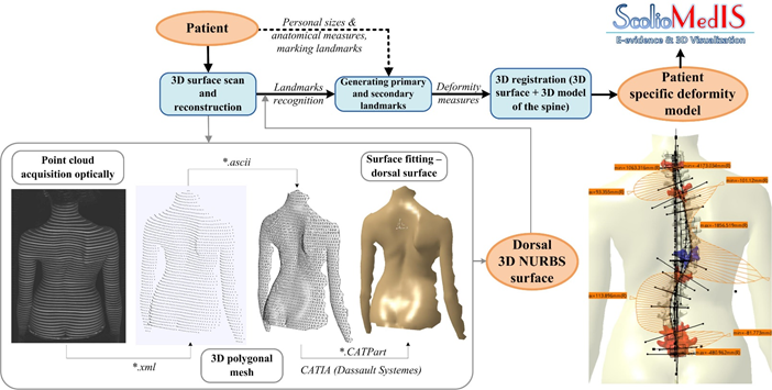
The dorsal side of 372 adolescent individuals (231 females, 141 males) suffering from postural insufficiencies but not yet diagnosed with AIS, were optically scanned using structured-light in a contactless and markerless manner. After 3D image acquisition and point-clouds processing, we applied KAx technologies to simulate the 3D spinal topology, generating over 100 deformity indicators (Fig.1). 3D visualization of the patient-specific deformity model is a result of 3D registration of generic spinal model developed in Mimics and CATIA v5 and a dorsal surface.

Uncaptioned visual

Fig. 1: General methodology: Optical non-invasive 3D diagnosis of AIS

Results
The proposed solution generates internal and external indicators that describe the 3D nature of the deformity and dorsal surface topography, and elucidate clinical patterns allowing rapid, reliable, and accurate diagnosis. After statistical analysis, the most frequent type of scoliosis in the female group was dextro-convex (n=158, 68.4% of cases), while in the male group, the ratio of dextro-convex vs sinistro-convex scoliosis was equal. The mean Cobb angle of the primary deformity was 25.5±12.1° in 215 females and 19.9±8.9° in 118 males, and for the secondary curve was 20.6±8.8° in 183 females and 17.5±7.1° in 76 males. The prevalence of the main curve type was lumbar (13.0%), thoracolumbar (23.8%) and thoracic (63.2%) in females, and 15.6%, 33.3% and 51.1% in males, respectively.

Discussion
Despite controversies surrounding optical methods, our innovative solution is capable of evaluating disorders with adequate precision, thereby decreasing the need for repetitive radiography. We are planning its final validation towards implementation in clinical environment and a new 3D scoliosis classification scheme. This method is embedded in the information system ScolioMedIS, unique in clinical practice, appropriate for both local and global monitoring of scoliosis [3].

Acknowledgements
MPNTR-RS-III41007, Swiss_SERI_Excellence_Scholarship, BioEMIS-530423-TEMPUS.

References
[1] Lukovic, T., et.al., (2015). J Back Musculoskelet Rehabil, 28(4), p721-730.
[2] Ćuković, S., et.al., (2015). J Prod Eng, 18(2), p103-106.
[3] ScolioMedIS-V2.0, http://www.scoliomedis.mfkg.rs.


P1197 A Framework for Cervical Spine FE Model Development and Validation

Maxim Van den Abbeele, Pierre Coloma, Sebastien Laporte, Baptiste Sandoz, Dominique Bonneau, Cedric Barrey, Wafa Skalli
Arts et Metiers ParisTech, Institut de Biomecanique Humaine Georges Charpak, Paris, France

Abstract

Introduction

One often refers to finite element (FE) models to better understand spinal biomechanics and related issues. Moreover, their use in a clinical and industrial context becomes increasingly popular. However, their utility is determined by the level of validation. The scientific community and regulatory agencies proposed recommendations aiming to standardize the development process (1,2). Given the complex morphology and the high inter-subject variability, cervical spine FE model generation remains strenuous. This study aims to propose a framework for cervical spine FE model development and validation, illustrated with a generic C4-C7 model.     

Materials and Methods

The model consists of four cervical vertebrae (C4-C7), the intervertebral discs (IVD) and the spinal ligaments. Geometry was obtained from the averaged CT-based 3D reconstruction of six human cadaveric samples (61±4y, 4♂). Osseous elements, nucleus and annulus matrix were meshed with hexahedral elements, while tension-only cable elements represented ligaments and IVD fibers (figure 1a). Material properties were extracted from the literature and assumed elastic and isotropic. The properties of the annulus matrix, the reinforcing fibers and the ligaments were adjusted by minimizing the difference with the experimental moment load vs. angular displacement curves, obtained from in-vitro experiments performed on the same six samples. Flexion/extension, lateral bending and axial rotation motions were simulated under the application of a pure moment of 2Nm.   

Indirect validation was achieved by comparing the simulated ranges of motion (RoM) with the literature data. The model behavior was evaluated against a different experimental dataset (3), which allowed confirming its biofidelity.

Results

The typical non-linear load-displacement behavior was reproduced (figure 1b) and consistent with the experimental behavior and with literature data in all configurations. Mean RMSE for the principal motions with the in-vitro data was 0.7° (± 0.3°) and 0.7° (± 0.6°) with the supplementary dataset (3). Slightly higher RMSE values were noted for coupled motions.

Uncaptioned visualDiscussion

This study proposed the following framework for FE model development and validation: mesh generation is based on medical image data, material properties are obtained from literature and adjusted with experimental data and validation is performed with a different dataset. Note that it is important to choose appropriate parameters to define validation. Indeed, the RoM might correspond with literature and/or in-vitro data, while the load-displacement curves lack the typical non-linear trend. Furthermore, validation can merely confirm the model biofidelity in the tested configurations. Each modification requires re-validation.

Acknowledgements

The authors thank the BiomecAM chair program, financed by Societe Generale, Covea and Proteor, for supporting this study.

References

  1. Erdemir et al. (2012), J. Biomech., 46(4), p625-633
  2. Viceconti et al. (2005), Clin. Biomech., 20(5), p451-454
  3. Barrey et al. (2015), Eur. J. Orthop. Surg., 25(suppl. 1) p155-165

P1198 The next step in routine creation of subject-specific models: A hybrid radiograph-based musculoskeletal model.

MSc, Eng. Thomas Overbergh1,2, MSc Pieter Severijns1,3, MD Lieven Moke4,2, Dr. Mariska Wesseling5, Prof. Ilse Jonkers5, Prof. Lennart Scheys1,4
1Institute for Orthopaedic Research and Training (IORT), Pellenberg, Belgium. 2Department of Regeneration and Development, K.U. Leuven, Leuven, Belgium. 3Department of Rehabilitation Sciences, K.U. Leuven, Leuven, Belgium. 4University Hospitals Leuven, Leuven, Belgium. 5Department of Biomedical Kinesiology, K.U. Leuven, Leuven, Belgium

Abstract

Introduction

The importance of subject-specificity in musculoskeletal modeling and simulations of movement is continuously increasing towards more realistic functional biomechanical characteristics, a fortiori in pathologic cases. The added value of using a subject-specific (SS) musculoskeletal model (MSM) created from CT or MRI, opposed to the use of generic models, has been frequently demonstrated. However, the fact that existing workflows1 require constructing new models from scratch makes that increasing levels of subject-specificity typically lead to increasing (time) costs and required levels of expertise. Additionally, the use of motion analysis in e.g. the field of spinal deformities, has been complicated by difficult accessibility of through-skin palpable vertebral landmarks for marker placement, leading to erroneous marker-based scaling, static initialization and dynamic tracking of the spine2. Until now, this impeded their routine clinical use.

Methods

This work proposes a user-friendly application to introduce subject-specificity to any generic OpenSim3 model, based on state-of-the-art biplanar images. These images (EOS System, Paris) are taken at low radiation and in an upright, weight-bearing position. Application of the Direct Linear Transform Principle4 on both spatially calibrated images allows making 3D reconstructions. Iteratively matching the 2D projections of bone geometry on both radiograph planes, allows subject-specific redefinition of the bony geometry and intervertebral joints. Furthermore, the software allows radiograph-based definition of virtual markers relative to the underlying bones, eliminating marker placement errors. As an example, markers were attached to a spinal deformity subject’s (male, 54y, 82 kg) skin, whereafter stereo-radiographic images were acquired.

Results

Personalization of the spinal alignment was accomplished (Fig.1A: 2D bone/radiograph-overlay) in less than 1 hour, thereby eliminating the erroneous marker-based scaling, with the generic model by Bruno5 as a starting point. The identification of markers as stereo corresponding points (Fig.1B), allows to individualize the virtual marker placement, preventing palpation error from negatively affecting calculated kinematics6.

Discussion

Creating hybrid SS models using stereo-radiographic images, using the developed software, offers advantages in terms of time cost, marker-placement accuracy and resemblance to weight-bearing conditions. We presented a use case where the spinal alignment of a generic model was personalized based on the radiographic images of a spinal deformity patient. This tool allows to modify generic models, and thus create hybrid models, in a routine and user friendly way.

References

1Valente G., Comput Methods Programs Biomed.,2017;152:85-92
2Overbergh T., Accepted Asbstract ORS2018.(in press)
3Delp SL., IEEE Trans Biomed Eng.,2007;54(11):1940-1950
4Abdel-Aziz YI., Photogramm Eng Remote Sens.,2015;81(2):103-107
5Bruno AG., J Biomech Eng.,2015;137(8):81003
6Osis ST., PLoS One.,2016;11(1):1-13

Uncaptioned visual

Figure 1: Screenshot of the user interface. For optimal visibility, SS vertebrae were used, segmented from standard-of-care CT.


P1199 Age does not influence erector spinae muscle activity during treadmill walking and running in healthy adults.

Dr Stephanie Valentin1, Professor Theresia Licka2,3
1School of Science and Sport, Institute for Clinical Exercise & Health Science, University of the West of Scotland, Hamilton, United Kingdom. 2Equine Clinic, University of Veterinary Medicine Vienna, Vienna, Austria. 3Large Animal Hospital, Royal (Dick) School of Veterinary Studies, University of Edinburgh, Roslyn, United Kingdom

Abstract

Introduction: Changes in motor control patterns [1] and muscle synergies [2] are known to occur with ageing. Although the activation of trunk musculature during walking and running has been evaluated previously [3], the effect of ageing on muscle activity during locomotion has primarily focussed on the lower limb muscles. Therefore, the purpose of this study was to report activation of the spinal extensor muscles during walking and running in a group of young and a group of mature male and female healthy adults.

Methods: Twelve young (age 18-25 years) and 12 mature (age 45-60 years) adults were recruited. Kinematic data were collected using a 10-camera motion capture system and surface electromyography (sEMG) was obtained from the Erector Spinae longissimus muscle (left and right sides). sEMG and kinematic data were collected synchronously during treadmill walk and run (three trials of 10s each). sEMG data were full-wave rectified, de-meaned and re-sampled to match kinematic data. A 4th order 20Hz low pass Butterworth filter was applied to obtain linear envelopes. sEMG data were normalised to the dynamic peak EMG. Data were cut into motion cycles and maximum, minimum, range, and mean normalised sEMG amplitude were obtained and the two groups compared using independent t-tests.

Results: Mean, maximum and minimum muscle activity was greater in the mature group, but no significant differences were found.

Table 1 - Average (± standard deviation) of the Maximum, minimum, range, and mean normalised erector spinae muscle activity (%)

Gait Muscle side Group Maximum (%) Minimum (%) Range (%) Mean (%)
walk left young 75.03
(± 8.73)
36.23
(± 14.19)
38.80 (±15.41) 53.18 (±11.14)
    mature  79.51
(± 4.84)
45.05 (±13.22) 34.46 (±14.54) 61.11
(±7.64)
    p-value 0.071 0.536 0.710 0.257
  right young 78.74
(±7.10)
45.06 (±11.99) 33.68 (±11.23) 59.94 (±8.14)
    mature 83.26
(±5.07)
45.54
(±9.17)
37.73 (±10.46) 63.84 (±7.15)
    p-value 0.293 0.149 0.931 0.485
run left young 77.68
(±8.42)
34.25 (±16.29) 43.44 (±17.11) 55.77 (±9.46)
    mature 84.23
(±5.35)
48.70 (±16.42) 35.53 (±16.79) 66.23 (±8.89)
    p-value 0.222 0.894 0.925 0.624
  right young 79.18
(±8.32)
36.17 (±14.76) 43.01 (±16.70) 57.69 (±8.81)
    mature 83.47
(± 6.67)
41.21 (±14.91) 42.26 (±16.68) 62.18 (±8.01)
    p-value 0.416 0.833 0.869 0.881


Discussion:
Changes in muscle activity often observed with ageing were not identified in the spinal extensors in healthy mature adults in this study. Future work should assess whether this finding applies to adults older than 65 years.

Acknowledgements: Austrian Science Fund (P24020).

References

[1] Kanekar et al. (2014)  Exp Brain Res. 232: 1127-36.

[2] Wang et al (2015) J Neuroeng Rehabil. 12:10.

[3] Cappellini et al. (2006) J Neurophysiol. 95: 3426-3437.


P1200 The Role of the Facet Capsular Ligament in Guiding the L4-L5 Motion Segment

Emily Bermel, Dr. Victor Barocas, Dr. Arin Ellingson
University of Minnesota, Minneapolis, USA

Abstract

Introduction: The lumbar facet capsular ligament (FCL) has been considered a source of low back pain (LBP), which affects up to 80% of adults in the United States. One primary cause of LBP is the degeneration of the spine – both the disc and ligaments. A useful tool for quantification and simulation of the lumbar spine is finite element (FE) modeling. The objective of this study was to determine how the FCL failure alters the complex motion of the lumbar motion segment. We hypothesized that the failure of the FCL would result in abnormal motion profiles with altered arthrokinematics (facet joint space).


Methods: A previously validated 3D FE model[1] of the L4-L5 motion segment (Figure 1A) was used to simulate pure bending of 15Nm, representative of heavy lifting, during flexion/extension, lateral bending, and axial rotation. To investigate the affect the FCL has on the spinal motion the Young’s modulus was diminished from the previously validated properties[1] for healthy FCLs to a value considered negligible. The motion of the model was analyzed by calculating the helical axes of motion, the range of motion (ROM), the bending stiffness, the facet joint space (Figure 1B), and the FCL stretch (Figure 1C).


Results: In general, the FCL guides the motion of the facets during flexion, extension, lateral bending, and axial rotation. The minimum distance maps (Figure 1B) show that during flexion the FCL doesn’t have a big impact, but during extension the FCLs constrain the joint during motion and do not allow the surfaces to slide past each other. During lateral bending and axial rotation similar surface slippage was seen in the model without the FCL. The FCL stretch plots (Figure 1C) show that there is an asymmetry between the left and right FCLs, which is most likely related to the slight asymmetry of the model. Flexion caused about 100% stretch in the FCLs and extension caused a 150% stretch of the right FCL. Lateral bending and axial rotation both show a higher stretch in the ipsilateral FCL. However, during axial rotation the contralateral facet became a pivot point during the motion and the contralateral FCL shows a higher stretch then during lateral bending.


Conclusions: The presence of the FCL stabilizes the facet joint and helps guide spinal motion. Removal of the FCL noticeably changed the motion of the facet joints as seen by the change in the joint space and the FCL stretch.


References: 1. Ellingson AM +. Comput Methods Biomech BME, 2016.

Uncaptioned visual


P1201 Estimating spine loading using an advanced full-body musculoskeletal model

Ms. Wei Wang1,2, Mr. Thomas Overbergh3, Mr. Pieter Severijns3, Dr. Mariska Wesseling1, Dr. Lieven Moke3, Dr. Friedl De Groote1, Dr. Lennart Scheys3, Dr. Ilse Jonkers1
1KU Leuven, Leuven, Belgium. 2Shanghai Jiao Tong University, Shanghai, China. 3University Hospitals Leuven, Leuven, Belgium

Abstract

Introduction

Musculoskeletal modelling offers an efficient and valuable approach to analyse human biomechanics. However, the spine has been investigated to a limited extend due to its complexity. Recently, several spine models were introduced [1-2], which were validated based on simulating motions of previously published in vivo experiments. Currently no study used a full-body musculoskeletal model to estimate subject-specific spinal kinetics during realistic functional motions. The present work combines an integrated, novel spine model with a workflow to determine spine loading.

Methods

A full-body model, extensively described in [3], consisting of 85 segments, 243 degrees of freedom and 636 muscle lines of action was used. For pilot investigation, one healthy subject performed gait at self-selected speed, spine flexion/extension and sitting, while marker trajectories (VICON, UK), ground reaction forces (AMTI, US) and surface electromyography (Zerowire, Cometa) were measured. In OpenSim, scaling, inverse kinematics, static optimization and joint reaction analysis were used to determine intervertebral loading. Specifically, five asymmetrical clusters as well as seven anatomical markers positioned on spine were used for the spine scaling. Intradiscal pressure was estimated based on [1], where the pressure is related to the compressive loading and vertebral cross-sectional area. Muscle activations were compared to the corresponding EMG signals, and were normalized to the maximal value in the motion cycle.

Results

Compressive loading, normalized to the percentage of neutral standing, showed a good agreement with in vivo data measured at L1-l2 (normal gait: 189% vs. 199%, spine bending: 339% vs. 329%, sitting: 113% vs. 124% for the present results and in vivo data respectively) [4] and L4-L5 (spine bending at 30°: 380% vs. 357%) [5]. The pattern and magnitude of the intervertebral disc pressure during spine flexion/extension was comparable to [4-5] (Figure 1). The estimated spinal muscle activations also resembled the EMG signals during gait.

Discussion

Lumbar spine loading was comparable to previously reported in vivo datasets and EMG measurements. Using the musculoskeletal model presented, spine loading can be estimated for functional motions, allowing validation based on EMG measurements for different motions and subjects without invasive methods. No study had used realistic experiment data to drive and validate a spine musculoskeletal model, thus factors in reality like the coupled motions during spine bending might be neglected. In future research, the model will be further improved, by incorporating bushing elements to simulate the function of joint stiffness and ligaments.
Uncaptioned visual

Reference

1. Bruno+, (2015). J Biomech Eng.

2. Christophy +, (2012). Biomech Model Mechan.

3. Overbergh+, (2018). ORS Annual Meeting

4. Rohlmann +, (2008). Spine.

5. Takahashi +, (2006). Spine.


P1202 Experimental Testing and Computational Analysis of Fusion and Non-Fusion Spinal Implants

Mary Foltz1, Andrew Freeman1, Joan Bechtold1,2, Victor Barocas1, Arin Ellingson1, David Polly1
1University of Minnesota, Minneapolis, USA. 2Excelen Center for Bone & Research and Education, Minneapolis, USA

Abstract

Introduction
Severe scoliosis deformities require spinal implants to correct the curvature.  Posterior fusion implants are used in individuals with fully matured spines, while non-fusion (i.e., growing-rods) implants are used in children with growing spines.  High failure rates are associated with non-fusion implants, particularly near the rod-connector interface, which may be due to the different setup required for non-fusion constructs (i.e., the inclusion of the side-by-side connectors)1.  The ASTM F1717 standard has been utilized to quantify the mechanical performance of spinal fusion implants; an equivalent standard for non-fusion implants does not exist.  Hence, the purpose of this study was to evaluate mechanical performance for fusion and non-fusion implants, and develop and validate the respective computational models.

Methods
Mechanical performance of fusion and non-fusion implants was evaluated per ASTM F1717 recommendations (n=5/group) using polyaxial pedicle screws, set screws, rods, and test blocks; with the addition of connectors for non-fusion implants.  Pilot holes placement in non-fusion blocks were modified by an offset to incorporate connectors; placed at the mid-point between superior and inferior blocks.  Experimental test conditions were computationally simulated with ABAQUS/CAE; material properties were determined from ASTM recommendations and opensource material databases, with isotropic linear-elastic properties for blocks and isotropic linear elastic-plastic properties for the spinal instrumentation.  Mechanical characteristics – stiffness, yield strength, and maximum strength – were recorded from experimental testing and computational simulations.  Comparison between experimental constructs was done using unpaired Student’s t-tests.  Validation was determined when mechanical performance of the simulation was within 10% of experimental test.

Results
Non-fusion implants were significantly stiffer than fusion implants at 48±2N/mm and 43±0.3N/mm, respectively.  Non-fusion implants had a significantly greater maximum strength compared to fusion implants with 1080±9N and 1007±6N, respectively.  No statistical difference was observed between the yield strength of the non-fusion and fusion implants: 475±60N and 457±17N, respectively.  Fusion and non-fusion simulations were within 10% of the experimental for stiffness and maximum strength.  However, the yield strength of the simulations deviated from experimental tests.  Maximum stress occurred at the apex of the fusion implants and at the superior rod-connector interaction for non-fusion implants.

Discussion
Stiffness and maximum strength were in agreement between the experimental and computational analysis for both implant types.  Maximum stress locaiton in the non-fusion implants was consistent with common clinical rod failure, near the rod-connector interaction.  Proper utilization of computational simulations provides the ability to observe stress concentrations across the instrumentation to better understand failure mechanisms.

Acknowledgements
Thanks to Medtronic, Inc. for donating hardware and the Minnesota Supercomputing Institute.  Support from NIH/NICHD (K12HD073945) and the University of Minnesota Foundation.

References
[1] Bess S et al., J Bone Jt. Surg. 2010.

Uncaptioned visual


P1204 Improved biomechanical testing for nucleus augmentation devices

Ms Ruth Coe, Dr James Warren, Dr Danielle Miles, Dr Sebastien Sikora, Prof Ruth Wilcox
University of Leeds, Leeds, United Kingdom

Abstract

Introduction
Intervertebral disc (IVD) degeneration is one of the major causes of back pain. One potential therapy is nucleus augmentation, in which a biomaterial is inserted or injected into the degenerated nucleus to restore disc height. However a number of devices have failed or been withdrawn at clinical trial, and there remains an issue in being able to evaluate such treatments at an early stage in their development pathway. Preclinical testing of nucleus augmentation devices is challenging due to the need to replicate the interactions with the surrounding tissues that occur in-vivo. The aim of this work was to develop improved in-vitro testing methods to enable new therapeutic approaches to nucleus augmentation to be examined pre-clinically.

Our previous static testing on extracted IVDs have shown large inter-specimen variation in the measured stiffness when specimen hydration and fluid flow were not well controlled [1]. In this work, a method of normalising the hydration state of IVDs prior-to and during compressive testing was developed.

Methods
Excised adult bovine IVDs (n = 6) underwent water-pik treatment and a 24-hour agitated bath in monosodium citrate solution to maximise fluid mobility. Specimens were submerged in a saline bath and held under constant pressure for 24 hours, after which the rate of change of displacement was low. Specimens were cyclically loaded over 100 cycles, from which the specimen stiffness was determined. The specimens then underwent an enzymatic degradation process to simulate disc degeneration, followed by a further testing cycle. Control specimens (n = 6) were also examined in which repeated testing cycles were undertaken without degradation.  

Results
Compared to previous static tests, the improved method reduced the variation in the specimen stiffness, and enabled longitudinal testing of the same specimen before and after interventions (mean difference in stiffness in control group between tests <10%). In addition, statistically significant differences were seen before and after enzymatic degradation to simulate degeneration, thus providing controls against which to evaluate treatments (Figure 1).

Discussion
The new method provides an approach to evaluate the efficacy of nucleus augmentation over cyclic loading against statistically different positive and negative controls. It is now being applied to examine the biomechanical efficacy of hydrogel nucleus augmentation.


[1] Sikora et al, Journal of Engineering in Medicine, in press. 

Uncaptioned visual


P1206 The response of micromechanical cancellous bone model on the load model simulating the walking

Dr. Ing. Zdenka Sant1, Ms. Louise Mifsud2,1, Mr. Carl Muscat1,3
1University of Malta, Msida, Malta. 2Imperial College London, London, United Kingdom. 3University of Strathclyde, Glasgow, United Kingdom

Abstract

Introduction
The effect of daily life activity (DLA) cannot be measured directly on the bone therefore a widely accepted computational simulation is used to obtain the insight of bone behaviour. The quality of results generated via simulation is highly dependent on the created model of bone material, load model, and applied boundary conditions. The DLA can have various forms but the walking is a common activity for all people, concurrently the kinematical data is easy to measure. This paper focuses on the biomechanical response of a cancellous bone structure of the vertebrae during walk.

Methods
The CT scan images were segmented to create model of the lumbar spine segment. The finite element (FE) model of the cancellous bone L3 vertebrae was created as a micromechanical model [1] derived from CT scans, while the remaining vertebrae retained isotropic model of cancellous and cortical bone with uniform thickness. The adopted generic model with markers, in the open software AnyBody (AB), was optimized to fit the anthropometric data recorded in C3D database. The load model simulating forces and moments acting on the vertebrae during the walk was generated and transferred to the FE model of spinal segment.

Results
The computational results of the load model based on AB were compared with simulation results based on published data [2] acting at the centre of the nucleus of superior intervertebral disc (IVD). The significant difference of the response to each load models (AB and IVD) shows the reduction of displacement of endplate from 4.62 mm due IVD load to 0.13 mm by the AB load. Further analysis compared the behaviour of isotropic and micromechanical material model of L3 vertebra showing the variation of stress intensity from 0.008 to 0.172 MPa, and 0.017 to 0.676 MPa respectively as shown in the diagram ‘Comparison of stress intensity variation among the segment L3-L4 with varying material and load models’.

Discussion
The major disparity of the results originates from the load distribution; 84 loaded points exported from AnyBody in contrast to IVD load at a single point. The analysis of the stress and strain intensity demonstrates the effect of the two load models, suggesting that the IVD load predicts nearly doubled stresses compare to AB load.
Uncaptioned visual

Acknowledgements
Work was supported by Radiology at MDH Malta with UREC permission ENG02/2014, and the cooperation between Malta and Vienna as an outgrowth of COST Action MP1005, NAMABIO.

References
[1] Blanchard, R., Dejaco, A., Bongaers, E. & Hellmich C. (2013) Intravoxel bone micromechanics for microCT-based FE simulations. Journal of Biomechanics, 46(15), pp. 2710-2721

[2] Arjmand, N. and Shirazi-Adl, A. (2006) Model and in vivo studies on human trunk load partitioning and stability in isometric forward flexions. Journal of Biomechanics, 39, pp. 510-521 


P1207 Internal fixation of spinal metastasis: a comparative finite element analysis of a new technique

Ph.D. Luigi La Barbera1, M.D. Alessandro Cianfoni2,3, M.Eng. Andrea Ferrari1, M.D. Daniela Distefano2, M.D. Giuseppe Bonaldi4, Ph.D. Tomaso Villa1,5
1Politecnico di Milano, Milano, Italy. 2Dept. of Neuroradiology, Neurocenter of Southern Switzerland, Lugano, Switzerland. 3Dept. of Neuroradiology, Inselspital, UniBern, Bern, Switzerland. 4Neuroradiology, Pope John XXIII Hospital, Bergamo, Italy. 5IRCCS Galeazzi Orthopaedic Institute, Milano, Italy

Abstract

The evolution of tumour metastasis often involves the formation of lithic regions within the vertebral bone, where the healthy tissues degenerate towards a fibrotic structure. Invasive posterior external fixation represents the first surgical option to shield the metastatic vertebra, otherwise highly exposed to collapse. A new internal fixation technique has been recently proposed, based on vertebral body stents, bone cement injection to prevent vertebral collapse and a monolateral cannulated pedicle screw anchoring the anterior column to the neural arch.
The first aim of the current study is to investigate how the biomechanics of an intact spine segment is altered due to the presence of a lytic defect. The second aim is to compare whether the new internal fixation technique can restore the strain on the surrounding bony structures compared to traditional posterior fixation.
A validated L1-S1 finite element model (Ottardi, 2016) was loaded to simulate standing (Rohlmann, 2012). The lithic defect was described in L3 as a low-modulus region, involving different bony structures: 2/3 of the middle part of the vertebra, 100% of the trabecular bone with and without total cortical bone disruption. The new internal fixation technique with monolateral pedicle screw and bipedicular bone cement injection at L3 was simulated considering two cement filling (7 and 20ml) and compared with a rigid posterior fixation with pedicle screws at L2 and L4. The axial stiffness of the lithic vertebra was calculated to evaluate the capability of each technique in providing adequate stability. The principal strains distribution on the surrounding bony structures (endplates, posterior and anterior walls) were analysed.

The intact vertebra exhibits an axial stiffness of 12.8kN/mm and the strains remain relatively low on every bony structure (range: -0.01 to 0.01%). A lithic defect involving 100% of the trabecular volume significantly decreases the stiffness of L3 vertebra to 3.3kN/mm; while the strains significantly increase on the endplates (range: -0.08 to 0.08%), on the posterior wall (-0.07 to 0.04%), as well as on the anterior cortical wall (-0.04 to 0.22%). The disruption of the cortical shell produces a dramatic loss of vertebral stability (287N/mm) with a general strain increase beyond the bone strength (0.8%).
The new internal fixation technique effectively restores the axial stiffness of L3, especially with the higher cement injection (7.8 and 33.9kN/mm, respectively with 7 and 20ml); posterior fixation shields the lytic vertebra, resulting in a lower axial displacement than with the internal fixation. The new technique is effective in reducing the strains on every structure (-0.03 to 0.02%), more than posterior fixation alone (range: -0.06 to 0.15%).
The present study provides a solid rationale to support the usage of the new internal fixation technique for the treatment of highly unstable lithic vertebrae.


P1208 A parametric study of pedicle screws to improve its mechanical performance in lumbar spine instrumentation

Dr. Ching-Lung Tai1,2, Dr. Po-Liang Lai2, Mr. Mu-Yi Liu1, Dr. Lih-Huei Chen2
1Graduate Institute of Medical Mechatronics, Chang Gung University, Taoyuan, Taiwan. 2Department of Orthopedic Surgery, Bone and Joint Research Center, Chang Gung Memorial Hospital, Taoyuan, Taiwan

Abstract

Background: Robust fixation of pedicle screws remains challenge. Previous studies have demonstrated that numerous factors such as screw types (perforation or expansion), screw designs (thread, thread pitch, shaft diameter) are related to screw anchoring strength. However, literatures addressing the influence of screw design including outer/inner diameter shape (cylindrical/ cylindrical, cylindrical/conical, conical/conical) and thread type (square or V-thread) on the screw fixation strength is lacking. This study aims to explore the mechanical performance of the aforementioned screws in two-fold: (1) Effect of bone quality on screw fixation strength; (2) Association among pilot-hole size, screw insertion torque and screw fixation strength.

Methods: Two types of synthetic bones (test blocks, 20 pcf and 30 pcf) were used to mimic human vertebrae with different bone qualities. Six kinds of pedicle screws were recruited. These include three different outer/inner diameter shape (conical/conical, cylindrical/conical and cylindrical/cylindrical) equipped with two different thread types (square or V-thread). Pilot hole in diameter of 2.7mm, 3.2mm and 3.7mm were prepared for screw insertion. During screw insertion, the insertion torque was measured using a torque gauge. Following specimen preparation, screw pullout tests were then conducted using MTS testing machine.

Results: (1). Regardless of bone quality, V-shape groups have higher pullout strength as compared to square-shape groups. Additionally, V-type pedicle screws with cylindrical/conical shape exhibited the highest pullout strength among groups (p < 0.05). (2). No significant difference was found between thread type (V- or Square) and insertion torque. (3). Regardless of screw types, a smaller pilot hole let to an increase in insertion torque and pullout strength. (4). Enlargement of pilot hole significantly reduced pullout strength, particularly for screw with a 3.7 mm pilot hole.

Conclusion: We concluded that pedicle screws presented the most robust fixation under preparation with combination of V-thread, cylindrical/conical projected shape, smaller size of pilot hole and higher bone density.


P1209 Inverse dynamic analysis of postural reactions to lateral perturbations in seated humans

Yushi Mabuchi, Naomichi Ogihara
Keio University, Yokohama, Japan

Abstract

Introduction

When a vehicle moves on a curved path, makes a lane change, or passes each other the oncoming vehicle at high speed, the seated driver and passengers are exposed to substantial lateral perturbation or acceleration. This acceleration makes steering difficult for the driver and causes discomfort for the passengers because the vehicle is pushed sideway and compensatory body movements should be generated to cope with the perturbation. The aim of the present study was to biomechanically investigate innate postural movement and successive reactions of the trunk segments in seated humans due to lateral perturbations. This would hopefully provide a fundamental basis for designing a vehicle seat that can possibly improve the safety and comfort of the driver and passengers against lateral accelerations.

Method

Five adult male participants sat on a stool placed on a movable platform that was made to translate in the lateral direction by linear actuators. Ramp-and-hold perturbations (displacement = 0.256 m, peak velocity = 0.60 m/s, peak acceleration = 4.5 m/s2) were presented to the seated participants and three-dimensional kinematics of the trunk and pelvis segments with respect to the stool were measured using a motion capture system. To investigate possible muscle activities in response to the sideways acceleration, we calculated forces generated by trunk muscles based on an inverse dynamic analysis using a three-dimensional musculoskeletal model of the human trunk.

Results and Discussion

When exposed to a lateral perturbation towards right, the trunk and pelvis segments were tilted towards left because the upper part of the body tends to remain stationary due to its inertia. However, we found that the thorax was simultaneously tilted forward and contralaterally rotated in response to the perturbation. This complex movement of the trunk probably occurs due to the innate mobility of the vertebral column and the position of the center of mass of the thorax with respect to the vertebral column.

Due to the lateral perturbation, the right external and internal oblique muscles and the left external oblique muscle was found to be activated approx. 0.2 sec after the onset of the platform movement, in order to cope with the lateral tilt and the contralateral rotation of the thorax segment, respectively, and recover postural balance. Such characteristic movements of the trunk segments and the successive postural reactions of the external and internal oblique muscles occurring due to the lateral acceleration should be taken into account when designing a vehicle seat.


P1210 Accounting for task-related active muscle force distribution patterns in a FE model of the neck

Mr. Bertrand FRECHEDE, M. Maamir
Univ Lyon, Université Claude Bernard Lyon 1, IFSTTAR, LBMC UMR_T9406, F69622, Lyon, France

Abstract

Introduction
This abstract presents the ongoing development of a 3D active musculoskeletal FE model of the neck in the FE code LS-Dyna. It focuses on the evaluation of a single 3D active muscle model and on its implementation to model the combined action of the 29 muscle pairs of the full neck model.

Methods
Previous stages of the development and validation of the FE neck model were presented earlier2. Two methods had been evaluated to develop a single active 3D FE muscle model: a Hill based model (LS-Dyna *MAT_MUSCLE) and a thermomechanical based control model5. In both cases the single FE muscle model consisted of 1D active fibers embedded in a 3D matrix of passive hexahedral elements. The Hill model was chosen to model each muscle of the neck model and two test-conditions were then simulated: (i) simulations1 of an axial loading of the head were reproduced, without and with pre-contraction of the neck muscles (Cf. Figure). The second set of simulations (ii) reproduced experimental horizontal head-loading scenarios on volunteers reported by Siegmund et al.4. In both cases normalized muscle activation patterns provided by the authors were used as input for up to 20 different muscles in the FE model. Missing data were extrapolated from a Rigid-Body modelling approach3.

Uncaptioned visual

Results
Both muscle models allowed modelling the passive lengthening and active contraction (shortening and lateral swelling) of the muscle-tendon complex. The normalized isometric contraction force behavior6 was reproduced by the model after calibration, together with a transverse stiffening effect of the muscle. Simulations could be performed fully for case (i) and in sagittal loading conditions for case (ii), showing (i) a significant effect of the muscles’ contraction on the cervical spine loads and (ii) an influence of the muscles activation patterns on these loads.

Discussion
At this stage contacts were only implemented between muscles and not between muscles and the spine, thus precluding a full investigation of transverse stiffening effects. Also, all loading directions could not be simulated owing to stability issues in some of the loading directions. Nevertheless, as further verification and validation are in progress, results of these simulations strongly promote that active 3D muscle models will improve the predictive capabilities of spinal loads distributions for in-silico virtual testing.

References
1Chancey, V C et al. 2003. Stapp Car Crash J 47 (Paper 2003-22-0008):135‑53.
2Howley, S, et al. 2014. CMBBE 17(S1):74‑75.
3Lamouri, P and Fréchède, B. 2013. CMBBE 16(S1):170‑71.
4Siegmund, G P, et al. 2007. J Biomech Eng 129 (1):66‑77.
5Stelletta, J, et al. 2017. In Biomechanics of Living Organs, Y Payan and J Ohayon Eds, 497‑521.
6Zajac, F, 1989. Crit Rev Biomed Eng 17(4):359‑441.


P1211 Uniplanar pedicle screw for vertebral derotation of scoliosis - a biomechanical study

Dr. Po-Liang Lai1, Mr. Po-Yi Liu2, Dr. Chun-Li Lin2
1Department of Orthopedic Surgery, Chang Gung Memorial Hospital, Taoyuan, Taiwan. 2Department of Biomedical Engineering, National Yang-Ming University, Taipei, Taiwan

Abstract

Introduction: Direct vertebral derotation (DVD) and rod derotation are two major maneuvers used for surgical correction of scoliosis. Polyaxial pedicle screws provide more degrees of freedom on the screw-to-rod connecting interface to facilitate easier rod seating. However, this design may lead to inadequate apical vertebral reduction when performing the DVD maneuver. In contrast, monoaxial screws are immobile at the screw head and are thus superior for performing the DVD maneuver to reduce vertebral rotation. However, monoaxial screws have a potential disadvantage regarding the difficulty of seating the rod into the screw head. This study investigates the efficiency of a newly designed uniplanar screw to that of monoaxial and polyaxial screws in the DVD maneuver.


Methods: Six T7-T13 porcine thoracic spine segments were used as test specimens in this study. Pedicle screws were inserted in the left pedicles of the T9-T11 spinal segments and then connected with a rod. Three types of pedicle screws with different screw head designs (monoaxial, polyaxial and uniplanar) were employed in this study. The material testing system (MTS) machine generated a rotational moment through the derotational tube on the T10 (apical body) pedicle screw, which simulated the motion applied during the surgical vertebral derotational procedure. The kinematics of the vertebral body and derotational tube were recorded to evaluate the derotational efficiency of different pedicle screw head designs.

Result: For the monoaxial, polyaxial and uniplanar screws, the variances of the derotation for the monoaxial, polyaxial and uniplanar screws were 2.22° ± 1.43°, 32.23° ± 2.26°, and 4.75° ± 1.60°, respectively; the derotation efficiency was 0.65, 0.51, and 0.12, respectively, when the torques of the spinal constructs reached 3 Nm. The rotational variance of the polyaxial screw was statistically greater than that of the monoaxial and uniplanar screws (p < 0.05).

Conclusions: The screw head design played an important role in the efficiency and variance of the derotation during the DVD maneuver. The derotational efficiency of the newly designed uniplanar screw was closer to that of the monoaxial screw group than to that of the polyaxial screw group. The polyaxial screw was inferior for DVD due to a derotational variance between the derotational tube and the apical body that was correlated with the range of motion of the screw head.


Keywords: pedicle screw, scoliosis, uniplanar screw, vertebral derotation, vertebral translation


P1212 Using subject-specific 3D reconstructions of the spine in adolescent idiopathic scoliosis in order to understand the relationship between the deformity in the 3 planes

Dr Ayman Assi1,2, Mr Mohammad Karam1, Dr Claudio Vergari2, Mr Joeffroy Otayek1, Mr Aren Joe Bizdikian1, Mr Fares Yared1, Ms Nour Khalil1, Dr Anthony Kassab1, Dr Cyril Hanna1, Dr Ziad Bakouny1, Prof Ismat Ghanem1, Prof Wafa Skalli2
1Faculty of Medicine, University of Saint-Joseph, Beirut, Lebanon. 2Institut de Biomécanique Humaine Georges Charpak, Arts et Métiers ParisTech, Paris, France

Abstract

Introduction

Scoliosis is a 3D deformity of the spine, usually assessed on 2D frontal and sagittal radiographs. Surgeons tend to focus on correcting the scoliotic deformities in the frontal and sagittal planes during surgery. Although it is often neglected, the transverse plane is also affected by this deformity, and its relationship to the frontal and sagittal planes is unexplained. The EOS® biplanar X-ray technique with low dose of radiation, performed in standing position, allows for subject-specific 3D spinal reconstructions. The aim was to investigate the spinal deformity in the transverse plane in Adolescent Idiopathic Scoliosis (AIS) patients, with different types of curvatures, and its relationship to the frontal and sagittal deformities.

Methods

AIS patients who had undergone pre-operative EOS® biplanar X-rays were included. Usual spino-pelvic parameters as well as Cobb angle, apical vertebra rotation (AVR), intervertebral axial rotation at the upper and lower junction (UIAR, LIAR), regional kyphosis (RK:curvature magnitude of the scoliotic segment in its local sagittal plane, (+)kyphosis, (-)lordosis) and torsion index of the scoliotic segment (TI) were extracted from 3D spinal reconstructions [1]. Patients were divided into 3 groups depending on the type of scoliotic curvature (Thoracic:T-group, Thoracolumbar:TL-group, and Lumbar:L-group) [2]. Scoliosis parameters were compared between the 3 groups. In order to investigate the determinants of the deformity in the transverse plane, a stepwise ANCOVA was computed with TI as a dependent variable, and demographics, spino-pelvic and scoliosis parameters as independent variables. A linear regression model was then fitted between the 3 planes deformity.

Results

229 patients were enrolled (192F; age: 14.6±2.5years; Cobb: 38±20° [10-110°]) and classified as follows: T-group:N=117; TL-group:N=74; L-group:N=38. Cobb angle and TI were significantly higher in the T-group compared to other groups (T-group:44° vs. TL-group:34° vs. L-group:26°, p<0.001; T-group:15° vs. TL-group:8° vs. L-group:5°, p<0.001, respectively). RK was significantly different between the groups (p<0.001): T-group:13° vs. TL-group:-13° vs. L-group:-25°. A significant ANCOVA model (adjusted-R2=0.72; p<0.001) showed that TI was associated with Cobb angle (β=0.928, p<0.001) and RK (β=0.162, p<0.001). A significant linear regression model (p<0.001, figure1) was found to best fit the relationship between TI and Cobb angle (R2=0.63), as well as RK (R2=0.23).

Discussion

This is the first study to analyze the relationships between the deformity in the 3 planes in AIS patients with different types of curvatures. Subjects with major thoracic curvatures generally had an increased deformity in the frontal and transverse planes (Cobb angle and TI). TI was found to be highly associated and to increase with increasing Cobb angle and RK.

References 

[1]Skalli,Spine,2017; [2]Lenke,JBJS,2001


Uncaptioned visual

P1213 Differences between a box lift and full forward bending in the spine and hip angles using a multi-segmental kinematic model in healthy people

MSc Stefan Seerden1, Prof. Dr. Wim Dankaerts1, MSc Thijs Swinnen1,2, Prof. Dr. Kurt De Vlam2, Prof. Dr. Rene Westhovens2,1, Prof. Dr. Benedicte Vanwanseele1
1KU Leuven, Leuven, Belgium. 2UZ Leuven, Leuven, Belgium

Abstract

Introduction
Clinical evaluation of the spine and hips is common in musculoskeletal therapies, such as physical rehabilitation/medicine, physiotherapy clinics [1]. Spinal impairments are commonly only evaluated by practitioners via general thoracic or lumbar range of motion (RoM) tasks, however multiple thoracic and lumbar regions could show different movement patterns during these evaluation tasks [1-3]. The purpose of this study was to explore the difference in sagittal kinematics in five spinal regions, the trunk as a whole and the hips between a maximal forward bending of the spine and a functional box lifting task.

Methods

Motion capture analysis was done with 18 healthy subjects. Vicon Nexus 2.4 with 10 motion capture cameras and Vicon Bodybuilder 3.6.1 software were used for analysis. A full body plug-in-gait marker-set was adapted with addition of 6 clustermarkers on the L5, L3, L1, T10, T6, and T2 spinous processes, representing Lumbosacral, Lower Lumbar, Upper Lumbar,  Lower Thoracic, Mid Thoracic and Upper Thoracic spinal kinematics [4,5]. ROM was analysed for full forward bending with straight legs and a boxlift task (6kg). A repeated measures ANOVA was done as statistical analysis with SPSS 20.

Results
RoM in the box lifting task was less compared to forward bending, for all thoracic and lumbar regions and in total trunk however larger RoM was seen in the box lifting task compared to forward bending for hips and L5/S1 due to anatomically more possible posterior pelvic-tilt and knee flexion during box lift.

Table 1: Range of Motion, percentage of box lift compared to full forward flexion task and repeated measured ANOVA significant differences between both tasks for each anatomical region.
https://s3.amazonaws.com/oxabs-file-hosting-production-us-west-2-submission-images/stage_id=123-checksum=1bafe556ef263f116fcde90519a3792e53f1c254
PiG: Plug-in-Gait, RoM: Range of Motion, SD: Standard Deviation, ANOVA: repeated measures ANOVA test, significant values are marked with an asterisk (*).

Discussion
Contrasting to previous studies [3,6], the current results show differences between box lifting and forward bending kinematics in Upper- and Lower Lumbar spine. Previous research worked with lower lifting load (3 kg vs. 6 kg in current study), which could lead to lifting more from the back instead of the lower extremities. Next to that, differently defined upper lumbar (L1-L3) and Lower lumbar spine (L3-L5) and most of all separating L5/S1 in the current study compared to T12-L3 and L3-S1 for upper- and lower lumbar spine respectively in previous studies could be the reason for finding lumbar differences between box lifting and forward bending in the current study.

References
[1] Dankaerts, Spine. 2006;31:698-704. [2] Leardini, Clin Biomech. 2011; 26:562-571. [3] Parkinson, Manual Ther. 2013; 18:390-394. [4] Seerden, Gait & Pos. 2016; 49S:53. [5] Needham, Prosthet Orthot Int. 2015; 40:624-635. [6] Williams, Manual Ther. 2013; 18:130-135.


P1214 Muscle Forces controlling the Lumbar Spine during Flexion/Extension – an Experimental Study

Dr Daniel Baumgartner, BSc. Bruno Schmid, MSc. Roman Kuster
Zurich University of Applied Sciences ZHAW, Wintertur, Switzerland

Abstract

Introduction: The estimation of trunk muscle forces to stabilise the lumbar spine have only been partially evaluated. EMG measurements of the intramuscular activity is difficult to perform as well as a validation by 3D virtual models simulating abdominal and back muscles. An experimental model allows to display the force reaction  of specific trunk muscles to stabilise an external weight. Additionally, back implants for surgery may be tested by an experimental design.

The aim of the study was to built an experimental trunk muscle simulator which stabilises a lumbar spine including 4 intervertebral discs during flexion/extension.

Material & Methods: An artificial lumbar spine including 3D-printed vertebrae from S1 to L1 with homogenous, moulded silicone discs of specific shore hardness was built. Illiopsoas and abdominal muscles were used as agonists to flex the spine, longissimus and intraspinal muscles were used in an antagonistic setting applying a constant resistant force. The simulator acted vice versa for extension.

Flexion/Extension were measured up to a RoM of +30°/-15° for a given upper body weight of 20 kg (50% BW) in upright position. Muscle Loads were measured with load cells, intradiscal load with a 6 DoF sensor (Kistler AG Switzerland) in L3.
In order to control a semi-stable system including 5 discs with variable stiffness properties, PID controllers (NI, USA) were used in a force controlled way  [Humbert et al. 2002].

Results: Full continuously performed flexion extension cycles were evaluated. Resulting muscle force characteristics were evaluated. The measured muscle forces for the protagonists (ventral muscles) showed a maximum of about 325N - 430N for flexion movement whereas the antagonists (dorsal muscles) showed a maximum of about 95N - 130N. The force for extension movement showed about 65N to 70N (ventrally) and 50N – 75N (dorsally).

Discussion : The simulator is able to display acting muscle forces in realtime for a substantial speed (5 sec. for neutral position till full flexion).

First tests showed that flexion / extension movements of the spine were reproducibly performed for the first 3 cycles with a subsequent  viscoelastic bending of the pre-shaped artificial spine. The higher forces for the flexion in comparison to extension may therefore be explained by the predefined lumbar lordosis curvature in neutral position and continuous buckling effect from the lumbar spine due to the upper body load. To optimise the reproducibility, a feedback system of the current spinal position is necessary to control the lumbar posture.

Limitations : the present device provides realistic muscle forces for a given external load under dynamic motion and may be used for the testing of spinal implants. Nevertheless, additional important trunk muscles need to be considered such as m. transversus and intraspinal muscles such as the m. multifidii.


P1215 A comparison of methods to quantify control of the spine

Mr Eric Bourdon1, Dr Jaap van Dieën2, Dr Ryan Graham1
1University of Ottawa, Ottawa, Canada. 2Vrije Universiteit, Amsterdam, Netherlands

Abstract

Introduction

Low back pain (LBP) has been shown to affect motor control of the spine, while it is known that adequate control of one’s spine is required to safely bear loads and reduce injury risk1. This has led to the development of different motor control quantification techniques; however, it is unknown if these techniques are obtaining similar outcomes. Local dynamic stability (LDS) is a popular approach that uses kinematic data and nonlinear analysis techniques to assess trunk control2. Another popular method uses systems identification (SI) techniques to assess trunk motor control by measuring an individual’s response to perturbations applied to the trunk3. While both methods have inherent advantages and disadvantages, it is unknown if these analysis techniques observe similar results. The purpose of this study was to compare outcomes of LDS and SI to understand the relationship between these variables.

Methods

15 healthy participants (9M and 6F) were recruited to participate in this study. Participants completed two tasks (A and B) in a random order. For task A, participants were seated and ventrally perturbed with a pseudorandom force signal at the level of the 10th thoracic vertebrae by a magnetically-driven linear actuator. Participants were instructed to maximally resist perturbations (resist condition) or to relax but maintain an upright posture (relax condition). Admittance was represented as a frequency response function that describes the actuator displacement as a function of the contact force3. Task B involved participants completing 35 cycles of a repetitive trunk flexion/extension task. 3D lumbar spine angles of the last 30 movement cycles were collected and used to calculate the maximum finite-time Lyapunov exponent (λmax)2. Pearson linear correlations were used to assess the relationship between λmax and admittance gain at frequencies < 1Hz.


Results

At present, data for 6 out of 15 subjects have been analyzed. There is a weak negative linear correlation between admittance gain at frequencies < 1Hz in the relax condition and λmax (r = -0.368). There is a weak positive linear correlation between admittance gain at frequencies < 1Hz in the resist condition and λmax (r = 0.372).   


Discussion

Understanding the relationship between LDS and SI outcomes is integral to interpretation of data. These outcomes, although frequently compared within the literature, demonstrate a weak linear correlation. Therefore, this weak correlation should be considered when comparing literature. Additional analysis of these data will include more participants and explore nonlinear relations. This will lead to improved comparison of literature and methods could be used in concert to exploit certain advantages.


Acknowledgements

NSERC


References

1. Reeves et al.,2007.Clin Biomech,22(3),266-274.

2. Graham et al.,2012.J Biomech,45(9),1593-1600.

3. van Drunen et al.,2013.J Biomech,46(8),1440-1446.


Uncaptioned visual


P1216 Assessment of the biodynamic response of human body exposed to whole-body vibration using apparent mass and muscle activation level

Ms Suzan Cansel DOGRU1, Dr Yi QIU2, Dr Richard COLLIER3, Dr Yunus Ziya ARSLAN1
1Department of Mechanical Engineering, Faculty of Engineering, Istanbul University, Istanbul, Turkey. 2Human Factors Research Unit, Institute of Sound and Vibration Research, University of Southampton, Southampton, United Kingdom. 3Physical and Rehabilitation Health Programme, Faculty of Health Sciences, University of Southampton, Southampton, United Kingdom

Abstract

Introduction

During exposure of seated human body to vertical whole-body vibration, muscles are activated to maintain the stability of the musculoskeletal system in response to the perturbation of the vibration. Vibration transmitted to body may cause functional and structural degenerations on spinal region, especially at the lumbar region.[1] Electromyography (EMG) signals and apparent mass (APMS) can be used to reflect the fundamental biodynamic responses of the human body under vibration. The objective of this experimental study was to investigate the relationship between the APMS and EMG signals, if any, which can provide further insight regarding the evaluation of the degenerative effects of whole-body vibration on the spine of seated participants.


Methods

Twelve subjects participated in the experiments (ethical approval was obtained from University of Southampton). Postures of the seated subjects were normal upright on a rigid seat with hands in lap without the backrest contact, wearing a loose lap belt. Subjects’ feet were supported from an adjustable footrest.  Sinusoidal random vibrations in vertical direction with duration of 30-second, magnitudes of 0.5, 1.0 and 2.0ms-2 root mean square (rms) and frequency range between 0.5 and 20Hz were applied to the subjects through the rigid seat. A 60-second resting period was given before each trial.

Acceleration data at the seat base were recorded using accelerometers (SiliconDesigns2260-005), while the forces in the vertical direction were simultaneously recorded at the rigid seat pan using a force plate (Kistler9281B). Muscle activity was measured from longissimus thoracis between T6 and T10, iliocostalis lumborum between T11 and L2, and lumbar multifidus between L2 and L5 using 16-channel array EMG electrode (EMG-USB128, LISiN–OT,Bioelettronica). EMG signals during the maximum voluntary contractions (MVC) of the corresponding muscles were also recorded without vibration affect based on the Biering-Sorensen test motion.


Results

The calculated APMSs were normalized with APMS value at 0.5 Hz. The rms value of muscle activation data for all applied frequency were calculated and normalized with MVC.  Fig.1. shows the APMS and muscle activation for all magnitudes (A,B and C) and all muscles (D,E and F).


Discussion

It was found from Fig.1 (A, B and C) that the activity of spinal muscles increased when the APMS increased and the muscle activation increased with increasing the magnitudes of excitation. Fig.1 (D, E and F) indicated that during the excitation, the multifidus and iliocostalis were more affected than the longissimus.


Acknowledgments

This study was supported by the Research Fund of Istanbul University [grant number FDK-2016-21712] and by the Scientific and Technological Research Council of Turkey [1059B141501146].


References

[1] Dupuis H, Zerlett G. Whole-body vibration and disorders of the spine. Int Arch Occup Environ Health 1987;59:323-336.
Uncaptioned visual


P1217 Evaluation of Biomechanics of A Novel Elastomer Lumbar Total Disc Replacement:A Finite Element Study

Dr. Ali Kiapour1, Dr. Joseph M. Zavatsky2, Dr. Vijay Goel1
1Engineering Center for Orthopaedic Research Excellence, Department of Bioengineering, The University of Toledo, Toledo, USA. 2Spine & Scoliosis Specialists, Tampa, USA

Abstract

INTRODUCTION: Lumbar total disc replacement (TDR) devices are intended to preserve motion in the surgically treated segment, in an attempt to eliminate many of the shortcomings of traditional fusion techniques. Currently available TDR instrumentation does preserve motion in the treated spinal segment, but this motion does not exactly mimic natural spinal kinematics. This study investigates the biomechanics of novel polymer disc design using the finite element model.

METHODS: An experimentally-validated FE model of lumbar spinal model was used and modified to simulate placement of FREEDOM total disc replacement device at L4-L5 level following removal of entire nucleus and ALL ligament and partial removal of annulus. Properties of titanium alloy and CarboSil20-80A silicone rubber were assigned to the endplates and implant’s flexible component. The intact and instrumented spines were subjected to 400N compressive follower load plus 10Nm bending moments in anatomical planes (flexion-extension, left-right bending, left-right rotation). The range of motion, intra-discal pressure and facet loads were compared for intact and instrumented spines.

RESULTS SECTION: The kinematic data for index and superior adjacent levels were calculated. At the index level, the instrumented model had range of motion equal to 88% and 80% of intact in extension and flexion motions, respectively.  The ratios were 62% in left and right bending, and 86% in axial rotation. The extension-to-flexion center of rotation (COR) of the intact FE was very close to the in vivo COR reported by Pearcy et al. (Spine 1988). The motion at the superior adjacent segment increased slightly, following instrumentation at L4-L5. The increase in motion was not greater than 8% in flexion, and 20% in extension. The intra-discal pressure at the adjacent segment was very close in the instrumented and intact model with the difference not being greater than 12% in any of the loadings. Compared to the intact spine under extension loading, the loads at the facet joints at the index level increased by 1% in the instrumented model. However, in the instrumented model, adjacent level facet loads decreased approximately 5% with extension, compared to the intact spine.

DISCUSSION: This elastomer TDR did not result in significant alterations in spinal kinematics, quality of motion and biomechanics of the instrumented spine, as compared to the intact spine model.  Rohlmann et al. (Spine 2005) showed that ProDisc-L increased the ROM of the spine by 38% in Extension and decreased by 40% in Flexion. The elastomer disc resulted in change of motion not greater than 20% of intact in either motion. Also the elastomer disc had minimal effect on range of motion, intradiscal pressure and facet joint of at L3-L4 segment which is an indicative of lower risk of adjacent segment disc and facet degeneration in the long run.


P1218 The effect of the dimension of reproduced metastases on the vertebrae strain distribution

PhD Marco Palanca1, Mrs Maria Luisa Ruspi1, Mr Giovanni Barbanti-Bròdano2, PhD Luca Cristofolini1
1Dept of Industrial Eng., School of Engineering and Architecture, Alma Mater Studiorum - Università di Bologna, Bologna, Italy. 2Department of Oncological and Degenerative Spine Surgery, Rizzoli Orthopedic Institute, Bologna, Italy

Abstract

Introduction
The vertebral metastases are an increasing problem for the healthcare due to the increasing risk of fracture.  A series of classifications[1]guide the clinician to manage the metastasis but, due to a series of complications, this problem is not fully solved. Identifying the critical dimension and location of the metastasis beyond which the spine becomes instable is, today again, a challenge for clinicians and biomechanicists. 

The aim of this work is to define the effect of the dimension of reproduced lytic metastases on the strain distribution on human vertebra in the location where metastases are more frequent.

Methods
Five human spines were obtained from an ethically-approved donation program (IIAM,Jessup,PA,USA). To investigate the effect of a metastasis in L4, three-vertebra segments(L3-L5)were tested.  The soft tissues were removed and a white-on-black speckle pattern was prepared on its surface to evaluate the strain field with Digital Image Correlation[2].

The spine segment was aligned and potted in poly-methyl-methacrylate and loaded with an anteriorly-offset force. Each specimen was tested first with L4 intact; then a defect was drilled in L4 through both pedicles in order to reproduce lytic metastasis.  Defects were 30mm deep, and the diameter was increased from 6mm to 12mm in 2mm increments, (this corresponded to 25%-50% of the vertebral body volume)[3].  Tests were performed on each specimen in the elastic regime up to 1 body weight for each level of defect.  The maximum and minimum principal strains were measured on the anterior side of the vertebral body of L4 with DIC(Dantec-Dynamics,DE).

Results
The tensile strains were larger than the compressive strains(Fig.1).  The max principal strains were similar for intact L4 and with defects of 6mm. In case of defects of 8 and 10mm, the strains increased but were in the range associated to physiological loads (<2000 microstrain). Finally, for defects of 12mm, the strains exceeded 3500microstrain in front of the defect.

Discussion
The strain distributions were successfully evaluated in all conditions and the results confirmed that the larger the metastatic defects, the weaker the vertebra and, then, the higher the strains.  In particular, an alarming scenario was the one with metastatic defects larger then 12mm: the maximum principal strains were larger than the typical physiological strains.  This study suggests that the tensile stress maybe is responsible for the fracture of metastatic vertebra.  Moreover, it provides a first quantification of full-field strain related to metastatic defects.

References
[1]      Fisher,C.G., et al.,2010,Spine,35(22),pp.1221–1229.
[2]      Palanca,M., Marco,M., Ruspi,M.L., and Cristofolini,L.,MEP,in press.
[3]      Alkalay,R.N. and Harrigan,T.P.,2016,J.Orthop.Res.,34(10),pp.1808–1819

Uncaptioned visual


P1219 Development of a Parametric Subject-Specific Finite Element Model to Investigate the Lumbar Spine Response before and after One-Level Posterior Fusion

Professor Kinda Khalaf1, Dr. Mohammad Nikkhoo2, Dr. Marwan ElRich1, Dr. Zahra Khoz2, Dr. Ehsan Ghobadih3, Dr. Chi-Chien Niu4, Dr. Chih-Hsiu Cheng5
1Khalifa University, Abu Dhabi, UAE. 2Azad University, Tehran, Iran, Islamic Republic of. 3Bone and Joint Research Center, Chang Gung Memorial Hospital, Linkou, Taiwan. 4Department of Orthopaedic Surgery, Chang Gung Memorial Hospital, Linkou, Taiwan. 5Physical Therapy and Graduate Institute of Rehabilitation Science, College of Medicine, Chang Gung University, Taoyuan, Taiwan

Abstract


Introduction

Finite element (FE) modeling has emerged in the past two decades as a useful tool to study spinal biomechanics. Its unique capability to noninvasively evaluate stresses in structures of complex geometry, loading, and material behavior makes it quite advantageous for exploring the intricate biomechanics of the human spine. Although many existing FE models have the potential to yield clinically relevant results, they are typically time-consuming and constrained to the input geometry, where inter-subject variability may not be considered [1]. The objective of the current study was twofold: first to develop and validate a robust parametric subject-specific FE model that automatically updates the geometry of the lumbar spine for different individuals; and second to investigate its clinical applicability by studying the motion patterns and spinal biomechanics in pre and post 1-level lumbar posterior fusion patients.

Materials and Methods

The FE model, including the typical vertebrae (L1-L5), IVDs, facet joints, and ligaments, was developed using ABAQUS. The geometry of the lumbar spine (L1-L5) was automatically updated by inputting new values of independent parameters based on defined geometrical conditions and constraints obtained from X-Ray images (Fig. 1A). The validity of the model was evaluated using pure moments of 1 to10 N.m, subsequent to a compressive preloading with a follower load of 500 N mimicking the upper body weight. The results of the intersegmental range of motion (ROM) and intradiscal pressure (IDP) were compared to experimental studies from literature [2, 3]. The FE model’s clinical applicability was validated by using a case study of two patients pre and 3 month post-surgery (Fig. 1B). The lumbar spine models of the patients were simulated under the same loading and boundary conditions and the results were analyzed.

Results

The ROM results in the intact FE models were consistent with available experimental data in literature [2, 3]. The total ROM decreased by 28.93% and 34.28% for patient #1 and patient #2, respectively. Upon fusion, the average Von Mises stress in the adjacent level (L3-L4) significantly increased for both patients (Fig. 1C).

Uncaptioned visual

Figure 1. (A) Procedure of the parametric FE modeling of the lumbar spine, (B) Updated geometry of the FE model for patient #1  (Before surgery and 3M Post-Op), (C) Increased stress in the adjacent level for the patients after fusion.

Discussion

The present model provides an opportunity for clinicians to use quantitative data towards subject-specific surgical planning and informed rehabilitation. Ongoing and future work includes expanding the studied population to investigate individuals with different spinal disorders.

References

[1] Campbell et al, (2016), J Biomechanics, 49 (13) p 2593-9.

[2] Panjabi et al., (1994), J Bone Joint Surg., 76 p 413-24.

[3] Heuer et al., (2007), J Biomechanics, 40 (2) p 271-80.


P1220 Kyphoplasty vs Stentoplasty: FE analysis of the risk of cement leakage

Kévin Aubert1, Dr Tanguy Vendeuvre1,2, Adrien Lanel1, Dr Valéry Morgenthaler3, Dr Michel Rochette3, Dr Paul Brossard4, Dr Jean-Baptiste Pic4, Dr Simon Teyssédou2, Pr Philippe Rigoard1,2, Dr Arnaud Germaneau1
1Institut Pprime UPR 3346, CNRS – Université de Poitiers – ISAE-ENSMA, Poitiers, France. 2Department of Neurosurgery, Spine & Neuromodulation Functional Unit, Prismatics Lab, CHU, Poitiers, Poitiers, France. 3ANSYS, Villeurbanne, France. 4Department of Orthopaedic Surgery and Traumatology, CHU Poitiers, Poitiers, France

Abstract

Introduction

Kyphoplasty and stentoplasty are two minimally invasive surgery methods used for the reduction and the stabilisation of spine fractures. The principle of kyphoplasty consists in the restoration of the vertebral body anatomy by balloon expansion after a compression fracture [1]. Reinforcement of the anterior vertebral column is obtained from PMMA cement filling. Recently, the vertebral body stenting has been developed to reduce the loss of vertebral height after balloons removal. The objective of this work was to develop a Finite Element (FE) model to analyse mechanical effects linked to injection of cement and leakage risks linked to both techniques.

Materials and methods

A FE model was built from a demonstrator geometry (Figure 1 a-b) simulating a cavity involved by balloon augmentation where the cement has to be injected. Fracture was modelled by a space corresponding to a separation of vertebral body (cancellous bone and cortical wall), giving the possibility to the cement to flow along frontal or sagittal directions. FE model simulated two cases: with or without stent. Simulation of fluid injection was performed with a flow speed of 5mL/min and a cement with a viscosity of 20kPa.s, corresponding to the PMMA viscosity 10min after mixing [2].

Results

Figure 1 (c-d) shows simulation results of velocity flow with or without stent. Presence of stent modified orientation of fluid kinematics: flow was slowed down according lateral directions and was guided along anterior direction. Without stent, cement flow did not have specific orientation and diffused along lateral and anterior directions.

Uncaptioned visual

Discussion

This numerical analysis showed differences in cement flow following injection. The contribution of stent was studied showing a tendency to slow down the flow according to lateral directions. For simulation without stent, cement diffused in the fracture area along lateral or anterior directions that can involve a higher risk of leakage. This preliminary analysis confirmed clinical observations revealed trough a monocentric, prospective and continuous study performed on 60 patients (mean age: 47.3 years) during three years [3]. This clinical study highlighted a higher rate of cement leakage for specimens stabilized by kyphoplasty. To enrich the clinical/simulation comparison, the present work has to be completed with dynamic simulation of injection flow to analyse parameters linked to the cement behaviour (viscosity) or to the geometry of a real fracture following bone augmentation.

Acknowledgements

The authors would like to thank ANSYS.

References

[1] Oner, R., et al (2005), Injury, 36, pp. 82-89.

[2] Baroud, G., et al (2006), Spine, 31(22), pp. 2562-8.

[3] Vendeuvre, T., et al (2017), GRIBOI Meeting, Athens, Greece.


12:00 - 12:20

O0387 Non-linear dynamics of the intervertebral disc

Prof. Stephen Ferguson
ETH Zurich, Zurich, Switzerland

Abstract

Previously we have shown that trauma to the intervertebral disc can lead to disc degeneration [1]. Furthermore, we have found that impact induces high-frequency oscillations within the disc [2]. Accurate predictions of the dynamic response of the intervertebral disc to impact and vibration are therefore highly relevant for studies of disc injury, degeneration and ergonomics. Various models have been developed to investigate the spine's response to whole body vibration. However, these models assume a linear-elastic behavior of the intervertebral disc. Recently, we have reported on experimental measures of the unique nonlinear dynamic behavior of the human lumbar intervertebral disc [3]. This response was shown to be dependent on the applied preload and amplitude of the stimuli, and we hypothesize that this cannot be described by linear-elastic models.
Simulation models have been implemented, with the quasi-static, asymmetric tension-compression behavior of the disc described with a polynomial function, fitting both directions independently while maintaining continuity across the transition. The strain-rate-dependent dissipative properties of the disc are included. Our prior base-excitation experiments of human discs were simulated with the model for validation, over a frequency sweep from 1 – 60 Hz and 60 – 1 Hz.  The model with the new material description was compared to a model with conventional linear elastic material properties. For further insight, results of a base-excitation experiment were compared to a conventional dynamic compression experiment.
Only the model considering the fully asymmetric non-linear tension-compression properties captured the dynamic characteristics observed in our prior experiments, such as material softening or hardening, and most importantly, a non-uniform "jump" phenomena in the oscillation. Specifically, the disc enters and exits resonance at different frequencies on the upwards and downwards frequency sweep. Quasi-static tension-compression data alone were sufficient to predict full non-linear response. In contrast, a conventional linear elastic model predicted only a single, symmetric resonance point at one characteristic frequency.  Dynamic compression loading failed to demonstrate the non-linear, frequency-dependent response seen in the free vibration experiments.
Linear elastic material models fail to adequately capture the non-linear dynamic response of the disc to vibration. For dynamic analysis, the use of standard linear-elastic models should therefore be avoided, or restricted to study cases where the amplitude of the stimuli is relatively small. For a biofidelic and accurate simulation of the vibrational response of the disc, the tension-compression asymmetry of the disc should be incorporated. Dynamic compression loading is not equivalent to base-excitation loading; therefore results should be interpreted with caution when exploring the effects of vibration on the intervertebral disc.

References

  1. Dudli, S. et al, J. Orthop. Res., 2012, 30(5):809-16
  2. Marini, G. and Ferguson, S.J., Comput Methods Biomech Biomed Engin., 2014, 17(9):1002-11
  3. Marini, G. et al., J. Biomech., 2015, 48(3):479-88

12:20 - 12:40

O0388 Barycentremetry and subject specific spine modeling from biplanar X-Rays

Ms Wafa Skalli
Arts et Métiers ParisTech, Paris, France

Abstract

Spine disorders prevention is difficult because underlying mechanisms are not well understood, and the rate of spine surgery mechanical complications is still high, particularly for adult spine deformities. Subject specific spine modelling could help better understanding of the biomechanichal issues in relation with spine disorders, thus yielding better prevention and treatment, providing it tackles a global view of the spine. Indeed it is essential, although extremely complex, to  consider not only subject specific geometry and material properties, but also the spine loads that apply for each individual and the mechanisms of compensation that occur due to motor control. Low dose biplanar X-Rays system allows acquisition from head to feet in standing position and 3D reconstruction of the skeleton in clinical routine[1]. The analysis of a large set of patients allowed to show a quasi invariant parameter : Odontoid dens (OD), which is close to the head center of gravity, is at the vertical of the middle of the segment joining the two femoral heads, indicating a « head upon the pelvis » (HUP) characteristic of the erect posture [2]. The accurate reconstruction of the external envelope from bilanar X-Rays [3], together with bone segment densities, allowed to perform barycentremetry, i.e. estimation of the barycenter and inertial parameters of each body segment and of the global body, including gravity line without the need for a forceplate [4]. Barycentremetry yields new insights in various compensation strategies to maintain HUP even in presence of local disorders or after surgical correction. This talk will present basic methods for such subject specific modelling and clinical implications for aging people and for scoliotic patients.

  1. Dubousset, Charpak, Skalli et al. Bull Acad Natl Med. 2005 189(2) :287-97.
  2. Amabile et al. Eur Spine J. 2016 (epub ahead of print)
  3. Nerot et al. J Biomech, 2015 45:1-10.
  4. Amabile et al. J Biomech. 2016 49(7):1162-1169.


Acknowledgements To all students, reseachers and clinical partners that participated to the research, and to the ParisTech BiomecAM Chair program on subject specific musculoskeletal modelling.


12:40 - 12:50

O0389 Modelling the Fatigue Properties of Fractured Bovine Vertebrae Treated with Vertebroplasty using Experimental and Finite Element Simulation

Miss Ruth Coe, Dr Sebastien Sikora, Professor David Barton, Professor Ruth Wilcox
University of Leeds, Leeds, United Kingdom

Abstract

Introduction
Osteoporotic vertebral fractures are prevalent in post-menopausal women, and can result in pain and a reduced quality of life. Surgical interventions, such as vertebroplasty, have had varied outcomes with work still needed to optimise materials and procedures. Biomechanical analysis and computational simulation have previously provided insight into the mechanisms of vertebroplasty, however this typically investigated short term responses. Therefore the purpose of this study was to investigate the fatigue response vertebrae treated with vertebroplasty to gain further insight into its efficacy.

Methods
Bovine tail vertebrae were fatigue tested at high loads to provide insight into failure mechanisms, as well as providing data to inform and validate finite element models. Thirty-one vertebrae were loaded axially to 9.5kN or failure. Eleven specimens were then injected with PMMA bone cement, and 20 left untreated. Untreated vertebrae were cyclically tested in four groups at loads of 60%-90% of the initial failure load. Treated specimens were tested at 80% failure load, all were tested up to 10000 cycles or failure.

Specimen-specific finite element models were created from mciroCT data of un-cemented vertebrae. These were given density-based Young’s modulus values and an elastic-perfectly plastic material model was used. An optimisation technique was used to determine yield strain from experimental data. Models were validated against a separate set of experimental data for a static loading case. An iterative approach whereby each element undergoes a reduction in Young’s modulus and yield stress depending on the level of plastic strain in that element was used to model fatigue. The level of material property reduction was determined using equations described by Keaveney et. al (1) calculated from experimental data.

Results
Experimental tests showed a relationship between initial strength and fatigue performance, with significant differences between the 60% load group and other groups (p<0.05) (Figure 1A). No significant difference was seen in fatigue performance between treated and untreated specimens (p>0.05) (Figure 1B), however the augmented group had a smaller reduction in mechanical stiffness. MicroCT scan data provided information about the location and severity of fractures. Finite element models successfully show the plastic strain damage accumulating in the vertebrae over a number of cycles.

Conclusions
This study demonstrates variation in the outcome of vertebroplasty even in a comparatively controlled environment, and shows the difference in response of fractured vertebrae under different levels of loading. Finite element methods show the damage accumulation over a number of cycles and the equations can now be optimised to more accurately predict experimental output. These models can be used to investigate vertebrae treated with bone cement to better understand vertebroplasty treatment mechanisms.


References

1. Keaveny, T.M. et al. Mechanical behavior of human trabecular bone after          overloading. Journal of Orthopaedic Research. 1999, 17(3), pp.346-353.

Uncaptioned visual


12:50 - 13:00

O0390 Functional determination of a cervical spine joint coordinate system via an optimization approach

Dr. Matthew Moran1, Dr. Paul Ullucci, Jr.1, Dr. Frank Tudini2
1Sacred Heart University, Fairfield, USA. 2Campbell University, Buies Creek, USA

Abstract

Determination of a subject-specific fixed cervical spine joint coordinate system (JCS) is challenging due to several anatomical constraints.  Cervical spine motion (flexion/extension, axial rotation, and lateral bending) is the combined contribution from the occipito-atlanto-axial complex and 2nd cervical vertebra through the 1st thoracic vertebra.  Additionally, there is a lack of accessible bony landmarks aligning with rotation axes.  Several methods of quantifying total cervical spine motion from three-dimensional kinematic data have been previously reported, however, none of the JCS determination methods have utilized an optimization approach.  PURPOSE: The purpose of this study was to determine the feasibility and accuracy of a numerical optimization approach in determining subject-specific cervical spine JCS through a series of coordinated movements. METHODS: Twenty-eight participants (15F; 21.4 ± 5.4 yo), free from any musculoskeletal limitations in the cervical spine, granted informed consent and participated in this IRB approved study.  While in a seated position, all participants performed the following sequence of ordered head movements representing their full active neck range of motion (ROM): (1) flexion-extension, (2) axial rotation left-right, (3) lateral flexion left-right.  Five reflective markers (dia.=12.5mm) were rigidly fixated to the head via a tight fitting elastic band and a 4-marker reflective marker cluster was rigidly fixated to the upper thoracic region.  Marker coordinates were measured with an 8-camera three-dimensional motion capture system (Qualisys OQUS 100; 120 Hz) with a residual accuracy < 0.3 mm.  All three-dimensional coordinates were filtered with a 4th order Butterworth lowpass filter with cutoff frequency of 25 Hz.  A custom MATLAB program utilized a Nelder-Mead simplex optimization routine to determine axes parameters that minimized the difference between the measured joint motion and the actual joint motion. Off-axis angle deviations were computed over the entire ordered movement sequence and for each joint rotation direction respectively.  Computed ROMs were compared against published norms from healthy participants [1].  RESULTS:  Averaged across all participants and over all rotations, off-axis angle deviations were 8.1±2.5o.  Off-axis deviations were smallest for flexion-extension rotations (3.5±2.3o) and largest for lateral bending rotations (14.0±5.1o). Aggregate active ROMs compared favorably to previous reports (Flexion 67.7±12.3o, Extension 68.4±12.3o, Rotation Left 74.7±8.7o, Rotation Right 73.5±6.6o, Lat. Bending Left 47.6±7.8o, Lat. Bending Right 46.4±6.2o) DISCUSSION:  Despite anatomical limitations that prevent traditional joint axes determination, the reported methodology adequately fit a set of ordered head motions with an optimized fixed cervical spine joint coordinate system.  An advantage of the current methodology is that body fixed reflective markers do not need to align with either the lab-based coordinate system or a segment coordinate system.        

REFERENCE: 1. Houck et al. Title. Gait & Posture. 1997;5(2):184.

13:00 - 13:10

O0392 The Subject-Specific FE Modeling of the Inferior Cervical Spine

Maxim Van den Abbeele, Pierre Coloma, Sebastien Laporte, Baptiste Sandoz, Dominique Bonneau, Cedric Barrey, Wafa Skalli
Arts et Metiers ParisTech, Institut de Biomecanique Humaine Georges Charpak, Paris, France

Abstract

Introduction

Neck pain, together with lower back pain, is the second most important cause of invalidity in high-income countries (1). Finite element (FE) modeling is widely used, complementary to in-vitro experiments, for the study of spinal biomechanics and related clinical or mechanical issues. However, due to the complex cervical spine morphology and the high inter-subject variability, developing and validating subject-specific models remains a challenge. The aim of this study is to propose and evaluate an FE model of the inferior cervical spine with subject-specific geometry.

Materials and Methods

The load-displacement behavior of five human cadaveric C3-T1 samples (61±4y, 3♂) was recorded in-vitro in flexion/extension, lateral bending and axial rotation. CT-based and biplanar X-ray-based 3D reconstructions were obtained for each sample.

A generic FE model was computed from the averaged reconstruction and composed of two functional spinal units, i.e. C4-C5 and C5-C6. Vertebrae, nucleus and annulus matrix were meshed with hexahedral elements, while tension-only cable elements represented the ligaments and IVD fibers. The elastic and isotropic material properties were derived from the literature and adjusted to minimize the difference with the mean experimental load-displacement curves. For each specimen, subject-specific mesh geometry was obtained by automatically deforming an initial parameterized mesh (2) to match the CT-data, while optimizing mesh quality (figure 1a). Material properties were set equal to those of the generic model. A pure moment of 1.4Nm was applied to simulate the in-vitro tested configurations. A one-to-one comparison between the numerical and experimental curves was performed.

Results

The five FE meshes respected the element quality criteria, with less than 0.2% of the elements beyond the warning limit and none beyond the error limit. The corresponding simulations converged in about 10min for each configuration.

The models predicted the typical non-linear load-displacement behavior and consistent coupled motions. The samples were properly ranked  in terms of range of motion with an average difference of 1.44° (std. 1.07°) (figure 1b). The RMSE between the numerical and experimental curves was on average 0.97° (std. 0.57°). The highest differences were noted for flexion/extension.

Uncaptioned visualDiscussion

This study proposes an experimentally evaluated subject-specific C4-C6 FE model. The model was able to differentiate between samples with varying geometry. The deviation from the experimental data is most likely due to differences in material properties, as these were held constant in each model. Although still preliminary, this model offers a promising basis for the study of spinal biomechanics and for implant design.

Acknowledgements

The authors thank the BiomecAM chair program, financed by Societe Generale, Covea and Proteor, for supporting this study.

References

  1. Institute for Health Metrics and Evaluation, 2017
  2. Laville et al. (2009), J. Biomech., 42(10), p1409-1415

13:10 - 13:20

O0393 Combined musculoskeletal and structural finite element modelling of the lumbar spine

Mr Clément Favier, Prof Alison McGregor, Dr Andrew Phillips
Imperial College London, London, United Kingdom

Abstract

Introduction

Lower back pain (LBP) is a common debilitating condition related to spinal biomechanics and often associated with altered lower limb stability strategies [1]. To investigate this condition, a simulation workflow combining musculoskeletal (MSK) and structural finite element (SFE) modelling techniques is being developed. The study presented here is focused on lifting activities.

Methods

Six healthy male volunteers with no history of back pain were recruited for a high-resolution full-body MRI scan.  Motion capture and electromyography (EMG) signals of a complete range of everyday life activities were also recorded for each subject.

A full body MSK model composed of 566 muscle actuators for the lower limbs and lumbar spine was developed (Figure 1a). The spine is composed of five articulated lumbar vertebrae and a three-segment thoracic and cervical spine. The complete bone geometry, muscle insertions and moment arms are adapted from the MRI scan. Muscle and joint reaction forces for the recorded activities are obtained from MSK simulations and used as loading conditions with the SFE model.

This second model is based on the same bone geometry. At first, cortical bone is modelled with standardised shell elements and trabecular bone with a random network of beam elements (Figure 1b). The structure is then optimised to resist the loading conditions using an adaptation algorithm [2].
Uncaptioned visual

Results

The MSK model has been evaluated for quasi-static movements against in-vivo measurements [3,4,5]. The dynamic assessment using EMG recordings suggests the model is suitable for modelling the lifting tasks. The SFE adaptation method used with a broad range of everyday life activities gives a structural bone architecture in agreement with clinical observations.

MSK simulations are now run for lifting tasks including spine twisting and flexion. These results will quantify the load applied on the spine and describe the muscle recruitment patterns for these activities. Applied to the previously optimised vertebrae, the computational workflow will give an estimation of the spine structures’ deformations for such tasks. The SFE adaptation method is also used here on its own to study how each lifting task can influence trabecular bone architecture.

Discussion

This study will give a first understanding on how different lifting techniques applied in various scenarios can trigger LBP. Future studies will investigate LBP mechanisms by modifying the models in the simulation workflow. Muscle weakness and structural deficiencies will be introduced to simulate altered biomechanics and understand how it can lead to pain. Other activities such as balance recovery following a perturbation will also be investigated.

References

[1] McGregor AH, et al., JBack Musculoskelet Rehabil, 2009.
[2] Phillips ATM, et al., Int Biomech, 2015.
[3] Rohlmann A, et al., Spine, 2008.
[4] Schultz A, et al., JBone Joint Surgery, 1982.
[5] Wilke HJ, et al., Spine, 1999.