To save this page as a PDF, click this button and choose the PDF destination.

Biomechanics of vascular tissue engineering

15:00 - 16:30 Monday, 9th July, 2018

Wicklow MR3

Track Tissue Engineering

Posters for this session are on display on Monday 9th July in the Forum.

Chair: Anne Robertson and Joshua Hutcheson

P1370 Visco-Elasto-Plastic modeling of Small Intestinal Submucosa (SIS) for its application as a vascular graft

Paolo Francisco Sánchez1, Mr Juan Carlos Briceño1,2
1Universidad de los Andes, Bogotá, Colombia. 2Fundación Cardioinfantil Cardioinfantil Cardioinfantil Cardioinfantil Instituto de Instituto de Instituto de Instituto de Cardiología Cardiología Cardiología, Bogotá, Colombia

Abstract

There is a clear necessity for functional vascular grafts to satisfy the high demand caused by cardiovascular disease worldwide. The development of new tissue engineered vascular grafts (TEVGs) has shown promising results because they can overcome the limitations presented by the synthetic materials currently used. TEVGs can provide an environment akin to the host cells as found in native vessels, exemplified by similar conditions such as the mechanical environment. The assessment of the mechanical properties of the material and their evolution once implanted is of utmost importance, because among different causes, compliance mismatch has been considered a determinant of graft failure.

In this project we analyze the dynamic properties of the small intestinal submucosa (SIS), which as collagen-based extracellular matrix, has shown promising results for its use as a vascular graft. Furthermore, we propose and validate a constitutive model to fit the material behavior and use it to simulate the SIS under the conditions found in native vessels.

 A uniaxial creep and recovery test was performed on 5 mm diameter SIS tubes (n=8) immersed in water at 37 °C. All the samples showed creep and plasticity even at smaller loads. A one-dimensional elasto-visco-plastic constitutive model was used to describe the material. It is composed by an elastic element in series to two Kelvin-Voigt solid elements and a plastic slider. The first elastic component was defined using Mooney-Rivlin strain energy function, while the plastic component was defined using a third-degree polynomic function of the plastic stress. The viscoelastic behavior was defined using the creep compliance formulation for the Kelvin-Voigt model. The identification of the material parameters was done using a custom-made minimization code. The parameters for the plastic and non-lineal elastic elements showed a normal behavior, while the spring and dashpot constants of the visco-elastic element had a linear dependence on the load applied. Furthermore, the model was validated comparing its results to a set of different experimental tests.  

Using the constitutive model, the material was simulated as a vascular graft. Assuming thick wall behavior, SIS tubes were simulated under physiological ranges of pressures for 1000 cycles at 60 cycles per minute. Tubes vary in radio and thickness. The simulation showed that the dissipative elements might cause a change in radio and a change in the range of strain between the initial cycle and the steady state, depending on pressure range. From the cases simulated a performance curve chart was obtained in terms of the compliance of the material for its use as a predictive tool of the graft behavior based on its geometry. This study showed the importance of the assessment of the dynamic mechanical properties of TEVGs to avoid possible compliance mismatch failure.


P1371 Fabrication and characterization of an aligned poly(glycerol sebacate) fibrous membrane

Ms Hsin-Ju Wu, Mr. Chia-Chun Lo, Dr. Jin-Jia Hu
National Cheng Kung University, Tainan, Taiwan

Abstract

Although we have successfully fabricated poly(glycerol sebacate) (PGS) fibrous membrane by coaxial electrospinning, it remains PGS prepolymer leak-out issue during the fabrication process. In this study, we tended to reduce the ratio of PEO to PLA in the shell solution or use more intensively crosslinked pPGS. On the other hand, one of the advantages of using fibrous scaffolds in tissue engineering is that cell alignment and hence tissue microstructure may be manipulated by the mechanism of contact guidance. Therefore, we focused on fabricating highly aligned PGS fibrous membrane. The core-shell fiber structure was confirmed by TEM. From the SEM images, we successfully mitigated pPGS leak-out from the inner core. Both of PEO and PLA were removed from the PGS fibrous membrane, which were verified by FTIR and DSC. The alignment index of PGS fibrous membrane was 2.06±0.21 indicating that PGS fibers were highly aligned. From the results of cyclic stretching test, we further affirmed that PLA was removed. PGS fibrous membrane also showed anisotropic mechanical properties examining by uniaxial tensile test. It’s Young’s modulus was 0.52-0.98MPa which was similar to soft tissues. Anisotropic PGS electrospun fibers promoted human umbilical smooth muscle cell guidance. We discovered that the degradation rate of PGS fibrous membrane was slower than PGS solid sheet, indicating that the crosslinking degree of PGS fibrous membrane might be higher than PGS solid sheet. The in vitro cytocompatibility of the electrospun membranes was confirmed. The aligned PGS fibrous membrane has the potential to be used for soft tissue engineering applications.

P1372 Fabrication of an anisotropic poly(glycerol sebacate) tubular scaffold for vascular tissue engineering

Dr. Tai-Hua Yang, Mr. Chen-Yu Li, Dr. Jin-Jia Hu
National Cheng Kung University, Tainan, Taiwan

Abstract

Poly(glycerol sebacate) (PGS) has been shown to be a promising biodegradable elastomer for engineering soft tissues. Fabrication of an anisotropic PGS scaffold, however, remains challenging. In our previous study, we developed a method based on the use of sacrificial fibers to fabricate an anisotropic PGS porous membrane. Briefly, aligned poly(vinyl alcohol) (PVA) membranes were prepared by electrospinning. The membrane was then embedded in PGS prepolymer to form a composite upon drying. After crosslinking of PGS, the embedded PVA fibers were removed by water, leaving numerous cylindrical pores in the PGS membrane. There were two limitations in the previous study, however. First, PVA could react with PGS, leading to incomplete removal of PVA. Second, the PVA electrospun fibers were nano-sized, and hence the resultant pores were in the nanoscale, which not be capable of guiding cell alignment. The purpose of this study was to deal with the two limitations. Polylactide (PLA), which does not react with PGS, was used in replacement of PVA for the preparation of the sacrificial fibers, which was then removed by chloroform upon crosslinking, leaving aligned grooves on the surface of the PGS membrane and cylindrical pores within the membrane. The porous PGS membrane was shown to be mechanically anisotropic. The membrane was not cytotoxic and the grooves on its surface were capable of directing cell alignment via contact guidance. With the same approach, we fabricated a PGS tubular scaffold with uniform wall thickness and examined their mechanical properties. The PGS anisotropic scaffold have potential in the applications of vascular tissue engineering.

P1373 A bioreactor for conditioning tissue engineered heart valves

Francisco Almeida-Gonzalez1,2, Prof. Fergal J. O'Brien2,3,4, Dr. Claire M. Brougham1,2
1School of Mechanical and Design Engineering, Dublin Institute of Technology, Dublin, Ireland. 2Tissue Engineering Research Group, Royal College of Surgeons in Ireland, Dublin, Ireland. 3Advanced Materials and Bioengineering Research (AMBER) Centre, RCSI & TCD, Dublin, Ireland. 4Trinity Centre for Bioengineering, Trinity College Dublin, Dublin, Ireland

Abstract

Introduction

Globally over 100 million people are affected by heart valve (HV) diseases and 300,000 HV replacements are required annually (Namiri et al., 2015). Mechanical and bioprosthetic HVs are unable to remodel, making replacement surgery the final choice for paediatric patients. Tissue engineered heart valves (TEHVs) promise full integration and remodelling in vivo, eliminating paediatric patients’ need for revision surgeries.

Our TE approach combines autologous cells with a fibrin-collagen-glycosaminoglycan scaffold (Brougham et al., 2017, 2015), conditioned using a bioreactor which simulates the biomechanics of the heart. This encourages directional cell proliferation and extracellular matrix development. The ideal bioreactor parameters to create functional TEHVs remain unknown. Our main goal is to create a pulmonary TEHV suitable for in vivo trials and the first milestone is to design a bioreactor capable of conditioning TEHVs.

Design Requirements

The bioreactor must mimic adult and neonate hearts (30-200 bpm, flow of 25-100 ml/beat and pressure drop of 13-25 mmHg throughout the HV). It must be fabricated from non-cytotoxic materials, sterilisable using ethylene oxide and/or autoclaving, allow observation of leaflets, while promoting gas and media exchange and remaining sterile.

Design Concept

In this design (Figure 1), the HV is fixed aseptically inside the pulmonary chamber of the bioreactor. Media is pumped through the HV by displacing a silicone membrane, using a magnetic actuator, secured in a stainless steel base. Silicone tubing will allow displaced media to circulate between polypropylene connectors (a-b). A peristaltic pump will pump media between connectors (c-d). The bioreactor itself will be fabricated from PMMA allowing clear observation of the HV and leaflets. A compliance chamber and a syringe filter will be attached to connector (e) to simulate arterial expansion and promote gas exchange.

  Uncaptioned visual

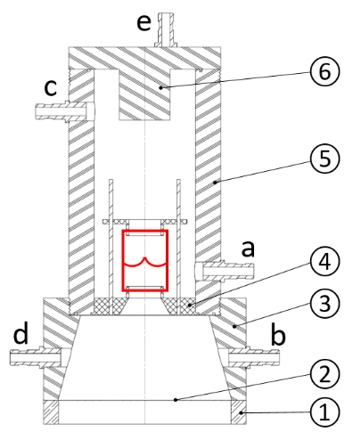
Figure 1.  Bioreactor design showing HV in red. Contains a base (1), silicone membrane (2), ventricular chamber (3), HV support (4), pulmonary chamber (5), lid (6) and connectors (a-e).

Discussion

Our bioreactor is loosely based on the conceptual design proposed by Hoerstrup et al. (2000), and the bioreactor designed by Moreira et al. (2014). Unlike those designs, the ventricular chamber and HV support act as nozzle directing the flow towards the HV. Operating parameters across the HV will be validated using pressure transducers and flow sensors. Cytotoxicity and sterility tests will validate materials selected. In time, this bioreactor will be employed to condition TEHVs using a variety of conditioning regimes.

References

Brougham et al., Advanced Healthcare Materials 6: 21 2017.

Brougham et al., Acta Biomaterialia 26: 205-214, 2015.

Hoerstrup et al., Tissue Engineering 6: 75-79, 2000.

Moreira et al., Tissue Engineering 20: 741-748, 2014.

Namiri et al., Journal of Tissue Engineering and Regenerative Medicine 11(5): 1675-1683, 2017.

Acknowledgments: DIT Dean of Graduate Research School Award 2017-2021


P1374 Stability study of hydrogels to prevent cerebral aneurysms rupture

Oriane Poupart1, Magalie Matray1, Dr. Pierre-Etienne Bourban2, Prof. Dominique P. Pioletti1
1Laboratory of Biomechanical Orthopedics, EPFL, Lausanne, Switzerland. 2Laboratory for Processing of Advanced Composites, EPFL, Lausanne, Switzerland

Abstract

Introduction

Brain aneurysms are a major health concern and affect 3.2 % of the population [1]. Rupture of intracranial aneurysms is the main complication because it leads to subarachnoid hemorrhage, which induces a mortality of 40 to 50 % and a severe morbidity of 10 to 20 % [2]. The two current treatments are open clipping surgery which is highly invasive and endovascular coiling therapy which is only short-term efficient. The use of a photoactivated hydrogel filling the aneurysm cavity has the potential to overcome these drawbacks due to their suitable biological properties and tunable mechanical properties.


Methods

The stability of photoactivated polyacrylamide hydrogels was studied in order to determine if they could be stable during at least one year of life.

Static stability was first tested by submerging hydrogels in PBS and weighing, measuring dimensions change and elastic modulus at different time points.

An aneurysm in vitro model was developed to be able to dynamically test the properties of the hydrogel. PBS flow at 300 mL/min was applied with a peristaltic pump on the model and microscopy images were taken at different time points to observe erosion or cracks formation.

A Wohler curve was also performed in order to estimate the damages caused by fatigue for a number of cycles.


Results

Static results show no statistical difference of dimensions and weight of PAAM hydrogels for 10 days immersion in PBS. The hydrogels did not swell therefore they will not apply additional pressure on the aneurysm wall. Elastic modulus is constant over time and no hysteresis is noticed for 3 cycles.

The aneurysm in vitro model was idealized and consisted of two main parts which can be connected to a peristaltic pump. The first part is the base in PMMA which represents the artery. The second part is the openable aneurysm part in PDMS to be able to assess the properties of the hydrogel.

Discussion

Photoactivated polyacrylamide hydrogels are stable over two weeks in PBS. These results are encouraging and must be performed for a longer period. However, these tests should be done in plasma and serum in order to be closer to blood properties. Others polymers hydrogels might also be tested, such as polyethylene glycol dimethacrylate.

In order to prevent cerebral aneurysms rupture, the hydrogels must also fulfill several requirements, such as adhesion to the aneurysm wall or biocompatibility.


References

[1] Monique H M Vlak et al. “Prevalence of unruptured intracranial aneurysms, with emphasis on sex, age, comorbidity, country, and time period: A systematic review and metaanalysis”. In: The Lancet Neurology 10.7 (2011), pp. 626–636.

[2] Coil embolization for intracranial aneurysms: an evidence-based analysis. Vol. 6. 1. 2006, pp. 1–114.

P1375 The role of mechanical environment in regulating vascular network formation

M.Sc. Fabian Stein, Dr. Nasim Salehi-Nik, Dr. Jeroen Rouwkema
University of Twente, Enschede, Netherlands

Abstract

Introduction

The integration of engineered tissues after implantation is limited due to the lack of a vascular network. When vascular networks are included, they generally are not organized, or lose their initial organization fast1. Due to the ability of cells to sense their environment by mechanotransduction, signaling, maturation, organization and cell survival is regulated. The objective of this study is to analyze the combinatory effect of substrate stiffness and fluid flow on vascular organization and maturation to find the basic parameters for pre-vascularization of engineered tissue.

Methods

A microfluidic PDMS framed system was used, which makes it possible to investigate different hydrogel compositions in parallel. Pre-glycation by D-(-)-Ribose allowed for the use of one type of hydrogel in the same concentration but with different modulated mechanical properties. During the polymerization process of the hydrogels, needles were integrated into the gels to create hollow channels by their removing afterwards. The down and top side of the system were sealed with thin cover glasses to ensure the visibility of the inner system. The fluid-flow channels were coated with 0.1% Gelatin to improve the cell attachment and seeded with Smooth muscle cells. Afterwards Human Umbilical Vein Endothelial Cells (HUVECs) were seeded on top of the smooth muscle cells to mimic the physiological blood vessel structure. An additional channel was filled with VEGF (50 ng/mL), which is known as one of the main angiogenic factors which diffuse into the hydrogel over time2. Different fluid-flow profiles were applied to the cell seeded channels. The newly formed capillary network was analyzed by ImageJ.

Results

The pre-glycation by incubation of different concentrations of D-(-)-Ribose resulted in an increase of stiffness of the same type of hydrogel by additionally crosslinking of the different hydrogels components. The use of different modulated hydrogels allowed for the simultaneous analysis of the effect of fluid flow on the vascular sprouting into the hydrogels triggered by the diffused VEGF. Different mechanical properties in combination with different fluid flow patterns affected the ability of HUVEC to migrate and organize into the hydrogels and show differences in the sprouting morphology.

Outlook

To mimic the physiological state, different Endothelial cell types (e.g. HUVECs, HMECs, HIAEC) will be integrated into the fluid flow channels. This will allow us to see if different endothelial cell origins leads to a different sprouting behavior or if the already described endothelial plasticity leads to similar results.

Acknowledgement

This work is supported by an ERC Consolidator Grant under grant agreement no 724469.

References

  1. Levenberg et. al., (2005). Nat Biotechnol, 23(7): 879-84.
  2. Rivron, N.C.,  et. al., (2012). Proc Natl Acad Sci U S A, 109(18): 6886-91.
  3. Mason, B.M., et. al., (2012). Acta Biomater. 9(1): 4635–4644


P1376 Biomechanical properties assessment of decellularized carotid arteries for blood vessel replacement

Dr. Elena López-Ruiz1,2, Dr. Seshasailam Venkateswaran3, Dr. Macarena Perán2,1, Dr. Gema Jimenez4,2, Dr. Salvatore Pernagallo3, Dr. Juan José Díaz-Monchón5, Dr. Olga Tura-Ceide6, Dr. Francisco Arrebola4, Dr. Juan Melchor4, Mr. Juan Manuel Soto7, Dr. Guillermo Rus4, Dr. Pedro Real5, Dr. Maria Diaz-Ricart8, Dr. Antonio Conde-Gonzalez9, Dr. Mark Bradley10, Dr. Juan Antonio Marchal4,2
1University of Jaén, Jaén, Spain. 2Biopathology and Regenerative Medicine Institute (IBIMER), Centre for Biomedical Research, Granada, Spain. 3School of Chemistry, EaStCHEM, Edinburgh, United Kingdom. 4University of Granada, Granada, Spain. 5Pfizer-Universidad de Granada-Junta de Andalucía Centre forGenomics and OncologicalResearch (GENYO), Granada, Spain. 6University of Barcelona, Barcelona, Spain. 7Complutense University of Madrid, Madrid, Spain. 8Department of Hemotherapy and Hemostasis, Hospital Clinic, Centre de Diagnostic Biomedic (CDB), Institute of Biomedical Research August Pi iSunyer (IDIBAPS),, Barcelona, Spain. 9School of Chemistry, EaStCHEM, University of Edinburgh, Edinbourgh, United Kingdom. 10School of Chemistry, EaStCHEM, University of Edinburgh, Edinburgh, United Kingdom

Abstract

Introduction

Decellularized vascular scaffolds are promising materials for vessel replacements [1,2]. However, despite the natural origin of decellularized vessels, issues such as biomechanical incompatibility, immunogenicity risks and the hazards of thrombus formation, still need to be addressed. In this study, we coated decellularized vessels obtained from porcine carotid arteries with poly (ethylmethacrylate-co-diethylaminoethylacrylate) (8g7) with the purpose of improving endothelial coverage and minimizing platelet attachment while enhancing the mechanical properties of the decellularized vascular scaffolds. Moreover, the coating of decellularized vessels with 8g7 improved biomechanical properties by increasing the ultimate tensile strength and load.

Methods

Samples were divided into three groups: native arteries, decellularized arteries and polymer-coated decellularized arteries. The biomechanical properties of all the groups (n = 3, in each group) were determined by conducting uniaxial tensile tests using an adhoc and lab-made device. The controlled static load was generated using a peristaltic pump that controls a fluid-filled at weight scale with a pause of 20 seconds each load increment of 8 grams per cycle, while keeping the vessels continuously hydrated. The strain was measured by image correlation techniques from a high-resolution video.

Results

The mechanical testing analyses of the arteries revealed significant differences in burst pressure between the native arteries (1330±135 mbar) and decellularized arteries (1115±108 mbar) while polymer-coated decellularized arteries (1153±138 mbar) showed marginal improvement (Figure 4). Moreover, the polymer-coated arteries were able to withstand a maximum load of 19.2±12.4 N and an ultimate tensile strength for circumferential stress of 4.6±0.4 MPa, which resembled the mechanical properties of native vessels (maximum load 18.5±1.8 N and ultimate circumferential tensile strength = 3.9±0.9 MPa). However, decellularized arteries showed a significantly lower maximum load (15.2±1.7 N) and ultimate tensile strength (2.8±0.4 MPa) than the polymer-coated decellularized arteries and native arteries (Figure 1).

Uncaptioned visual
Discussion

Our study demonstrates a novel method of coating decellularized vessels with poly (ethylmethacrylate-co-diethylaminoethyl acrylate). This material is biocompatible and readily synthesized. The polymer coating enhances attachment of ECs promotingendothelium regeneration, while it is able to reduce platelet attachment and improve the mechanical properties of decellularized vessels. This work suggests the combination of the natural vascular architecture of decellularized arteries with the properties of 8g7 appear to be ideal for the development of chimeric tissue engineered vessels aiming to promote endothelialization and prevent thrombosis.

Acknowledgements

    This work was supported by Junta de Andalucía, excellence project number CTS-6568, Ministry of Education DPI2014-51870-R and UNGR15-CE-3664, Ministry of Health DTS15/00093 and PI16/00339, and P11-CTS-8089 projects.

References

1.    Chlupác, J., et al., (2009). Physiol Res 58, p119

2.    Seifu, D. G., et al., (2013).  Nat. Rev. Cardiol. 10, p410


P1377 Cell-mediated matrix recruitment underlies endothelial cell network formation

Christopher Davidson, William Wang, Dr. Brendon Baker
University of Michigan, Ann Arbor, USA

Abstract

Vasculogenesis involves the formation of an interconnected multicellular network from individual endothelial cells (ECs), and can be modeled and studied in vitro using EC network formation assays [1]. These assays have aided in the identification of various biochemical and genetic factors that regulate EC network formation, but the contribution of mechanical cues from the extracellular matrix (ECM) is far less understood. Such an understanding is particularly critical to the design of biomaterials that facilitate rapid vessel formation and host integration, an important outstanding challenge in the field of tissue engineering [2].

To examine the role of mechanical cues from the ECM on vasculogenesis, we first utilized Matrigel, a reconstituted basement membrane matrix known to promote the rapid formation of EC networks in vitro [1]. Through time-lapse imaging of gel-embedded fluorescent microspheres, we found that human umbilical vein endothelial cells (HUVECs) actively recruited ECM concurrently with network assembly (Fig. A). Increasing ECM stiffness via glutaraldehyde crosslinking abrogated network formation and matrix recruitment (data not shown). Due to the challenges of orthogonally modulating individual biophysical attributes of Matrigel, we adopted a previously developed synthetic matrix with definable mechanical and biochemical properties to further explore cell-ECM interactions underlying EC network formation [3]. Seeding HUVECs onto matrices composed of electrospun dextran methacrylate fibers, we found that at low matrix stiffness (E = 1.5 kPa) cells rapidly adhere, reorganize ECM fibers, and assemble into networks within 24 hours (Fig. B). To first confirm that the observed material deformations and resulting networks required cell-generated forces, we inhibited actomyosin contractility via blebbistatin treatment and noted full repression of matrix reorganization and network formation (Fig. C-E). Next, we modulated the physical properties of the fibrous ECM. Increasing fiber/matrix stiffness, fiber density, and the interconnectedness of ECM fibers (via inter-fiber welding) all led to diminished fiber reorganization and impaired formation of EC networks (Fig. C-E).

In this work, we employed both natural and synthetic materials to better understand how physical attributes of the ECM influence the assembly of EC networks. Across these structurally and biochemically distinct settings, we consistently found that cell-mediated matrix recruitment underlies EC network formation. In particular, the use of a synthetic fibrous matrix with tunable structure and mechanics enabled the observation that matrix recruitment and network formation are exquisitely sensitive to not only matrix stiffness, but also fiber density and fiber interconnectedness. Taken together, these results support a central role for the physical environment in regulating multicellular morphogenetic processes and suggest that biomechanical complexity beyond bulk stiffness should be considered in the design of vasculogenic biomaterials.


References:
[1] Kubota+, J Cell Biol 1988. [2] Auger+, Annu Rev Biomed Eng 2013. [3] Baker+, Nat Mater 2015.

Uncaptioned visual


P1378 Computationally optimizing the compliance of biopolymer tissue engineered vascular grafts

Mr. Ehab Tamimi1, Mr. Scott Harrison2, Dr. Jonathan Vande Geest1
1University of Pittsburgh, Pittsburgh, PA, USA. 2University of Arizona, Tucson, AZ, USA

Abstract

Cardiovascular disease (CVD) is the most common underlying cause of death (17.3 million out of 54 million deaths globally in 2013) [1]. Coronary artery bypass grafting (CABG) remains one of the main intervention therapies for CVD. Tissue engineered vascular grafts (TEVGs) offer an alternative to existing graft options which often fail due to compliance mismatch among other reasons. TEVGs can be designed to have appropriate mechanical properties like compliance and suture deliverability. In this study, we used an experimental/computational optimization method to develop and fabricate biopolymer TEVGs compliance-matched to rat aorta.

In an effort to mimic the alternating collagen/elastin layered geometry of native aorta, the TEVGs were made of alternating layers of electrospun porcine gelatin and human tropoelastin crosslinked in a glutaraldehyde (GLUT) vapor phase. Each biopolymer layer was mechanically characterized individually at different GLUT-crosslinking times using an in-house optomechanical biaxial tensile testing device [2]. For each biopolymer, the generated stress-strain data were used to develop a predictive model that could predict the mechanical response of the biopolymer at any GLUT-crosslinking duration within the experimental range. This predictive model was used as part of an optimization program that was used to determine crosslinking duration, number of layers, individual layer thickness of a TEVG that would compliance match rat aorta [3]. Briefly, initial guesses of these parameters were used to generate a geometry which was submitted to ABAQUS with material properties generated by the predictive model. The scheme was iterated using MATLAB until the finite elements model compliance-matched the target compliance. This scheme is shown in Figure 1A. The optimized parameters were then validated experimentally by comparing the experimental compliance to the predicted value.

Our research group has successfully predicted the compliance of single layered gelatin constructs with a relative difference of 9.2% [3]. For this study, the target compliance was 0.00071 mmHg -1. The optimized gelatin/tropoelastin layered constructs were found to have an experimental compliance 0.0004 ± 0.0002 mmHg -1. A cross-sectional multiphoton image of an optimized gelatin/tropoelastin construct is shown in Figure 1B. Future studies will investigate modulating the compliance of individual layers by adding synthetic polymers like polycaprolactone, which could provide more accuracy in predicting compliance values of layered TEVGs to compliance-match native arteries.
Uncaptioned visual


This research was funded by the NIH Grant No. NHLBI- 1R21HL111990to JPVG

1. Benjamin, E.J., et al., Heart Disease and Stroke Statistics—2017 Update: A Report From the American Heart Association. Circulation, 2017.

2. Tamimi, E., et al., Biomechanical Comparison of Glutaraldehyde-Crosslinked Gelatin Fibrinogen Electrospun Scaffolds to Porcine Coronary Arteries. J Biomech Eng, 2016. 138(1).

3. Harrison, S., et al., Computationally Optimizing the Compliance of a Biopolymer Based Tissue Engineered Vascular Graft. J Biomech Eng, 2016. 138(1).


P1379 Assessment of Saint Venant principle in Soft Biological Tissues

Prof David Durban, Dr Baruch Karp, Mrs Neta Blum
Technion, Haifa, Israel

Abstract

Assessment of Saint Venant Principle

 in Soft Biological Tissues


Neta Blum, Baruch Karp, David Durban

Faculty of Aerospace Engineering

Technion, Haifa, ISRAEL


Abstract


Saint Venant principle is commonly accepted as a central and useful idea in structural mechanics, particularly in engineering practice. However, while considerable research on the validity of this principle is available for standard structural materials, like metals, relatively few studies have examined the validity of the principle in biological tissues. This lacuna is surprising since there are numerous practical situations, like local defects of blood vessels, onset of aneurysm, stents insertion, skin injuries and membrane perforation that impose local self-equilibrating loads on bio-tissues. It is important to understand how stress and strain fields induced by such local irregularities diffuse and decay with distance from perturbed zone.


Another practical context is the analysis of end effects, in experiments with bio-tissues, induced by applied loads distribution and details of boundary fixation. It is noted that unlike common metals, biological tissues admit large strains, are not isotropic and material response varies with age.


The present research aims at an initial theoretical analysis of diffusion with distance of self equilibrating loads applied to biological tissues. Formulation is within the framework of finite strains, incompressible, continuum mechanics, employing laboratory verified hyperelastic constitutive relations. Results are presented over a range of tissues, including arteries, skin, brain and liver.


We start with the simple case of symmetric fields due to internally pressurized cavities (simulating solid tumors growth) and examine intensity of near interface (Saint Venant zone) boundary layer build up as deformation progresses. Decay rates are sensitive to levels of stretch and strong gradients develop within the boundary layer (with possible influence on cell migration).


The second and main part of the lecture addresses decay of incremental end loads imposed on an axially pre-stretched strip, in the style of Papkovich-Fadle classical linear elastic analysis, predicting exponential decay. Considerable sensitivity of decay rate is exposed, as influenced by material properties and initial strain. A direct connection is established between convexity of strain energy function and very low decay rates at high strains. In fact, it can be safely argued that above moderate stretch Saint Venat principle is not valid. Another interesting finding is that the presence of transverse stretch (say due to high blood pressure in arteries) further lowers axial rates of decay.


While this research is only an initial step in assessing the validity of Saint Venant principle in soft bio-tissues, it provides new and challenging observations that call for further study on the applicability of that fundamental principle in biomechanics.   


P1380 Validating Microspheres for Use in Porous Scaffolds

Ms. Katherine Lorentz1, Dr. Jeffrey Krawiec1, Dr. Darren Haskett2, Dr. Justin Weinbaum1,3, Dr. Morgan Fedorchak4,1,5, Ms. Liza Bruk1,4, Dr. Abhinav Acharya5, Dr. Antonio D'Amore1,2,3, Dr. William Wagner1,6,3, Dr. Steven Little1,5,3, Dr. David Vorp1,6,2
1University of Pittsburgh Department of Bioengineering, Pittsburgh, USA. 2University of Pittsburgh Department of Sugery, Pittsburgh, USA. 3University of Pittsburgh McGowan Institute for Regenerative Medicine, Pittsburgh, USA. 4University of Pittsburgh Department of Opthalmology, Pittsburgh, USA. 5University of Pittsburgh Department of Chemical and Petroleum Engineering, Pittsburgh, USA. 6University of Pittsburgh Department of Cardiothoracic Sugery, Pittsburgh, USA

Abstract

Significance  

               Cardiovascular disease is the number one cause of death in the US and treatment of this disease often requires the use of a vascular graft. In response, the burgeoning field of vascular tissue engineering has been working towards the development of a clinically-viable tissue engineered vascular graft (TEVG). Despite the promising results of previous studies utilizing autologous cell-based TEVGs, the use of this cell type poses two problems: regulatory concerns with regards to in-vitro cell expansion, and cell variability within different patient demographics (e.g., diabetics) precluding their use in TEVGs.   


Objective

            The purpose of this study was to determine if the pro-remodeling and anti-thrombotic properties of cell-based TEVGs could be replicated in a biodegradable, synthetic graft seeded with microspheres that release bioactive factors.


Methods

            Bioactive factors known to promote remodeling were loaded into biodegradable microspheres and seeded into poly(ester urethane) urea (PEUU) scaffolds. The seeded scaffolds were incubated in a saline solution for 21 days, and samples of supernatant were withdrawn daily. Total protein in each supernatant sample was then measured with a commercial bicinchoninic acid assay, vascular endothelial growth factor (VEGF) and IL-8 ELISA kits. To ensure successful release from a loaded scaffold, microspheres were seeded into a PEUU scaffold and released in the same manner. A total protein assay was used to quantify cargo release from within the scaffold. These supernatant samples were also used to test any potential toxicity of the microspheres in vitro using a LIVE/DEAD assay. To test for uniform loading and retention in vivo, microspheres were loaded with fluorescein isothiocyanate (FITC), a fluorescent marker, and seeded into tubular PEUU scaffolds, sectioned and the seeding density was quantified. The FITC seeded scaffolds were then implanted interpositionally within the abdominal aorta of a rat. The scaffolds were then explanted after 3 days, sectioned, and imaged for FITC fluorescence.


Results

            A linear release of cargo from the scaffolds over a 10 day period was observed from the BCA and ELISA assays. No cellular death was observed due to the microsphere releasates over the course of 24 hours. Preliminary studies also showed that the microspheres remain within the graft after exposure to physiological flow in vivo after 3 days. FITC-loaded microspheres alone were insufficient to prevent acute clotting, validating the need for pro-remodeling agents within synthetic scaffolds. 


Conclusion

            These pilot studies suggest that our bioactive factor-loaded microsphere approach to TEVGs could be a viable alternative to current cell-based TEVGs.


P1381 Optimizing collagen thin film thickness for development of biomimetic microvascular network for lung engineering

Dr. David Hoganson, Ms Kimberly Rich, Dr. Peter Hammer
Boston Children's Hospital, Boston, USA

Abstract

  1. Introduction

    We have developed a microfluidic engineered lung with a biomimetic vascular network and adjacent alveolar chamber which supports robust gas exchange [1].  Development of a microvascular network utilizing biomimetic extracellular matrix proteins is the essential hurdle for creating an engineered lung scaffold.  Gas exchange can be accomplished across an ECM based respiratory membrane seeded with endothelial cells [2].  Minimizing the thickness of the ECM scaffold will maximize gas exchange efficiency and minimize scaffold size.  We have developed thin collagen films as a substrate for ECM vascular network and evaluated their strength as a vascular scaffold material. 

    Methods

    Collagen gels of 2 and 4mg/ml concentration were made on paper frames (1.65µl/mm2) supported by silicone blocks.  The gels were air dried to create a thin film and tensile strength was measured on a biaxial tensile tester (n=4 for 4 mg/ml and n=3 for 2 mg/ml).  Collagen film thickness was imaged using light microscopy of cut edge.  An engineered lung vascular network with smallest channels of 100µm diameter and inlet channels of 765 µm diameter was utilized as the model network for calculations to determine optimal collagen ECM film strength.

    Results

    The collagen films were 1.4 µm and 2.9 µm thick for 2 mg/ml and 4 mg/ml gels, respectively.  The collagen films were isotropic with peak forces of 47 ± 17 mN for the 2.9 µm films and 48 ± 21 for the 1.4 µm films.  This equated to average stress of 782,000 N for the 2.9 µm films and 1,840,000 N for the 1.4 µm films.  The stress in the 100 µm channels with a pressure of 15 mmHg will be 76,000 N.  This equates to a safety factor of over 10 if the 1.4 µm collagen film is used for these channels and suggests a 0.35 µm collagen film would still result is sufficient strength.  For the inlet channel of 765 µm with a pressure of 30 mmHg the pressure would be 505,000 N and the 2.9 µm film would result in a safety factor of 1.5.

    Conclusions

    The 1.4 µm thin films have sufficient strength for the smallest channels of the vascular network and could be made less than 0.5 µm thick and still have adequate strength.  The largest inlet channels of the network could be constructed with the 2.9 µm film and have a strength safety factor of 1.5X.  A thin collagen film based microfluidic vascular network scaffold for lung engineering may support cell growth and have sufficiently strength and thin scaffold to have effective gas exchange.

    References

    1. Hoganson DM et al, (2011). Lab Chip; 11: 700-7.
    2. Lo JH et al, (2015).Tissue Eng Part A;21: 2147-55.

    Uncaptioned visual


P1382 On Geometric and Biomechanical Measures for Rupture Risk Assessment of Abdominal Aortic Aneurysms: a Classification Analysis

Dr. Wei Wu1, Balaji Rengarajan1, Mrs. Mirunalini Thirugnanasambandam1, Raymond Gomez1, Shalin Parikh2, Prof. Ender Finol1
1Department of Mechanical Engineering, University of Texas at San Antonio, San Antonio, USA. 2Department of Biomedical Engineering, University of Texas at San Antonio, San Antonio, USA

Abstract

Introduction
Abdominal aortic aneurysm (AAA) is a prevalent cardiovascular disease characterized by the focal dilation of the abdominal aorta and a survival rate of 20% after rupture. A maximum diameter of 5.5 cm is the most important factor in its clinical management. However, other measures of AAA geometry and mechanical behavior can provide a more individualized approach to AAA rupture risk assessment. In this work, we consider a combination of geometric indices and biomechanical parameters to assess the classification accuracy of AAA and determine which measures are the most accurate determinants of rupture risk.

Materials and Methods
Fifty-six geometric indices and three biomechanical parameters (peak wall stress, spatially averaged wall stress, and 99th percentile wall stress [99thWS]) were used to quantify the overall classification accuracy of AAA based on their ruptured or unruptured status. This was based on a retrospective review of existing medical records of 100 AAA patients (50 electively repaired and 50 emergently repaired). Using a machine learning classification algorithm implemented in RStudio, C5.0 decision trees were used to train a model using 70% of the dataset and test it on the remaining 30% for five classification analyses that ran for 1000 iterations each. The analyses were based on using (I) all geometric indices and biomechanical parameters; (II) all geometric indices; (III) all biomechanical parameters; (IV) maximum diameter only; and (V) maximum diameter and 99thWS.

Results
The average and maximum classification rates for the aforementioned analyses are summarized in Table 1. These represent the percentages of correctly classified AAA as either ruptured or unruptured. It can be seen that using all 56 geometric indices yields the lowest accuracy (67.8%). When geometric indices and biomechanical parameters are combined, the average accuracy increases to 84.0%, although this is lower than using only biomechanical parameters (85.9%), where 99thWS was the main contributor to the classification. Using only the maximum diameter can provide a higher average accuracy (73.9%) than using all geometric indices. The combination of maximum diameter and 99thWS yields the highest average accuracy (87.0%). The highest maximum accuracy was 100% and achieved by using only the biomechanical parameters or combining the maximum diameter and 99thWS.

Table 1. C5.0 decision tree classification accuracy for the five classification analyses

Classification Analysis Average accuracy (%) Maximum accuracy (%)
(I) 84.0 96.7
(II)  67.8 93.3
(III)  85.9 100.0
(IV)  73.9 93.3
(V) 87.0 100.0

Discussion

Our results indicate 99thWS is the most important biomechanical parameter to evaluate AAA rupture risk. The choice and combination of the geometric and biomechanical measures is critical in performing the classification analysis. The importance of this work lies in the creation of decision tree model that uses non-invasive measures of geometry and biomechanics for rupture risk assessment of AAA.


15:00 - 15:20

O0540 Designing mechanically heterogeneous scaffolds for cardiovascular tissue engineering

Dr. Jane Grande-Allen
Rice University, Houston, USA

Abstract

Introduction

The essential function of many connective tissues is made possible by the unique microstructural arrangement of fibrous extracellular matrix proteins, but these structure-function relationships have not been translated into the next generation of tissue engineering investigations and for in vitro analyses of cell biology and disease. For example, the primary microstructural attributes of heart valves are their anisotropic nature and their interconnected, layered structure. These characteristics are not always provided by the polymer mesh scaffolds being investigated for tissue engineered heart valves (TEHVs), and there is not uniform consensus about optimal strategies to produce acellular leaflet scaffolds. Strategies such as electrospinning can produce layered structures and anisotropy, but this approach can be sensitive to operating parameters.


Methods

We aspire to integrate these heterogeneous structure and material characteristics of heart valves into hydrogel biomaterials. Hydrogel biomaterials are appealing for use as TEHV scaffolds because they have tunable structure and mechanics, can be readily bio-functionalized, and can easily encapsulate cells. Research concerning these materials, however, has generally been focused on their biological activities, as opposed to the development of advanced material behavior. Our goal has been to apply novel patterning and layering methodologies to generate advanced 3D hydrogels that mimic the complex microstructure and material behavior of aortic valve tissues.


Results

To mimic the layered structure of heart valves, an anatomy characteristic shared by many cardiovascular tissues, we devised a novel sandwich method to create quasilaminates with layers of varying stiffnesses. Flexural testing showed that the bending modulus of acellular quasilaminates fell between the bending moduli of the "stiff" and "soft" hydrogel layers.  To mimic the effect of the stiffer fibrosa layer, we have embedded either isotropic or anisotropic electrospun scaffolds within these hydrogel layers, and found that the VICs encapsulated in the hydrogels are highly sensitive to the presence of the nearby stiffer material, particularly in regions of local curvature. To provide anisotropic behavior, we have added unidirectionally-oriented reinforcements into the material through the use of photolithographic patterning of a secondary hydrogel material prepared from a lower molecular weight polymer. Finally, to mimic the non-linear nature of the valve’s biological stress-strain curve, with its characteristic “toe region,” we have incorporated sinusoidally-shaped reinforcements into the hydrogel matrix.


Discussion

In conclusion, it is possible to prepare hydrogels and hydrogel-mesh hybrids that mimic the microstructure and mechanical behavior of heart valves and other cardiovascular tissues. These structures will have tremendous impact on the next generation of TEHV scaffolds and could also be used as more faithful biomimetic platforms for 3D investigations of valvular cell biology and disease mechanisms.


Acknowledgements

Thanks to Chris Durst, Xing Zhang, Hubert Tseng, Dan Puperi, and Shail Mehta. Funding sources: NIH R01 HL107765, NSF CMMI 1463432.

15:20 - 15:40

O0541 Retention of seeded mesenchymal stem cells within an implanted elastomeric vascular scaffold

Dr David Vorp1,2,3, Ms Katherine Lorentz1,4, Dr. Darren Haskett3, Dr. Justin Weinbaum1,4, Dr. Antonio D'Amore4, Dr. William Wagner1,3,4
1University of Pittsburgh Department of Bioengineering, Pittsburgh, USA. 2University of Pittsburgh Department of Cardiothoracic Surgery, Pittsburgh, USA. 3University of Pittsburgh Department of Sugery, Pittsburgh, USA. 4University of Pittsburgh McGowan Institute for Regenerative Medicine, Pittsburgh, USA

Abstract

Introduction 

Cardiovascular disease is the number one cause of death in the US and treatment of this disease often requires the use of a vascular graft. Of the strategies designed by tissue engineers to address this need, many include the seeding of stem cells in or on a tubular scaffold. Often these seeded scaffolds are then matured into a tissue engineered vascular graft (TEVG) within a bioreactor or in vivo. In our work, we have observed that mesenchymal stem cell (MSC)-seeded scaffolds implanted in vivo remodel after 8 weeks into a patent native-like TEVG containing endothelial cells, smooth muscle cells, collagen, and elastin. However, these TEVGs no longer contain the seeded MSC. In order to better understand the role of the seeded MSCs in the remodeling of our TEVG, we assessed the timecourse of retention of the cells in implanted constructs.


Methods

Human MSCs (RoosterBio) were seeded into porous bilayered poly(ester urethane)urea scaffolds using a custom rotational vacuum seeding device. The scaffolds were incubated in dynamic culture for 48 hours to allow for cell binding then implanted as an infrarenal aortic graft in Lewis rats. After 1 or 4 weeks in vivo (n=1 and 3, respectively), patency was tested with angiography and the graft and surrounding aorta was harvested. The grafts were sectioned and stained using immunofluorescent chemistry for human nuclear antigen (HNA) to detect any remaining human cells, alpha smooth muscle actin (aSMA) and calponin to detect any smooth muscle cells, and von Willebrand factor (vWF) to positively stain any endothelial cells present within the explant.


Results

All TEVGs were observed to be patent at explant. Immunostaining for HNA was positive at both 1 and 4 weeks post-implant, with no significant decrease in the percentage of  seeded cells that were HNA positive from 1 to 4 weeks. Positive vWF staining was observed at both the 1 and 4 week timepoints along the lumen of the graft indicating the formation of an endothelial lining at the blood interface. Additionally, a decrease in aSMA staining was observed from 1 to 4 weeks along with increase in calponin staining indicating the infiltration and maturation of vascular smooth muscle cells within the graft.


Discussion & Conclusion

Our findings that the seeded MSCs remain in the implanted constructs for as long as 4 weeks may indicate a more prolonged role for the seeded cells than first thought – e.g., as active modulators of host cell remodeling or preventing thrombosis. Further analysis at multiple timepoints is ongoing and will provide a more robust picture of seeded cell retention and their role in TEVG remodeling and regeneration.


15:40 - 15:50

O0542 Physiologic flow reduces pathologic indications of microvessels cultured in vitro

Dr. Kristina Haase1, Roberta Visone2,1, Dr. Roger Kamm1,3
1Massachusetts Institute of Technology, Cambridge, USA. 2Politecnico di Milano, Milan, Italy. 3Singapore MIT Alliance for Research & Technology, Singapore, Singapore

Abstract

Introduction
A major advantage of capillary networks generated using human cells is the potential for elucidating the onset and progression of vascular pathologies, such as ischemia. Various platforms capable of producing perfusable microvessels exist [1, 2]; however, whether luminal flow can curb the inflammatory state of statically cultured in vitro vessels remains to be seen [3]. Here, we generate fully perfusable 3D human vasculature and demonstrate flow-induced proliferation and remodelling, in a comparison of physiologic flow and ischemic-like (static) microvessels.

Methods
Human umbilical vein endothelial cells (HUVEC) and normal human lung fibroblasts (nhLF) were co-cultured in fibrin under fluid flow and static conditions. Interstitial flow (IF) was generated using a pressure head applied across a gel in a micro-scale device (Aim Chip, Aim Biotech), while continuous shear flow was applied in a macro-scale PDMS device to established (7 day) microvessels. Continuous flow was generated by a hydrostatic pressure head, maintained by an in-house pump.

Uncaptioned visual

Fig.1: Flow promotes connectivity and network remodelling. (A) Confocal images demonstrate morphologic differences between static and IF conditions. VE cadherin: green. (B) Microvessel area is maintained when exposed to IF, while significant changes in (C) number of branches and vessel length are observed. *P<0.05, **P<0.01, ***P<0.001 (t-test)

Results
During early vasculogenesis, IF enhances connectivity and proliferation of endothelial cells, as shown by immunostaining and increased vessel area (Fig. 1A, B). Furthermore, following cell coalescence and formation, observable changes in lumen alignment and diameter can be seen (Fig. 1A), corresponding to significant geometric (Fig. 1C) and tight junction remodelling. To investigate vascular function under normal and pathologic conditions, we applied continuous flow (~1 mm/s) and no flow, respectively, to co-cultures following network formation. Cytokine analysis reveals that static culture results in inflammatory ischemic-like microvessels. Microvessels exposed to shear stress (~1-3 Pa) result in significant changes in vascular geometry and endothelial barrier function within 24-48hrs. Moreover, we demonstrate that nitric oxide production goes up immediately following flow applied to static-cultured networks, akin to reperfusion injury. Our results confirm the functional adaptability of in vitro microvessels and significant impact of early luminal flow on vascular homeostasis.

Discussion
Nascent microvessels exposed to physiologic levels of flow demonstrate prolonged longevity and increased barrier function. In vitro capillaries, like ours, are capable of recapitulating rapid changes in vascular function caused by reduced luminal flow, leading to a greater understanding of human vascular pathologies, in particular ischemia.

Acknowledgements
KH is supported by NSERC and RV by a Rocca fellowship.

References
1. Whisler, J.A., et al., (2014). Tissue Engineering Part C-Methods, 20(7): p. 543
2. Moya, M.L., et al., (2013). Tissue Engineering Part C-Methods, 19(9): p. 730
3. Holloway, P.M., et al., (2016). Stroke, 47(2): p. 561


15:50 - 16:00

O0543 Vascular Redundancy and Damage Tolerance in Microvascular Networks

Dr Gabriel Gruionu1, Dr. James Baish2, Dr Lucian Gruionu3, Dr Despina Bazou1, Mara Onita-Lenco1, Sean McMahon2, Dr Nir Maimon1, Dr Lance Munn1
1Harvard Medical School and Massachusetts General Hospital, Boston, USA. 2Bucknell University, Lewisburg, PA, USA. 3University of Craiova, Craiova, Romania

Abstract

To survive sudden changes in blood flow and vascular damage such as in myocardial infarction or stroke, microvascular networks respond by acute or structural remodeling (i.e. changes in flow direction and vessel diameter, revascularization of blocked pathways and collateral formation). We hypothesize that redundancy of a vascular network (defined collectively by the number of vascular loops, the ability to change flow direction and remodel alternative pathways) determines the resilience to vascular injury. To test this hypothesis, we have induced blood flow changes by selective interruption of flow via multi photon laser ablation of individual segments of arterioles and venules in peripheral normal microvascular beds in murine dorsal skin chamber preparations. We used a mathematical network model and a simulated annealing method (SAM) to predict the observed flow changes in an incomplete microvascular network based on the experimental results. We also created a generic honeycomb lattice network to simulate the observed networks and test new hypotheses related to the effect of blood flow and metabolic demand on topology (i.e. interconnected loops vs. dichotomous tree networks). The laser ablation caused vessel wall disruption and complete flow cessation in the targeted but not the neighboring microvessels. The laser microsurgery caused significant remodeling in both arterial (mean: 151%, max: 229%) and venous (mean: 153%, max: 581%) sides of the circulation as compared to pre-ablation values. As early as 3 days post ablation the venous circulation was reestablished via a transverse venule which enlarged 500% to match the original vein. The arterial segments remained either unconnected or connected through a meshwork of capillaries. The result of the SAM application is a distribution of intravascular pressure that corresponds to the observed flow direction. The lattice model revealed that the ablation of random vessel segments has stronger, more distant effects when the loop density is low – demonstrated on a two-outlet loop, a multi-outlet loop, binary trees with a few arcades, lattices and pruned lattices which mimic injured networks. Adaptation to the shear stress caused by variable demand for blood flow can sustain redundant segments that would otherwise regress when the metabolic demand is steady. Steady demand yields loop-less trees, whereas highly variable demand retains a high density of loops that can serve as collaterals. Unknown boundary pressures can be reconstructed approximately from known flow directions by simulated annealing method of optimization. Network connectivity plays a major role in flow patterns and vascular remodeling. The vessels in which the flow is most uncertain seem to be most likely to undergo flow reversal after others are ablated. This research was sponsored by UEFISCDI Romania, project “Innovative portable insufflation device to stop uncontrolled abdominal bleeding in military and civilian trauma”, contract no. 244PED/2017, PN-III-P2-2.1-PED-2016-158.

16:00 - 16:10

O0544 Effect of age on biomechanics of in situ engineered small diameter vascular grafts

Piyusha Gade1, Keewon Lee1, Yadong Wang2, Anne Robertson1,3,4
1Department of Bioengineering, University of Pittsburgh, Pittsburgh, USA. 2Meinig School of Biomedical Engineering, Cornell University, Ithaca, USA. 3Department of Mechanical Engineering and Materials Science, University of Pittsburgh, Pittsburgh, USA. 4McGowan Institute for Regenerative Medicine, University of Pittsburgh, Pittsburgh, USA

Abstract

Lack of small diameter vascular grafts for treating atherosclerosis of coronary and peripheral arteries remains an unsolved clinical problem. Tissue engineered alternatives suffer from suboptimal remodeling and have been tested only in young, healthy animals. Yet most grafts are implanted in patients above the age of 66 who possess different remodeling abilities.  In this work, we have translated successful neoartery formation through a degrading acellular synthetic graft in young rats to senescent rats. The objective of this work is to elucidate the effect of age on the biomechanics of neoartery formation and compare the neoarteries formed in young (YN) and old (ON) animals with the gold standard vein graft.

Poly-glycerol (sebacate) (PGS) grafts (n=4) with inner diameter of 900μm were implanted in young (2 month) and old (18 month) rats and compared with extra-jugular vein graft (EJV) as the positive control (Fig. 1A). Remodeled neoarteries and EJV’s were explanted at 90 days and their mechanical structure-function relationship assessed using biaxial inflation testing. Collagen fibers were imaged using second harmonic generation under increasing pressures at respective in vivo axial strains. Our constrained mixture model-based, growth and remodeling (G&R) computational tool, was tailored using YN data and then used to compare the coupling between graft degradation and collagen deposition in YN and ON.

The YN’s were similar in patency to the EJV’s at 90 days with different collagen remodeling mechanisms from EJVs. While the collagen fibers in YN’s were crimped and reoriented circumferentially upon loading, similar to native arteries, EJV collagen fibers did not display recruitment or reorientation (Fig. 1 B,D). For the first time, we also translated healthy remodeling to ON’s at 90 days with 80% patency. The collagen microstructure of ON’s was dense, with straightened fibers not exhibiting preferential orientation. Our G&R tool captured qualitative features of remodeling such as a 50 fold increase in collagen content at 90 days. The other major finding was PGS degradation was slower in old rats. Using our G&R tool we demonstrated this delay leads to prolonged collagen deposition (Fig. 1E), a finding consistent with biochemical evaluation (Fig. 1C).

This is one of the first studies that successfully engineered small diameter vascular grafts in situ in a senescent model and showed comparable remodeling to the clinical gold standard EJV. Successful collagen remodeling, with fibers exhibiting reorientation and recruitment plays a major role in healthy YN remodeling. Our simulations suggest that rates of graft degradation and collagen synthesis are important host-specific contributors to long-term biomechanical response and can be tuned to achieve desired remodeled properties. Our G&R tool offers a platform for mechanism-led graft design to further optimize the graft properties in older, clinically relevant animals.

Uncaptioned visual
Acknowledgements: NIH/1R21HL124479-01


16:10 - 16:20

O0545 Computational modeling of the inflammatory response to implanted polymeric scaffolds to improve tissue engineering of vascular grafts

Jason Szafron1, Ramak Khosravi1, Dr. Abhay Bangalore Ramachandra1, James Reinhardt2, Cameron Best2, Dr. Christopher Breuer2, Dr. Alison Marsden3, Dr. Jay Humphrey1
1Department of Biomedical Engineering, Yale University, New Haven, CT, USA. 2Tissue Engineering Program, Nationwide Children's Hospital, Columbus, OH, USA. 3Department of Pediatrics (Cardiology), Bioengineering, and ICME, Stanford University, Stanford, CA, USA

Abstract

Introduction

            Inflammation plays critical roles in both the development and failure of tissue-engineered vascular grafts arising from implanted polymeric scaffolds, with possible stenosis of these conduits driven by exuberant matrix production [1]. Amongst other approaches, modification of scaffold microstructure has potential to mitigate the excessive inflammatory phenotype [2]. Here, we seek to use computational modeling to understand emerging phenotypic differences in two murine models with known inflammatory differences for a given scaffold microstructure to enable rational redesigns to improve graft outcomes.

Methods

            Two murine models, a normal C57BL/6 wild-type and a CB17 SCID/bg knock-out with an altered innate and adaptive immune response, were implanted with identical tubular polymeric scaffolds, which were then explanted over 6 months for biomechanical and histochemical characterization. A previously described constrained mixture theory-based growth and remodeling (G&R) model incorporating direct microstructural effects on host inflammation was then coupled to a Surrogate Management Framework (SMF) optimization method to fit the biomechanical data sets [3, 4]. Initial graft mechanical behavior, degradation, and microstructure were quantified in vitro for inclusion in G&R. Optimization of graft microstructure to match in vivo biomechanical cues was then similarly performed using the SMF method.

Results

            The G&R fitting of biomechanical data identified increased intensity and earlier onset of the inflammatory response in C57BL/6 mice consistent with qualitative biological observations of the graft phenotypes. Furthermore, graft compliance, an important clinical marker of normal hemodynamic function, was found to evolve towards the native compliance in SCID/bg but not C57BL/6 mice (Figure 1A). Using a compliance-based objective function, the microstructure of the graft was then numerically optimized using the SMF / G&R framework to identify possible long-term matching of graft and SCID/bg native biomechanical behavior (Figure 1B). Similar results were less clear in the more inflammatory C57BL/6 model.

Discussion

            This model-based approach to graft design allows in silico hypothesis testing to direct potential experimental testing. The difference in inflammatory time course and graft outcome for the different mouse models suggests that earlier biological data on C57BL/6 mice will be critical for understanding and preventing stenosis. Additionally, the utility of the model for predicting potential graft designs with native biomechanical behavior at long times via optimization methods promises to reduce the traditional experimental burden necessary to iteratively identify potential improvements in function.

References

  1. Hibino et al. (2015) FASEB J, 29(6) p2431.
  2. Garg et al. (2013) Biomaterials, 34(18) p4439.
  3. Miller et al. (2015) Acta Biomater, 11 p283.
  4. Ramachandra et al. (2015) J Biomech Eng, 137(3) p031009.


Uncaptioned visual

Figure 1. Predicted evolving compliance of experimentally-tested grafts based on G&R model fits (A) and predictions for a numerically optimized graft with long-term compliance matching (B).


16:20 - 16:30

O0546 Arteriole-Scale Human Tissue-Engineered Blood Vessel Models of Healthy and Disease States

Dr. George Truskey1, Ms. Leigh Atchison1, Dr. Cristina Fernandez1, Dr. Xu Zhang1, Dr. Thomas Povsic2
1Duke University, Durham, USA. 2Duke University Medical Center, Durham, USA

Abstract

Introduction To overcome limitations of animal models that imperfectly replicate key features of cardiovascular diseases, we established functional human endothelialized tissue-engineered blood vessels (eTEBVs) using primary and induced pluripotent stem cells (hiPSCs).

Methods  The eTEBVs were prepared with collagen gels containing human neonatal dermal fibroblasts (hNDF), mesenchymal stem cells (hMSCs) or hiPSC-derived smooth muscle cells (SMCs)1,2, and strengthened using plastic compression3. Removal of mandrels (600 µm or 800 µm diameter) produced the lumen. The resulting dense collagen gels with cells were mounted in a perfusion chamber, and the luminal surface was seeded with human endothelial colony forming cells (ECFCs) or hiPS-derived endothelial cells (ECs)1. Continuous, steady laminar flow produced shear stresses between 3-7 dyne/cm2. Vessel vasoactivity was measured as the percent change in eTEBV diameter after exposure to phenylephrine or acetylcholine at doses between 0.001-100 µM.

Results  Vasoactivity was  stable for five weeks after eTEBV fabrication and perfusion.  When exposed to 100 U/ml TNFα for 4.5 hours, ECFCs expressed E-selectin, VCAM-1 and ICAM-1.  Vasoconstriction was unaffected, but vasorelaxation was inhibited. Over the following 7 days, vasorelaxation returned to baseline levels. Inhibition of vasorelaxation by TNFα was blocked by exposure to 1 µM Lovastatin, Atorvasatin or Rosuvastatin 48 h prior to TNFα addition.  When fabricated with ECFCs from individuals with coronary artery disease, exposure to TNFα had a greater effect on vasoactivity than that observed with ECFCs from healthy individuals.

After one week of perfusion, eTEBVs with primary ECs and either hMSCs or hNDFs exhibited greater vasoactivity than vessels prepared with iPS-derived SMCs using the protocol of Xie et al.3.  The protocol of Patsch et al.2 produced better differentiation of SMCs than did the protocol of Xie et al.3, as judged by the presence of SMC myosin heavy chain, improved vasoconstriction (-0.31±0.10% for the Xi et al. protocol and -3.2±0.1% for the Patsch et al. protocol) and burst pressures (1498 ±52 mm Hg) close to those of eTEBVs using primary cells (1778±16 mm Hg).  However, eTEBVs with iPSC-derived ECs had 40% less vasorelaxation relative to eTEBVs with ECFCs.

Discussion These arteriolar-scale eTEBVs maintained mechanical properties, exhibiting key functions of small arteries, with similar dose-response curves to phenylephrine and acetylcholine to those observed in vivo4. These eTEBVs can model disease states and test drug efficacy, such as progeria and early stage atherosclerosis.

Acknowledgements Supported by NIH Grants UH3TR000505, UG3TR002412, and 1R01HL138252.

References

1.  Patsch, C. et al. Nat Cell Biol 17, 994-1003 (2015)

2.  Xie, C-Q. et al. Arterioscler. Thromb. Vasc. Biol. 27, e311-312 (2007)

3.  Ghezzi, C.E. et al. Biomaterials 34, 1954-1966 (2013)

4.  Tillig, L.  Cardiology Research and Practice 2014, 367537 (2014)