To save this page as a PDF, click this button and choose the PDF destination.

ASME BED PhD Student Paper Competition - Musculoskeletal Mechanics

11:20 - 12:50 Tuesday, 10th July, 2018

Wicklow MR2

Track Society

Chairs: Brendon Baker and Mariana Kersh

11:20 - 11:30

O0867 Exploring mechanisms for functional changes in the major bundles of the anterior cruciate ligament of the knee during skeletal growth

Stephanie Cone1,2, Dr. Lynn Fordham1, Dr. Jorge Piedrahita2, Dr. Jeffrey Spang1, Dr. Matthew Fisher1,2
1University of North Carolina, Chapel Hill, USA. 2North Carolina State University, Raleigh, USA

Abstract

Introduction: Anterior cruciate ligament (ACL) injuries are increasingly prevalent in children and adolescents. Known changes in ACL orientation in humans [1] suggest a potential need for age-specific clinical treatments. Yet, much is unknown regarding ACL function during growth at the joint and tissue levels. Our recent data has shown age-specific force distribution within the anteromedial (AM) and posterolateral (PL) ACL bundles [2]. The current objective was to determine if observed age-dependent joint-level changes in function correspond to tissue-level changes in ACL size and function. We hypothesized that the size and stiffness of the AM and PL bundles would undergo bundle-specific changes during growth.

Methods: Hind limbs were collected from 36 female Yorkshire-cross pigs at ages ranging from birth through skeletal maturity (6 groups, n=6/age). Each limb was imaged in a high-field MRI scanner and scans were processed using image segmentation software [3]. Cross sectional area (CSA) data were calculated for the AM and PL bundles. Functional testing was performed with a 6 degree-of-freedom force sensing robotic system to apply defined loads and moments simulating clinical exams to the joints and determine joint kinematics and tissue contributions to load resistance [4]. Statistical analysis included one-way or two-way ANOVA testing and Tukey’s post-hoc analysis.

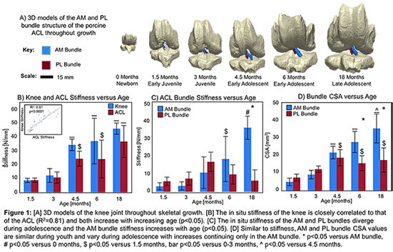
Results: Mean knee stiffness increased by 483% from 1.5 months to 18 months under applied loading (Fig. 1B). ACL stiffness increased by 468% over the same age range. Knee and ACL stiffness were closely correlated across all age groups (R2:0.81-0.93). AM bundle stiffness also increased by 383% by 18 months, whereas the PL bundle stiffness reached a plateau at 4.5 months of age (Fig. 1C). Structurally, the CSA of the AM and PL bundles were similar in young groups (p>0.05), while the CSA of the AM bundle was significantly greater in mid- and late-adolescent groups (p<0.05) (Fig. 1D).

Discussion: Although the porcine ACL plays a primary role in stabilizing the knee under anterior drawer throughout post-natal growth, the stiffness and CSA of its AM and PL bundles undergo bundle-specific changes during growth. These changes explain age-dependent differences in the bundle contribution to resisting anterior drawer loads and valgus torques as observed previously [2]. These changes occur alongside previously noted changes in ACL orientation [3]. Ongoing work is focusing on additional intrinsic bundle mechanical and biochemical properties. Overall, these data highlight underappreciated, bundle-specific changes to the ACL during post-natal growth and should be considered when establishing clinical interventions for ACL injuries in young patients.

Acknowledgements: Funding provided by NSF GRFP (DSE-1252376) and NIH (R03 AR068112).

References: [1] HK Kim+, J Pediatr Orthop, 2012, [2] SG Cone+, SB3C 2017, [3] SG Cone+, J Orthop Res, 2017, [4] MB Fisher+, J Orthop Res, 2010.


Uncaptioned visual


11:30 - 11:40

O0868 Shear stress in bone marrow is correlated to altered gene expression in in situ culture

Kimberly Curtis, Dr. Glen Niebur
University of Notre Dame, South Bend, USA

Abstract

Introduction

            Mechanical loading elicits a cellular response that serves to adapt bone mass and architecture. Osteocytes are essential mechanosensory cells [1], but many marrow cells are also mechanosensitive [2]. Indeed, stimulation of the marrow with minimal strain in the bone tissue induced bone formation without altering osteocyte signaling, while marrow cells exhibit increased expression of mechanoregulatory genes [3]. The objective of this study was to determine whether mechanical regulation by marrow cells was dependent on shear stress in the marrow. Specifically, a bioreactor that imparts shear stress to marrow cells via low-magnitude mechanical stimulation (LMMS) was used to culture porcine trabecular bone explants and 1) bone formation was quantified and 2) shear stress in the marrow was calculated using CFD models and correlated to expression of cFOS and cyclin-D1.

Methods

            Eight trabecular explants were harvested from porcine cervical vertebrae and cultured for 28d in a bioreactor. LMMS was applied to four explants, with an acceleration of 0.3g at 50Hz in two 30min bouts per day. The other four were not stimulated. Explants were imaged by μCT before and after culture for image based histomorphometry. Gene expression was measured in marrow cells taken from eight additional explants cultured for 5d using RT-qPCR. CT images of the stimulated explants were converted to CFD models, which were used to quantify the mean shear stress in the marrow.

Results

           LMMS induced a higher net change in bone volume in stimulated explants, which was positively correlated with mean shear stress in the marrow (Fig. 1a,b). The ratio of BFR/BRR was also positively correlated with the mean marrow shear stress (Fig. 1c).

            Both cFOS and cyclin-D1 expression increased with mean marrow shear stress in stimulated explants (Fig. 1d).
Uncaptioned visual

Figure 1. LMMS increased bone volume fraction (A;p=0.02) compared to controls. BV/TV and BFR/BRR were positively correlated to mean marrow shear stress (B,C) as were expression of cFOS and cyclin-D1 genes (D).


Discussion

            These data suggest that gene expression in marrow cells is affected by shear stress imposed by loading. Upregulated cFOS expression is a common mechanobiological response in cells. Cyclin-D1 is a downstream target of the AP-1 transcription factor, which is a dimer of cFos and cJun proteins. Although mean shear stress was used as a measure of stimulation here, the shear stress is greater at the bone surface, and may preferentially affect cells in that location. Treatments that target mechanobiological pathways in both osteocytes and marrow cells may be more effective for diseases characterized by bone loss.

Acknowledgements

NSF CMMI-1453467, CMMI-1100207

References

[1]Weinbaum et al.,J.Biomech.27(1994)339–360. [2]Castillo & Jacobs, Curr.Osteoporos.Rep.8(2010)98–104. [3]Curtis et al.,Bone.107(2018)78-87.


11:40 - 11:50

O0869 ERα deletion from mature osteoblasts increases severity of load-induced osteoarthritis in female mice

Sophia Ziemian1, Dr Olufunmilayo Adebayo1, Amanda Rooney1, Dr Natalie Kelly1, Derek Holyoak1, Dr F. Patrick Ross2, Dr Marjolein van der Meulen1,2
1Cornell University, Ithaca, USA. 2Hospital for Special Surgery, New York City, USA

Abstract

INTRODUCTION: The onset of menopause is associated with increased osteoarthritis (OA) incidence and severity1. Subchondral bone mass decreases with estrogen loss during menopause2; however, the direct effect of reduced subchondral bone mass on OA development is unclear. Osteoblast-specific estrogen receptor-α knockout (ERαKO) mice generated via osteocalcin-Cre3 provide a targeted in vivo model to study bone-specific estrogen loss on OA development. We hypothesized that ERαKO mice would develop more severe load-induced OA relative to littermate control (LC) mice. Furthermore, we hypothesized that OA severity would correlate with differences in intrinsic subchondral bone properties.             

METHODS: Cyclic mechanical loading was applied to the left tibia of 26-week-old female ERαKO and LC mice at peak loads of 6.5N, 7N, or 9N for 2 weeks (4Hz, 1200 cycles/day, 5 days/week)4. Right limbs served as contralateral controls. Subchondral bone morphology was analyzed using microCT. Cartilage damage was assessed histologically using the OARSI scoring system5. Cartilage thickness and medial tibial osteophyte size were measured from histological sections. Synovial pathology was quantified using histological scoring6. Effects of mouse genotype, loading, and load magnitude were determined using a linear mixed-effects model. Correlations between OA severity and intrinsic subchondral bone parameters were determined using Pearson correlation coefficient for bivariate analysis and linear mixed-effects model for multivariate analysis. Results are presented for p<0.05.

RESULTS: ERαKO control limbs had thinner subchondral bone (-9.91%) and lower bone volume fraction (BV/TV) in the epiphysis (-14.6%) and metaphysis (-32.0%). When pooled across control and loaded limbs, ERαKO had thinner cartilage (-4.11%). With loading, ERαKO had a greater increase in OARSI score (+138%, Fig1A,B), larger osteophyte size (+30.3%, Fig1A,C), and higher synovitis score (+11.4%). Subchondral plate thickness (-7.36%) and epiphyseal BV/TV (-8.67%) decreased with loading similarly between genotypes. Control limb subchondral plate thickness negatively correlated with cartilage damage (r = -0.439, Fig1D). Combined epiphyseal and metaphyseal BV/TV, and load magnitude explained 45% of variability in OARSI score (R2=0.455, Fig1E).

DISCUSSION: Bone-specific estrogen loss via osteoblast-specific ERαKO resulted in osteopenia of subchondral bone and more severe OA development following mechanical loading. Intrinsic subchondral bone mass negatively correlated with OA severity, indicating that individuals with reduced subchondral bone mass may have an increased risk of OA development and increased rate of OA progression. Our results suggest that bone-specific changes associated with estrogen loss, specifically reduced subchondral bone mass, may directly contribute to increased OA development following menopause, thus highlighting subchondral bone as a potential target for OA treatment and prevention in post-menopausal women.

ACKNOWLEDGMENTS: NIH-R21-AR064034 and NIH-T32-AR007281.

REFERENCES:  1Srikanth+ OAC 2005  2Brouwers+ JOR 2009  3Melville+ JBMR 2014  4Ko+. A&R 2013  5Glasson+ OAC 2010  6Lewis+ OAC 2011.

Uncaptioned visual


11:50 - 12:00

O0870 Comparison of knee joint mechanics in concurrent finite element musculoskeletal model of the healthy and mobile bearing prosthesis knee

Mr. Liming Shu1, Prof. Naohiko Sugita1, Miss. Jiang Yao2, Prof. Ko Yamamoto1, Prof. Mamoru Mitsuishi1
1The University of Tokyo, Tokyo, Japan. 2Dassault Systemes Simulia Corp., Johnston, USA

Abstract

Introduction

Understanding the kinematics and contact mechanics for in-vivo specific patient is considered as a significant facilitating factor in implementing successfully total knee replacement (TKR) prostheses. The in-vitro experiment and simulation have been widely used to evaluate the performance of prostheses, while highly standard boundary condition and dynamic input of simulator could not meet the subjective patients. Although in vivo experimental analysis have been implemented using instrumented knee prostheses and fluoroscopic measurement during daily activities in a limited number of patients, implementation of these devices is invasive, expensive, and could not be obtained during design phase of prostheses. In the paper, a systematically experimental evaluated concurrent finite element (FE) musculoskeletal (MS) model was developed and used to compare kinematics and contact mechanics differences between healthy and mobile bearing prosthesis knee joints. The goal of this study is to establish a framework to improve the design of total knee replacement to better restore the normal knee mechanics.

Materials and Methods

A novel subject-specific MS FE model, mainly including the musculoskeletal model and CT based subject-specific FE knee model with ligament constrain, was created and systematically evaluated by grand challenge dataset [1]. The detailed workflow of the simulator is shown in Fig.1.  The FE knee model was coupled to preoperative MS bone model based on iterative closest point algorithm. A stance phase of gait from a healthy male subject (BW-65 Kg, Height-183cm) was applied to demonstrate the knee joint kinematics and contact mechanics difference between healthy knee and implanted knee during gait cycle. The mobile bearing prosthesis components used in the framework were provided by Robert Reid Inc. (Japan). Prosthesis components were positioned in the FE knee model in a similar way to surgical procedure.  

Uncaptioned visual

Fig.1. The workflow of the subject-specific FE-MS simulator

Results and Discussion

A high predicted accuracy, quantified by the root-mean-square error (RMSE) and the squared Pearson correlation coefficient (r2), was found in the FE-MS model (RMSE = 177.2 N, r2 = 0.90) on the total tibiofemoral contact force on comparison with in-vivo experimental results from grand challenge dataset.  The results of comparison present the mobile bearing prosthesis has a stable and similar contact stress, while lateral-to-central pivoting motion can be found without being observed in healthy knee, which is consistent with previous clinical results [2].

Acknowledgements

This research is supported by JSPS KAKENHI Grant Number 16H05874. The authors also thank Dr. Kai Shin from Robert Reid Inc., Japan.

References

[1] Fregly BJ, et al. (2012) J Orthop Res, 30:503–13.

[2] Watanabe T, et al. (2004). J Orthop Res, 22:1044–9.


12:00 - 12:10

O0871 A combined statistical shape modelling and musculoskeletal simulation approach to investigate the effect of patellofemoral geometry on joint mechanics in tibial tubercle osteotomy

Allison Clouthier1, Dr Dan Borschneck2, Dr Darryl Thelen3, Dr Kevin Deluzio1, Dr Michael Rainbow1
1Queen's University, Kingston, Canada. 2Kingston General Hospital, Kingston, Canada. 3University of Wisconsin-Madison, Madison, USA

Abstract

Introduction

Variations in articular geometry affect joint function and can influence response to interventions. Tibial tubercle osteotomy (TTO) treats chronic lateral patellofemoral instability and pain through medial and/or anterior displacement of the tibial patellar tendon (PT) attachment. Though joint geometry varies in this population, shallow femoral trochlear grooves are common. Our goal was to examine how patellofemoral joint geometry interacts with PT displacement direction and magnitude in simulated TTO during dynamic loading. This was achieved using a combined statistical shape modelling and musculoskeletal simulation approach.

Methods

A whole-joint statistical shape model was created using surface models of the femur, tibia, and patella bone and cartilage from 14 asymptomatic participants. The shape model was used to generate new multibody knee models by varying the scores for the identified principal components (PCs). Ligament and muscle attachments and wrapping surfaces were fixed to nodes in the shape model to ensure these remained consistent with geometry. PC2 captured the depth of the femoral trochlear groove; therefore, variations from -3–3 standard deviations of PC2 were used in subsequent simulations.

A Monte Carlo approach was used to investigate the interaction between shape and surgical parameters. 750 new models were generated by randomly choosing one of seven geometries and perturbing the PT tibial attachment -1–2 cm medially and 0–2 cm anteriorly. These knee models were then integrated into a lower extremity musculoskeletal model and the same overground walking trial was simulated for all models. The tibiofemoral flexion angle was set based on the measured kinematics and the COMAK  simulation routine [1] was used to predict the patellofemoral and remaining tibiofemoral degrees of freedom, muscle and ligament forces, and cartilage contact pressures.

Results

There was a medial displacement of the PT that minimized contact pressure and maximized the contact area (Fig 1A); however, this varied based on geometry. Additionally, the shallow trochlear grooves were much more sensitive to medial displacement of the PT and the patella began to dislocate medially for large medial PT displacements in these geometries. Anterior displacement reduced the maximum contact pressure but also reduced the contact area, regardless of geometry (Fig 1B).

Uncaptioned visual
Figure 1: Contact area (indication of groove engagement) for the various trochlear groove depths (-3SD = shallow, +3SD = deep). Effect of medial PT displacement is shown for early stance (A) and anterior displacement for swing (B).

Discussion

Contact pressure and groove engagement (contact area) were dependent on joint geometry, with the knees with shallow trochlear grooves being more sensitive to medialization. The framework employed here can be extended to investigate how joint morphology can influence outcomes of other interventions and development of pathologies.

References

[1] Smith CR, et al. J Knee Surg 29(02):099-106, 2016.


12:10 - 12:20

O0872 Changes in bone tissue strength caused by disruption of the gut microbiome depend on stage of skeletal growth

Marysol Luna1, Jason Guss1, Remy Walk1, Dr. Marjolein van der Meulen1,2, Dr. Christopher Hernandez1,2
1Cornell University, Ithaca, USA. 2Hospital for Special Surgery, New York, USA

Abstract

INTRODUCTION: The gut microbiome influences bone morphology and density [1-2]. Recently, we demonstrated that disruption of the gut microbiome throughout life (4-16wks of age in mice) reduces the mechanical performance of whole bone at skeletal maturity by modifying bone tissue material properties [3]. However, it is unclear if disruption of the gut microbiome alters all bone tissue or only affects bone tissue at the time of formation. To test the idea that the microbiome influence bone tissue at the time of formation, we disrupted the microbiome at different stages of growth and determined the effects on whole bone strength at skeletal maturity. We hypothesized that alterations in the microbiome at earlier stages of life, when more bone tissue is formed, would have the greatest influence on adult bone mechanical performance.

METHODS: Male mice (B6.129S1-Tlr5tm1Flv/J) were divided into three groups (n=11-15/group, 37 total): disrupted microbiome from 4-10wks, disrupted microbiome from 10-16wks, and untreated.  Disruption of the microbiome was performed using chronic oral antibiotics (ampicillin + neomycin) in drinking water. Animals were euthanized at 16wks of age. µCT images of the femoral mid-diaphysis were taken to determine the section moduli (Moment of Inertia/c).Whole bone strength was determined with 3-point bending to failure. Tissue strength was estimated from whole bone strength per unit section modulus (Whole Bone Strength / (Moment of Inertia/c)).                                   

RESULTS: Disruption of the microbiome from 4-10wks resulted in a 17.2% reduction in tissue strength as compared to untreated mice (Fig 1B). Disruption of the microbiome from 10-16wks resulted in an intermediary tissue strength (but not noticeably different from untreated animals). Section moduli were less in animals with a disrupted microbiome compared to untreated animals (p<0.05) (Fig. 1A). Section moduli among treated groups were not different.
                                                                                                 

DISCUSSION:  Although the microbiome was only disrupted for 6 weeks, disruption during rapid bone growth (4-10wks) had a greater effect on tissue strength. The antibiotics used are not absorbed in the gut and do not cause gut inflammation, hence we attribute the effect of the treatment to changes in the microbiome and state of the microbiome. The difference in tissue strength among groups was likely due to the amount of bone tissue formed during disruption of the microbiome: on average 24% of the total bone tissue in the femoral diaphysis originated between 4-10wks, while only 10% was formed from 10-16wks [4] (Fig. 1B, inset). Our findings demonstrate that an impaired gut microbiome at a crucial point in skeletal growth can have a long lasting effect on bone tissue strength at skeletal maturity.                                                                

REFERENCES: [1] Hernandez +, JBMR, 2016. [2]Yan+, CurrOsteoporosRep, 2017. [3] Guss +, JBMR, 2016. [4] Ferguson+, Bone, 2003.

ACKNOWLEDGMENTS:  DoD-CDMRP(W81XWH-15-1-0239), NSF-GRFP

Uncaptioned visual