To save this page as a PDF, click this button and choose the PDF destination.

Session 5: Thoracic

09:00 - 10:20 Thursday, 8th July, 2021

Chairs Adil Aslam, Janet McNally, Fiona Metcalf

Free paper session


MINIMAL ACCESS SURGERY FOR CONGENITAL DIAPHRAGMATIC HERNIA: A SINGLE INSTITUTION'S 10-YEAR EXPERIENCE

Hemanshoo Thakkar1, Abigail Morbi1, Martin Sidler1, Dhanya Mullassery1, Stefano Giuliani1, Simon Blackburn1, Kate Cross1, Paolo De Coppi1,2, Joe Curry1
1Great Ormond Street Hospital, London, United Kingdom. 2Institute of Child Health, University of London, London, United Kingdom

Abstract

Aims:

Minimal access surgery (MAS) for the repair of Congenital Diaphragmatic Hernia (CDH) is well described in both neonatal and late presentations. The recurrence rate for this approach has been reported to be as high as 20% in the literature. We sought to review our institutional experience with this approach. 

Methods:

A 10 year (2009-2019) retrospective review of all patients undergoing primary MAS repair of CDH was conducted. Outcome measures included conversion, length of surgery, complications, patch utilisation and recurrence rates. Any patients on ECMO, poor ventilatory parameters or significant pulmonary hypertension were not offered MAS and underwent open surgery. 

Results:

132 patients underwent surgery during the study period of which 80 (60%) were performed open due to the exclusion criteria above. 52 (40%) patients underwent MAS surgery of which 36/52 (69%) were neonatal. The median gestational age for neonatal patients undergoing surgery was 38 weeks (32-42) with a median weight of 2800g (1300-4280). 83% of cases were left sided. The median anaesthetic time was 190 minutes (60-343). 13/36 (36%) of neonates were on inotropes pre-operatively. 12/52 (23%) cases were converted to open. 

Overall there were 9 complications; intra-operative supraventricular tachycardia (1), pneumothorax requiring a chest drain (1), chylothorax (1), bowel perforation (2), chest drain site requiring formal closure under GA (1), sepsis (1), wound site collection (1) and post-operative chest infection (1).

21/52 (40%) underwent a patch repair using a woven polyester synthetic material. The overall recurrence rate was 3/52 (6%) with a median follow-up of 23 months (1-95).  

Conclusions:

In our experience, MAS repair of CDH is safe with few complications. Patient selection is extremely important when employing this technique. Our low recurrence rate may be attributable to having a low threshold in using a patch to achieve a tension-free repair.  


PROSTHETIC PATCH REPAIR OF CONGENITAL DIAPHRAGMATIC HERNIA (CDH): REASONS FOR RECURRENCE.

Maria Asimakidoo, Niyi Ade-Ajayi, Mark Davenport
Kings College Hospital, London, United Kingdom

Abstract

AIMS: 

Primary repair is not always possible in congenital diaphragmatic hernia (CDH) with the usual alternative being a prosthetic patch repair. We performed a multifactor analysis using recurrence as the end-point in those children with patch repairs.


METHODS: 

Retrospective single-centre review in surgical unit co-located with large fetal-medicine centre. Follow-up included routine chest X-ray. Birth weight, gestational age, fetoscopic treatment and patch type were independent variables. Univariate and then logistic regression analysis was used with statistical significance defined as P≤0.05.


RESULTS: 

180 infants were managed between 1994 and 2017 and of these 45 died at ≤9 months leaving n=135 for core analysis. FETO had been used in 49/135(36.3%); prosthetic patches used in 56/135(41.5%) [“biologic”, n=34(61%) and non-biologic, n=22(39%)]. There was increased need for patch in the FETO group [40/49(81.6%) vs. (16/86–38%)(P<0.0001]. No infant with a post-natal diagnosis required a patch. 


Median follow-up was 59 (1-194) months.  There were 18 recurrences of which 17/34(50%) were in the "biologic" group (P<0.0001). Use of FETO (P=0.007)and gestational age (P=0.005) (15/24-62.5%) were also significantly different (but not birth weight IP=0.45).  


Multivariable analysis showed that type of patch was the key factor influencing outcome.


Variables

P-value

Odds Ratio (95% CI)

Gestation at birth

0.13

0.8 (0.60–1.1)

BW

0.20

2.9 (0.56– 15.5)

Patch type

0.01

30.3 (2.2–412)

FETO

0.77

0.7 (0.12 – 4.8)


 CONCLUSIONS:

  • FETO intervention in CDH increases the need for prosthetic patch repair. 
  • "Biologic" patches increase the risk of recurrence.


PREOPERATIVE AADO2 AS AN INDICATOR FOR PHYSYOLOGIC STABILIZATION FOR REPAIR IN  INFANTS WITH CONGENITAL DIAPHRAGMATIC HERNIA: A MULITICENTER RETROSPECTIVE STUDY.

Akiko Yokoi1, Shunsuke Fujii2, Akiyoshi Nomura3, Masaya Yamoto3, Satoko Ofuji4, Masato Shinkai2, Naoto Urushihara3
1Kobe Children's Hospital, Department of Pediatric Surgery, Kobe, Japan. 2Kanagawa Children's Medical Center, Department of Pediatric Surgery, Yokohama, Japan. 3Shizuoka Children's Hospital, Department of Pediatric Surgery, Shizuoka, Japan. 4Osaka City University Graduate School of Medicine, Department of Public Health,, Osaka, Japan

Abstract

Aim

Appropriate timing of surgery for neonates with congenital diaphragmatic hernia (CDH) has been generally considered after cardiopulmonary stabilization. However, few reports clearly showed the definition of physiologic stabilization for surgery, while many studies have addressed hours of life for optimal timing of repair. To aim of this study is to elucidate an optimal respiratory condition for repair in CDH.

Method

A retrospective chart review of 81 isolated CDH patients who were born and treated in the three children’s hospitals during Jan. 2009 through Sep.2018 was conducted. To see an optimal respiratory condition for repair, alveolar-arterial oxygen difference (AaDO2) and oxygenation index (OI) right before surgery was investigated as well as prenatal prognostic factors (o/e LHR and liver up), patients’ demographics (sex, gestational weeks, birth weight, Apgar scores) and the 30-day survival. Multivariate logistic regression analysis adjusting for the antenatal severity combined with o/e LHR and liver position was performed.

Result

Among 81patients, 75 patients underwent surgery. The data of AaDO2 and OI right before surgery were available in 73 patients. Among73 patients, 6 patients died in total, and 2 out of 5 patients with ECMO died. The 30-day mortality in the subset of the fourth quartile of AaDO1 (340 mmHg) was significantly higher (OR 9.23 95% CI 1.18072.11 P=0.03), while mortality of the fourth quartile of OI (8) was also higher with marginally significant. (OR 6.52 95% CI 0.93-45.59 p=0.06).

Conclusion

AaDO2 less than 340 mmHg could be a useful stabilization index for safe surgery in neonates with CDH.


INTRABDOMINAL HYPERTENSION IN NEONATES FOLLOWING CONGENITAL DIAPHRAGMATIC HERNIA REPAIR: CORRELATION TO EARLY POSTOPERATIVE RESPIRATORY AND GASTROINTESTINAL OUTCOMES

ELANGO THAMBUSAMY1, CR THAMBIDORAI1,2, Srihari Singaravel1, Shireen Anne Nah1, Anand Sanmugam1
1UNIVERSITY MALAYA MEDICAL CENTRE, KUALA LUMPUR, Malaysia. 2

Abstract

AIM:

Increased intra-abdominal pressure (IAP) is seen in patients after congenital diaphragmatic hernia (CDH) repair due to reduction of thoracic content into the relatively smaller abdominal cavity. In infants, IAP>10 mmHg is considered Intra-abdominal hypertension (IAH). 

This study aimed to determine the incidence of IAH in neonates post CDH repair. We also evaluated the relationship between IAH, and duration of respiratory support and the early return of gastrointestinal function respectively.


Methods: We prospectively recruited all neonates who had CDH repair in our institution from June 2019 to October 2020. Intravesical pressure was used as a proxy for IAP and was measured using a closed system transducer for 5 consecutive days post-surgery. Daily median values were used for analysis. We categorised IAP as <11mmHg (no IAH), 11-15mmHg (IAH) and >15mmHg (severe IAH).

The study was ethically approved (No 201951-7379).

Spearman correlation analysis was used to measure the strength of association between nonparametric data. A value of p < 0.05 was considered statistically significant.

Results: There were 24 neonates included in this study, operated between 1-6 days of life (median age day 4 of life). No patient recorded IAP>15mmHg. The intra-abdominal pressure trend is shown in the figure below: 

Uncaptioned visual

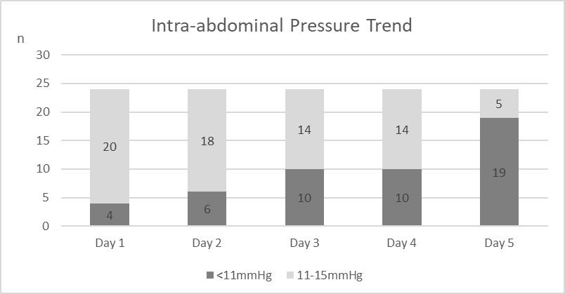
Figure: Trend of Intra-abdominal Pressure Post Congenital Diaphragmatic Hernia Repair

Those requiring ventilatory support for 4-7 days post repair formed the largest proportion (n=10,41.7%); 7(29.2%) were ventilated <4 days, the remainder >7 days. There was strong correlation between days of IAH and duration of ventilation (p-value<0.001, r=0.704). There was moderate correlation between days of IAH and duration taken for full enteral feeding (p-value 0.005, r=0.704).

 Conclusion: IAP measurement is a safe and useful adjunct in post CDH monitoring to detect IAH and in predicting ventilatory support requirements and the time required for feeding establishment.


PHARMACOKINETICS AND PHARMACODYNAMICS OF SILDENAFIL IN FETAL LAMBS ON EXTRACORPOREAL SUPPORT.

Felix De Bie1,2, Francesca Russo2, Pieter Van Brantegem3, Zexuan Yang1, James Moon1, Barbara Coons1, Pieter Annaert3, Karel Allegaert2, Marcus Davey1, Jan Deprest2, Alan Flake1
1Center for Fetal Research, Children's Hospital of Philadelphia, Philadelphia, USA. 2Department of Development and Regeneration, KU Leuven, Leuven, Belgium. 3Department of Drug Delivery and Disposition, KU Leuven, Leuven, Belgium

Abstract

Purpose: To assess the pharmacokinetic profile of sildenafil in an artificial womb model for fetal lambs, to define the pharmacodynamic dose-effect relationship on the pulmonary vasculature and to evaluate short-term hemodynamic fetal tolerance to the drug.


Methods: Healthy fetal lambs (Gestational age 106-124; term 145) in an artificial womb environment were exposed to different doses of continuous intravenous (IV) sildenafil (0.3 – 0.5 – 0.7 mg/kg/24hr) for a period of one to seven days. A wash-out period of two to six days was scheduled before another dose was tested in the same animal. Blood sampling and Doppler ultrasound were performed at fixed timepoints. Primary outcomes were sildenafil pharmacokinetic profiles generated using NONMEM modeling and changes in the Doppler waveform of the right pulmonary artery (RPA). Secondary outcomes were proxies for fetal tolerance (blood gas and hemodynamic parameters).


Results: A total of eight administrations were tested in five lambs. Sildenafil concentrations reached plateau after six days, with only the 0.5 and 0.7 doses reaching therapeutic concentrations (cf. figure 1). In 62.5% (n=5/8) of cases, sildenafil administration was associated with a temporary increase in the acceleration/ejection time of the RPA >10% and a temporary decrease in both mean arterial pressure and circuit flow. The magnitude of the effects were not dose-dependent. No biochemical toxicity was observed.


Conclusion: In 62.5%, administration of sildenafil caused a temporary, non-dose-dependent pulmonary vasodilation in fetal lambs on extracorporeal support. A daily 0.5 mg/kg IV dose of sildenafil allows to achieve therapeutic concentrations and can safely be administered to fetal lambs on EXTEND.


Figure 1: Pharmacokinetic profile

Uncaptioned visual

Pharmacokinetic profile obtained through NONMEM modelling. The red quadrangle represents the therapeutic range of sildenafil (47 – 500ng/mL).


COMPENSATORY SWEATING AFTER THORACOSCOPIC SYMPATHECTOMY FOR PRIMARY FOCAL HYPERHIDROSIS IN CHILDREN: ARE THERE PATIENT-RELATED RISK FACTORS?

Catarina Carvalho, Ana Sofia Marinho, Joana Barbosa-Sequeira, Mário Rui Correia, José Banquart-Leitão, Fátima Carvalho
Centro Materno-Infantil do Norte, Oporto, Portugal

Abstract

Aim of the Study: Evaluate and identify potential risk-factors for compensatory sweating (CS) after thoracoscopic sympathectomy for primary focal hyperhidrosis (PFH) in children.

Methods: Retrospective, single-center review of all bilateral thoracoscopic sympathectomies performed between January/2017 and July/2019. All patients were evaluated at the 3rd and 6th month post-operative, with an inquiry and physical examination.

Results: Over the 30-month period, 41 patients were submitted to thoracoscopic sympathectomy; 92.6% had severe palmar hyperhidrosis. 30 were females (73.2%). A T2-T4 sympathectomy was performed in all patients, with selective lung intubation. Mean age at surgery was 14.9 years.

CS was identified at the 3rd month in 23 patients (56%), most in the dorsolumbar region (DL; 56.5%). By the 6th month, there was a significant CS reduction (56% to 36.6%, p<0.05). In DL and abdominal CS, the probability of CS resolution by 6 months was around 50% (p<0.05).

Patient-related factors evaluated were age, gender, z-score of body mass index (average 0.52), family history (5%), concomitant illnesses (14.6%) and pre-operative severity of hyperhidrosis, none of which influenced CS (p>0.05), except for pre-operative axillary hyperhidrosis, that is associated with dorsolumbar CS (p<0.05).

Conclusions: Thoracoscopic sympathectomy for PFH is a safe and effective procedure to perform in the pediatric population. Compensatory sweating is a possible and non-negligible complication. Patient-related factors, namely age, gender and body mass index, do not appear to influence CS; pre-operative axillary hyperhidrosis is related to dorsolumbar CS. In one third of patients, CS appears to resolve after 6 months.


MULTIDISCIPLINARY APPROACH TO DYNAMIC COMPRESSION BRACING FOR PECTUS CARINATUM IMPROVES COMPLIANCE AND OUTCOMES IN CHILDREN

Gillian Mullervy1, Gillian Connolly2, Brice Antao1
1Paediatric Unit, Beacon Hospital, Dublin, Ireland. 2Physiotherapy Department, Beacon Hospital, Dublin, Ireland

Abstract

AIM OF THE STUDY: Bracing for pectus carinatum (PC) has emerged as an alternative to surgical correction. This study describes treatment protocols and outcomes and evaluates contributory factors for a successful dynamic compression bracing (DCB) program for PC in children. 


METHOD: Prospective data were collected on patients enrolled in a DCB program from September 2018 to January 2020. Our DCB program includes two phases [Treatment phase (TP) and Maintenance Phase (MP)], complimented by a customised exercise program, physical therapy, close monitoring and follow-up. All patients had thoracic height (TH) assessed with 3-D Scans. 


RESULTS: Sixteen patients [13(81%) male, 3(19%) female] were enrolled into DCB program at a median age of 15 years (range, 12-19 years). All patients had a spectrum of other musculoskeletal problems, such as kyphosis, rib flaring and flattening and suffered low self-esteem and impaired body image. All 16 patients completed TP at a median duration of 6 months (range, 6-7months) and achieved a complete correction of their deformity (mean pre-treatment TH =204.10±13.80mm, mean post-treatment TH =181.20±20.56mm, p<0.01). Eight of these patients also completed MP at mean duration of 6 months (range, 5-6 months). There was a good correction of musculoskeletal problems in all patients. The overall success and compliance rate was 100%, with no complications or recurrences. 


CONCLUSION: Dynamic compressive bracing results in a significant improvement in PC appearance and should be used as a first-line treatment for PC in children. For a successful pectus bracing program, treatment protocols need to be individualised based on severity of the deformity, patient’s age and associated musculoskeletal problems and requires a multidiciplinary approach with intensive involvement of a Clinical Nurse Specialist (CNSp) and a physiotherapist. A customised exercise program and physical therapy appears to significantly improve compliance, shortens duration of bracing therapy and improves associated musculoskeletal problems. 


ULTRASOUND-GUIDED ERECTOR SPINAE PLANE BLOCK VERSUS THORACIC EPIDURAL ANALGESIA POSTOPERATIVE PAIN MANAGEMENT AFTER NUSS REPAIR FOR PECTUS EXCAVATUM

David P. Bliss, Jr., MD, MBA1,2, Thomas B. Strandness, MD1,2, Sarkis C. Derderian, MD3,2, Alexander M. Kaizer, PhD2, David A. Partrick, MD3,2
1Children's Hospital Colorado, Colorado Springs, USA. 2University of Colorado School of Medicine, Aurora, USA. 3Children's Hospital Colorado, Aurora, USA

Abstract

Aim of the Study

Postoperative pain management is a significant challenge in patients undergoing Nuss repair for pectus excavatum chest wall deformity.  Therapeutic anesthetic options primarily include patient-controlled analgesia, thoracic epidural analgesia (TEA), and cryoanesthesia.  However, TEA is limited to in-patient use and both TEA and cryoanesthesia can result in neurologic injury.  The novel technique of ultrasound-guided erector spinae plane regional analgesia has been employed recently in our patients with pectus excavatum undergoing the Nuss repair and has demonstrated impressive pain relief, but without the potential complications of other modalities.   Erector spinae plane block (ESPB) postoperative pain management outcomes were studied as compared to TEA.

Methods

Thirty consecutive patients with severe pectus excavatum undergoing Nuss repair and placement of ultrasound-guided ESPB were each paired to a cohort control patient with TEA postoperative pain management.  The cohort patient match was defined by age (+/- 2 years), gender, and CT pectus index (+/- 15%).  Study variables included hospital length of stay (LOS), pain scores, and pain medication usage.

Results

Pain scores and oral morphine equivalent (OME) pain medication usage were higher for the first two postoperative days in the ESPB group (p<0.05).  However, average hospital LOS was nearly one day shorter for ESPB patients who were discharged home with the catheter in place until removal at 5-7 days postoperatively (p<0.001).  

Conclusion

Ultrasound-guided ESPB is a feasible alternative to TEA in postoperative pain management after Nuss repair and results in decreased hospital stay.  

____________________________________________________

Covariate                   TEA                 ESPB           p-value

                                 (N=30)              (N=30)  __________

POD 1 Avg.            2.67 (1.28)        3.75 (1.78)       0.009

  Pain Score      

POD 1 OME/hr.      0.5 (0.2)            2.33 (1.34)     <0.001

POD 2 Avg.            2.76 (1.33)        3.8 (1.85)         0.016

 Pain Score      

POD 2 OME/hr.      0.61 (0.3)          1.95 (0.91)     <0.001

Hospital LOS          3.78 (0.82)        2.9 (0.87)       <0.001


NEXT DAY DISCHARGE AFTER THE NUSS PROCEDURE USING INTERCOSTAL NERVE CRYOABLATION, INTERCOSTAL NERVE BLOCKS AND PERIOPERATIVE ERAS PAIN PROTOCOL

John DiFiore, Surendrasingh Chhabada, Jason Robertson, Anthony DeRoss, Wai Sung
Cleveland Clinic Childrens Hopsital, Cleveland, USA

Abstract

Aim of Study: To assess the effectiveness of a multimodality treatment protocol for the Nuss procedure. The primary outcome is length of stay (LOS). Secondary outcomes include opioid use and pain scores.

Methods:  40 patients age 13-29 underwent the Nuss procedure over a one-year period. Intra-operatively, intercostal nerves T3-T8 were treated bilaterally with cryoablation and bupivacaine nerve blocks. Exparel was used locally on the incisions. ERAS (Enhanced Recovery After Surgery) protocol included preoperative treatment with gabapentin for 4 days, and Celebrex and Tylenol for 2 days. These were continued in-hospital and one week post-discharge. Oxycodone and valium were added immediately postop and continued 2 days after discharge. Intravenous hydromorphone patient-controlled analgesia (IVPCA) was used overnight (demand only/no basal rate) for the first 19/40 patients. 

RESULTS:   37/40 patients (93%) were discharged home the day after surgery; 3/40 (7%) were discharged post-operative day 2 (LOS= 1.1). In-hospital, the average number of PCA doses was 14.8 (0.2 mg/dose) and the average number of oxycodone tablets used was 6.4 (5mg/tablet) for a total oral morphine equivalent (OME) of 47.5 mg.  8/40 patients (20%) took no oxycodone at all.  Average pain score was 2.2/10.  After eliminating IVPCA, total OME decreased by 74% with no change in pain scores or discharge timing. At discharge, patients were given a two-day prescription for oxycodone; only 3/40 patients (8%) requested a refill. 

CONCLUSION: Intercostal nerve cryoablation combined with intercostal bupivacaine nerve blocks and a well-defined pre- and post-hospital ERAS analgesic protocol was successful in discharging patients one day after the Nuss procedure, eliminated the need for intravenous opioids and achieved excellent pain control. 


PAIN SCORES AFTER THORACOSCOPY VERSUS THORACOTOMY IN NEONATES. IS THERE A DIFFERENCE?

Kailas Bhandarkar1, Simon hannam1, Paolo decoppi2
1Great ormond street hospital, london, United Kingdom. 2Great ormond street hospital, United Kingdom

Abstract

Aim. 

Thoracoscopy is increasingly performed in neonates to treat congenital anomalies. We sought to assess if neonatal pain scores are significantly different between thoracoscopic (T) and open thoracotomy (O) approach.


Methods

This is a review of a single institutional retrospective study between January 2012 to December 2019. We included neonates (up to 40 weeks corrected gestational age) who underwent T procedure for congenital diaphragmatic hernia (CDH), and T or O Oesophageal atresia (OA) repairs. Exclusion criteria included conversion and simultaneous additional procedures.  The Neonatal PAT scores were used to assess and score postoperative pain.  Data are quoted as median (range) unless otherwise specified.


Results 

During the study period there were 54 neonates. Complete pain management data were available for total of 28 neonates, of which 13 were T and 15 in O group. Gestation age was 37 weeks (31-41 weeks) (O) compared 38 weeks (32-42) (T). Median birthweight was 2.49 kilogram (1.16-3.57) (O) and 3.15 (1.97- 4) (T). Time to extubation was similar in both (3 days), range being 1-13 days (O) and 1-11 days (T). Neonatal PAT scores were similar in both O and T groups. Morphine usage was similar between both groups. 


Conclusions

There is an increased used of MIS in neonate, however there are no data available regarding post-operative pain on this population. This preliminary retrospective data seems to indicate there is no difference in the postoperative score and in pain management between the 2 groups. Further prospective studies are needed.


OESOPHAGEAL ATRESIA WITH VERY LOW BIRTH WEIGHT - CLINICAL CHARACTERISTICS AND LONG-TERM OUTCOME

Antti Koivusalo, Janne Suominen, Annika Mutanen, Mikko Pakarinen
Childrens Hospital, Helsinki, Finland

Abstract

Aim of the Study: Assessment of the clinical data and outcome of patients with oesophageal atresia (OA) with very low birth weight (VLBW). 

Method: With ethical consent, we reviewed the records of 318 successive patients with OA from 1980 to 2019. Clinical data and outcome were compared between of patients with VLBW(≤1500g) and with BW>1500g. Main outcome measures were survival and oesophageal repair.  

Results: Thirty-seven (12%) patients had VLBW. Gross types of OA in patients with VLBW and with BW >1500g were: A(13%/7%), B(3%/3%), C(79%/81%), D(3%/4%), E(0%/7%), F(0%/1%) (p=0.14–0.99). In patients with VLBW the incidence of congenital heart disease (CHD) (46%) and trisomy 18 and 13 and Cri du Chat (14%) were higher than with BW>1500g (25% and 1%), (p = 0.01 and p=0.001). Early death (within 30 days) occurred in 10(27%) patients with VLBW and in 13(5%) in patients with BW>1500g (p<0.001). Cause of early deaths in VLBW patients were CDH w/wo chromosomal abnormality (n=6), cerebral haemorrhage (n=1), gastric perforation (n=1) and renal agenesia (n=2). Oesophageal repair was performed in 31(84%) patients with VLBW and in 275(98%) with BW>1500g (p=0.01). Repair in VLBW patients included early (n=21) or delayed (n=6) end-to-end anastomosis and reconstruction (n=4). Anastomotic complications occurred in 23% of patients with VLBW and in 15% with BW>1500g, (p=0.29). Overall five-year survival was 50% in VLBW and 91% in BW>1500g (p<0.001). From 1980–99 to 2000–2019 survival in VLBW changed from 26% to 76% (p=0.002), and early deaths decreased from 9/19 (47%) to 1/18 (6%) (p=0.01). During 2000–2019 all VLBW patients underwent repair.

Conclusion: OA with VLBW was associated with increased incidence of life-threatening associated anomalies and decreased survival. Recently survival and the rate of definite oesophageal repair have improved significantly. 


VALIDATION OF NOVEL 3D-PRINTED SIMULATION MODEL FOR OPEN OESOPHAGEAL ATRESIA AND TRACHEO-OESOPHAGEAL FISTULA REPAIR DEVELOPED FOLLOWING  UK-WIDE TRAINING NEEDS ASSESSMENT

Carmen Sofia Chacon1, Jonathan Neville1, Reza Haghighi-Osgouei2, Natasha Houghton2, Simon Clarke1
1Chelsea and Westminster, London, United Kingdom. 2Imperial College, London, United Kingdom

Abstract

Background: Simulation training in paediatric surgery is at an early stage, but its role is expanding. The aim of this study was to conduct a training needs assessment of UK paediatric surgeons to identify and prioritise procedures for simulation, and to validate a novel 3D-printed simulation model for oesophageal atresia and tracheo-oesophageal fistula (OA-TOF) repair. 

Methods: An anonymised online questionnaire was sent to UK paediatric surgeons surveying the availability and perceived utility of simulation. The operation ranked as most useful to simulate was OA-TOF repair. 3D-printing techniques were used to build an OA-TOF repair model. Open repair was validated by 20 surgeons. Participants rated the content and face validity of the model on a five-point Likert scale, and were assessed during the procedure to determine construct validity. 

Results: Thirty-four paediatric surgeons completed the survey; 79% had access to surgical simulation at least monthly, and 47% had access to paediatric-specific resources. Perceived utility of simulation (mean score [SD]) was 4.1/5 (±1.1). Validation of open OA-TOF repair was conducted by five experienced and 15 inexperienced surgeons. Participants rated the model as useful 4.9/5 (±0.4), and believed all trainees should have access 4.6/5 (±0.8). Anatomical realism was scored 3.9/5 (±0.7) and surgical realism 3.8/5 (±0.8). Time to complete procedural steps was not significantly different between the groups. However, oesophageal anastomosis quality was higher in the experienced group (4.4/5 versus 3.3/5, p=0.015), and fewer sutures were misplaced (1.4 versus 5.1, p=0.057). 

Conclusion: UK paediatric surgeons believe simulation is useful for their training, but fewer than half have access to paediatric surgery-specific resources. In response we have developed and validated a 3D-printed simulation model for open OA-TOF repair. 


A SINGLE CENTER’S EXPERIENCE OF USING INTERNAL TRACTION SUTURES IN THE MANAGEMENT OF LONG GAP OESOPHAGEAL ATRESIA

Thejasvi Subramanian, Benjamin Martin, Ingo Jester, Max Pachl, Michael Singh, Giampierro Soccorso, Oliver Gee, Anthony Lander, Suren Arul
Birmingham Women and Children's Hospital, Birmingham, United Kingdom

Abstract

Aim

To review our experience and outcomes of using internal traction sutures (ITS)1 in patients with long gap oesophageal atresia (LGOA) with or without trachea-oesophageal fistula (TOF).


Method

The case-notes of all infants treated with ITS for LGOA (December 2015 to December 2019) were reviewed. The core steps of ITS placement involved ligation and division of the TOF with mobilization of the upper and lower pouches.  Gaps considered too wide for anastomosis had ITS placed between the two pouches. All patients were gastrostomy fed.  Patients returned to theatre with an expectation to complete the anastomosis or re-tighten the traction sutures.


Results

Seven patients with LGOA were managed using ITS — M:F 4:3, birthweight 2.28 kg, (1.2–3.6 kg),  pure OA (4),  OA/distal TOF (2), OA/proximal TOF(1). 

The median gap length was 4.5 (3–5.5) vertebral bodies.  ITS were placed thoracoscopically in 5 cases and via right-thoracotomy in 2 at a median 54 days (0–193) after birth.  A second thoracoscopic re-tightening of the ITS was attempted in 3 patients.  Patients had a median of 4 procedures during their first admission.

In all cases ITS were associated with significant intra-thoracic adhesions and 5 patients developed a leak due to the traction sutures, later complications included frozen chest (4) and recurrent TOF (2). 

Four patients had a delayed primary anastomosis performed at thoracotomy but 3 required a cervical oesophagostomy due to complications.  The median length of stay was 159 days (98–282).  No patient was managed exclusively using thoracoscopy. 


Conclusion

Our use of ITS was associated with significant complications.  Problems were caused by intra-thoracic adhesions and leaks.  Ultimately ITS did not improve the feasibility of thoracoscopic repair for long gap OA as we had hoped.

  1. Patkowski D, Thoracoscopic multistage repair of long-gap esophageal atresia using internal traction sutures. FrontiersinPeds2016. 


MANAGEMENT METHODS OF LARYNGOTRACHEAL SEPARATION IN PEDIATRIC AND ADOLESCENTS PATIENTS: 9-YEAR EXPERIENCE IN CHILDREN'S HOSPITAL

Toru Shimizu, Shigeru Takamizawa, Katsumi Yoshizawa, Tomoko Hatata, Junya Ishii
Nagano Children's Hospital, Azumino, Japan

Abstract

Aim of the study:

Laryngotracheal separation (LTS) is known to be the definitive solution for intractable aspiration pneumonia in neurologically impaired children. Postoperatively, tracheostomy cannula is usually required for home ventilator care and at school for suctioning. However, there are cannula related complications including trachea-innominate artery fistula (TIF) that could be life-threatening. We present our methods of LTS and the result in our experience of 57 patients.


Method:

A retrospective review in a single center from 2011 to 2019 identified 57 cases treated with LTS. We divided them into two groups; no previous tracheostomy (n = 26) and previous tracheostomy (n = 31). The first group underwent traditional modified Lindeman’s procedure without laryngoesophagostomy. The second group received circle skin incision along the previous tracheostomy and transection of the trachea above the tracheostomy site in order to prevent the anterior deviation of trachea. In addition, for all LTS, it is critically important to select the proper length and the curve of cannula carefully in order to prevent damaging innominate artery by the tip of cannula through the tracheal wall. If it is apparent that innominate artery compresses trachea anteriorly and causes respiratory problems, transection of innominate artery should be considered before LTS.


Results:

Three patients (5.3%) deceased by unrelated cause of surgical treatment. Only one patient experienced postoperative TIF followed by LTS (3.8%, 0%). Other postoperative complications in two groups are as follows: wound infection (7.7%, 9.7%), intratracheal granuloma (11.5%, 12.9%), wound granuloma (53.8%, 29.0%), intratracheal minor bleeding (7.7%, 16.1%), and minor proximal tracheal stump leakage (0%, 6.5%). No one experienced revision of LTS.


Conclusion:

Both in these two groups, the success rates of LTS were high without major complications. With our methods, LTS can be done safely, and it could be the great solution for intractable aspiration.