To save this page as a PDF, click this button and choose the PDF destination.

Poster Exhibition Opens

09:00 - 09:10 Wednesday, 30th June, 2021

Poster Groups General

Poster Exhibition opens!  The poster Exhibition will be open for this week and during the 3 days of the congress.  You can interact with poster presenters by asking questions and engaging in discussion with colleagues.


A RARE COMPLICATION OF SPLENECTOMY IN A CHILD WITH HEMATOLOGIC DISORDER: CASE REPORT AND SYSTEMATIC REVIEW OF THE LITERATURE

Fatma Thamri, Sondes Sahli, Arije Zouaoui, Oussema Meherzi, Yasmine Houas, Yosra Kerkeni, Riadh Jouini
Department of Pediatric Surgery " A", Hospital of Children of Tunis, University Tunis El Manar, Tunis, Tunisia, Tunis, Tunisia

Abstract

Clinical History

A 13-year-old boy, followed for a major betathalassemia, had a splenectomy indicated for increased transfusion needs and hypersplenism. The preoperative blood formula count showed anemia and a normal platelet level. The aftermath of the surgery was simple and the patient was discharged on day three post-operatively. 

Ten days later, the child returned for isolated abdominal pain. Clinical examination was without abnormalities. The hemogram had not shown deglobulization but it showed thrombocytosis (655000 platelets / milliliter). Doppler ultrasonography and CT scan had concluded to an extended portal thrombosis. The upper mesenteric vein was permeable.

The patient had a complete hemostasis balance which was normal. He was put on subcutaneous low-molecular-weight heparin relayed by anti-vitamin K. He was discharged without anticoagulant treatment, within 45 days, after normalization of the Doppler.

Purpose of presenting the case 

Portal vein thrombosis is a rare but life-threatening medical complication of splenectomy in children with hematologic diseases. Its frequency is often underestimated because of the existence of sub-clinical cases. 

Our purpose was to analyze clinical and radiological manifestations of such complication and to assess the evolution after appropriate medical treatment. 

Main learning points 

Portal vein thrombosis after splenectomy should be systematically sought after post-operative abdominal pain and thrombocytosis. Treatment of such a complication is simple and rests on the heparin therapy. Its misdiagnosis is responsible of the upper mesenteric vein thrombosis and the portal hypertension.


PERCUTANEOUS SCLEROTHERAPY OF INTRA-ABDOMINAL CYSTS: PERISHING DRAINS AND ALCOHOL TOXICITY

Jessica Ng1, Paul Charlesworth1, Ashwini Joshi1, Samantha Chippington2,3
1Department of Paediatric Surgery, The Royal London Hospital, London, United Kingdom. 2Department of Paediatric Radiology, Great Ormond Street Hospital, London, United Kingdom. 3Department of Paediatric Radiology, The Royal London Hospital, London, United Kingdom

Abstract

Clinical history

An 8-year old boy presented with chest wall asymmetry, scoliosis and splenomegaly was found on USS and MRI to have a large simple splenic cyst (11 x 11 x 13cm). He underwent laparoscopic marsupialisation. Repeat USS at 1-month post-operation showed cyst recurrence (16 x 13x 13cm). 

After MDT discussion, he underwent sonographic-guided percutaneous drainage (1.6L of cyst fluid drained) and sclerotherapy with 95% ethanol using a 6Fr FleximaTM drain. During the procedure the catheter was removed and the drain had corroded leaving the tip of the drain within the cyst cavity, no extravasation was seen at the time of USS. Following extubation his GCS was persistently low and required reintubation. He was found to have elevated serum ethanol level 217mg/dL. He was extubated after 24-hours and discharged from hospital at day-2 post operation. Follow-up at 1-year post-operation he is asymptomatic, USS showed cyst reduction (6 x5 x 6cm) and the drain remnant remains within the cyst cavity.

To date, the senior author has experience of 10 children who underwent percutaneous high-dose ethanol sclerotherapy of intra-abdominal cyst. This includes 8 unilateral renal and 2 splenic cysts (median volume aspirated 117mL; range 9-1600mL). In 2 (20%) cases the drains perished; including 2 of 3 cases where FleximaTM drain was used, the remaining 7 cases utilised Cook® pigtail drains with no effect on drain integrity. All cysts have significantly reduced in size or resolved. Those complicated by drain tip retention remain asymptomatic.


Purpose

To present the risks of ethanol as a sclerotherapy agent for intra-abdominal cysts.


Main learning points

  • Not all catheters are compatible for use with alcohol and can adversely affect drain integrity. 
  • The complication of ethanol intoxication during percutaneous sclerotherapy is due to systemic absorption with or without extravasation. 
  • Stratification of drain type with sclerotherapy agent and ethanol dosing is required.

PATIENT SATISFACTION WITH A NEW PAEDIATRIC AND ADOLESCENT BREAST SERVICE

Chipo Mushonga1, Bhavna Lakhiani2, Jackie Dalton3, Dimitri Hadjiminas3, Clare M Rees1
1Department of Paediatric Surgery, Imperial College Healthcare NHS Trust, London, United Kingdom. 2Department of Paediatrics, Imperial College Healthcare NHS Trust, London, United Kingdom. 3Department of Breast Surgery, Imperial College Healthcare NHS Trust, London, United Kingdom

Abstract

Aim

To describe a novel paediatric and adolescent breast service and evaluate patient satisfaction.

Methods

A new breast service was set up in 2019, with pre-pubertal patients seen by a single paediatric surgeon, and pubertal patients in a dedicated clinic with a paediatric surgeon, adult breast surgeon and breast radiologist. Data was collected prospectively for all patients Jan-Dec 2019. Families were contacted by two authors (not involved in the clinic) and patient satisfaction evaluated with friends and family test questionnaires, with institutional approval.

Data are given as median [range].

Results

Twenty-nine patients were seen aged 13.2 years [1.9-15.9], 66% (19/29) were female. Half (15) were seen initially by paediatric surgery, one in a paediatric clinic and 13/29 in the breast clinic. Reason for referral and diagnosis after assessment are shown in Table 1. Only two patients required surgery, performed by the paediatric surgeon and adult breast surgeon together – mastectomy in a 9yr old boy with unilateral gynaecomastia and lumpectomy in a 15yr old girl with giant fibroadenomata.

Twenty-five families (86%) were contactable for feedback – 24 (96%) said they were likely or very likely to recommend the service to friends and family. They were reassured to be seen quickly and have one-stop imaging and found the service accessible and efficient. 22/25 (88%) said nothing could be improved.

Conclusions

Patient and parent satisfaction with a dedicated one-stop paediatric and adolescent breast service is very high and this model is recommended for future practice, combining expertise of surgeons and radiologists for the benefit of patients.

Table 1

Reason for referral

n

%

Diagnosis

n

%

Breast lump

16

55

Normal

9

31

Gynaecomastia

4

14

Gynaecomastia

7

24

Nipple discharge

3

10

Fibroadenoma

4

17

Abscess

3

10

Cyst

3

10

Pain

2

7

Asymmetry

3

10


Duct ectasia

2

7


Premature thelarche

1

3


PRELIMINARY UK STUDY REPORTING TREPHINATION FOR THE MANAGEMENT OF PILONIDAL DISEASE

Chiugo Ike, Dean Rex, Bruce Okoye, Katherine Burnand
St George's Hospital, London, United Kingdom

Abstract

Aim of the Study:  Traditionally, pilonidal disease has been treated by wide local excision with or without primary closure. This approach has been associated with a recurrent rate of 5-20% as well as the need for frequent dressing changes. With the growing popularity of less invasive techniques, the purpose of this study was to present our preliminary experience with trephination for pilonidal sinus surgery.

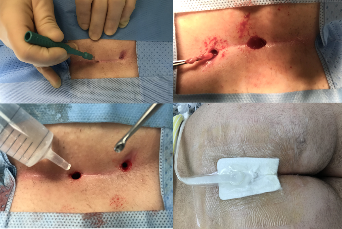
Method: The procedure includes a set of 4 steps :

a. Trephination of the sinus exit points using disposable punch biopsy kits (3-8mm)

b. Hair removal from and curettage of the sinus tracts

c. Irrigation with hydrogen peroxide: saline (50:50 mix)

d. Negative suction dressing applied (PICO) at end of procedure (for one week)

Single centre prospective review of pilonidal sinus surgery since April 2019. Patient demographics, operation notes and case notes were reviewed and analysed.

 Results: Eight children (4 male) had trephination to their natal cleft, the median age was 16 years. The mean BMI was 26.4 kg/m2. One of the patients had undergone a previous wide local excision (WLE) with primary closure and recurred. The median number of punch biopsies performed was three (range 2-7). Four (50%) children were done as day cases, compared with only 21% (4/19) in our historic cohort (WLE). There was one recurrence, this occurred at the most inferior location on a male who had undergone three punch biopsies at the procedure. The follow up time was a median of 3 months. There were no other complications.

Conclusion: Trephination for pilonidal surgery appears straightforward and feasible following this four step approach. The length of hospital stay is potentially shorter and can more often be day case. Longer term follow up will be required to ensure the recurrence rate is comparable.

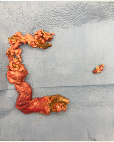
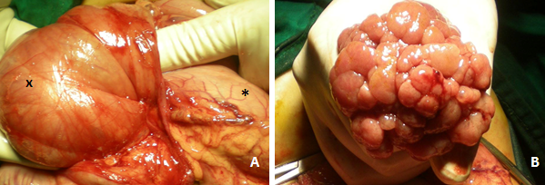
Figure 1: Minimally invasive trephination for pilonidal sinuses in pictures

Uncaptioned visual


DEVELOPING A MULTIDISCIPLINARY BREAST SERVICE FOR CHILDREN AND ADOLESCENTS

Jessica Ng1, Laura Johnson2, Jenette Kelsall2, Anthony Peel2, Paul Charlesworth1
1Department of Paediatric Surgery, The Royal London Hospital, London, United Kingdom. 2Department of Breast Surgery, St Bartholomew's Hospital, London, United Kingdom

Abstract

Aim

Adolescent and paediatric breast pathology lies at a crossover between breast and paediatric surgeons. At our centre breast surgeons are based in a non-paediatric setting. We identified a need for a multidisciplinary clinic comprising of a breast specialist, paediatric surgeon and paediatric radiologist to manage these patients. This ensures all patients, irrespective of age, have parity of care and expertise when compared with the adult breast service. The aim of this study is to share our experience of our joint paediatric breast service. 


Method

We retrospectively reviewed all patients who attended the joint breast clinic at our centre February 2017 to January 2020. Data collected included: patient demographics, presenting complaint/diagnosis, invasive management and follow-up. 


Results

Sixty patients attended joint clinic; 49 (82%) female, median age 14 years (3 months-16 years). Median number of clinic attendance per patient was 1 (1-4), 23 (38%) patients currently remain under joint-surveillance.

Presenting complaints: breast lump(s) (n=41;68%), gynaecomastia (n=9;15%), nipple discharge (n=7;12%), breast asymmetry (n=3;5%) and accessory nipple (n=1;2%). 

The diagnosis of the 41 patients with breast lump(s) included: normal breast tissue (22;54%); fibroadenoma (10;24%), breast abscess (6;15%), breast cyst (2;5%), sebaceous cyst (1;2%).

Five (8%) patients have undergone invasive procedure. Three had excision of enlarging breast lump (>5cm) after dicussion at the breast extended-MDT meeting, and histologically confirmed as fibroadenoma. One patient underwent core biopsy prior to excision. One patient had excision of accessory nipple. Of the remaining patients with radiologically suspected fibroadenomas, 3 are under joint-surveillance and 4 transitioned to our adult breast service.


Conclusion

This specialist service is of great value; concentrating expertise to improve patient experience and minimise cross specialty referrals. 8% had a surgical procedure and 7% transitioned to the adult service. A gold standard multidisciplinary approach would incorporate a one-stop service.


IS CROSS MATCHED BLOOD ESSENTIAL FOR  ALL NEONATAL SURGERY?

Leel Nellihela1, Charlene Furtado2, Ursula Wood2, Rosie O’Dea2, Susan Robinson2, Iain Yardley1
1Department of Paediatric surgery, Evelina London Children’s Hospital, London, United Kingdom. 2Department of Haematology, St Thomas’ Hospital, London, United Kingdom

Abstract

Background and Aim: When performing neonatal surgery, it is common practice to cross match blood products to ensure they are immediately available if needed. However, not all of these products are used, and their preparation has significant resource implications and reduces the pool of products available for general use. We aimed to review the usage of blood products in neonatal surgery to determine if universal cross matching was necessary. 

Methods: The medical records of all neonates undergoing major surgery in the six month period January - June 2019 were reviewed. The hospital haematology database for the same period was also accessed to cross-reference blood product preparation and usage. Data were collected on patient demographics, surgery undertaken, blood product preparation, and blood product usage. 

Results:  Thirty-four neonates (18 M, 16 F) were included. All had packed red cells cross matched prior to surgery. Twenty-five neonates (73.5%) received a transfusion of blood or blood products in the perioperative period. Babies receiving transfusions were significantly younger and smaller than those not receiving a transfusion (Table 1). Of the transfusions given, 5 (20%) were pre-operative, 15 (60%) intra-operative and 5 (20%) post-operative. Neonates undergoing surgery for Necrotising Enterocolitis (NEC) all received a transfusion with 9/15 (60%) requiring red cells, platelets and fresh frozen plasma (FFP) and 6/15 (40%) requiring red cell transfusion only. Babies undergoing surgery for less acute conditions frequently did not require transfusion peri-operatively.

 Conclusion:  In our cohort, 73.5% of neonates undergoing surgery required blood and blood products transfusion peri-operatively, the remaining blood products were returned to the laboratory unused. Transfusion was universal in babies undergoing surgery for NEC whilst older, larger babies with stable conditions often did not require transfusion. There is potential for a criteria-based peri-operative cross-matching policy to significantly reduce resource utilisation whilst maintaining safety.

Table 1.

Uncaptioned visual


THE OPTIMAL RESOURCES FOR CHILDREN’S SURGERY (ORECS) DOCUMENT: A PROOF OF CONCEPT STUDY

Kathryn Ford1, Annabel Killen2, Krupa Ravi2, Marie Lucas2, James Moran2, Leenah Abuelgasim2, Soham Bandyopadhyay2, Jessica Prince2, Kokila Lakhoo1,2
1Oxford University Hospital, Oxford, United Kingdom. 2Oxford University, Oxford, United Kingdom

Abstract

Aim of the study:

The Optimal Resources for Children’s Surgery (OReCS) document provides a strategy for integrating paediatric surgical care into national plans. We aimed to validate its use.

Method:

This is a cross-sectional study design analysing paediatric surgery capacity within a regional hub-and-spoke network in the United Kingdom. Data were gathered from one teaching hospital (TH) and three district general hospitals (DGH) using an electronic survey. Questions were based on the OReCS document and World Health Organisation (WHO) tool for assessing surgical care. Hospitals were classified by level (as per OReCS specification): level 1 (health centre), level 2 (DGH), level 3 (TH), National Children’s Hospital (NCH). Data were collected between January 2016-September 2019 as part of a pre-existing research collaboration of medical students. 

Results:

The three DGH were level 2 facilities (Table 1). Exclusive paediatric operating theatres and on-site paediatric surgeons were resources at the TH only. Paediatric surgeons visit DGH C to conduct outreach clinics.

The TH performed at NCH level for all specialities except cardiac surgery. The TH did not treat Tetralogy of Fallot or coarctations (level 3 procedures). 

In terms of general surgery procedures, DGH A and B performed inguinal herniotomy and orchidopexy for palpable testes and DGH A also performed some urology, including corrective hypospadias procedures, at level 3. Ophthalmology in DGH B was performed at level 3 and otolaryngology in DGH A and C was also performed at level 3. Plastic surgery was performed at level 1 in each of the DGHs and burns at level 1 across all hospitals. DGH A and C performed neurosurgery at level 1, but neither performed exploratory burr holes. 

Conclusion:

The OReCS document facilitated assessment and comparison of paediatric surgery services in a high-income setting and is a useful document for commissioning of services.


Uncaptioned visual


Should we explore systematically explore the contralateral side in unilateral inguinal hernia in children ? A systematic review and meta-analysis

Yasmine Houas, Fatma Thamri, Nesrine Chebil, Ghada habbachi, Nada Sghairoun, Mourad Hamzaoui
Pediatric surgery department "A", Children Hospital " Bechir Hamza", Tunis, Tunisia

Abstract

Aim of the study

Systematic evaluation of contralateral inguinal ring in children with unilateral hernia is controversial.

Our goal was to systematically review the literature to evaluate the incidence of initially asymptomatic metachronous hernias as well as the effectiveness of the various methods reported by the authors.

Method

We reviewed all the english and french literature published on MEDLINE, EMBASE, and Cochrane between 1940-2018 using as keywords "metachronous inguinal hernia children" or "pediatric metachronous inguinal hernia". Articles reporting patients who underwent unilateral inguinal hernia repair were assessed of the risk of contralateral hernia.

Mean age, Sex, incidence of metachronous hernias, time to onset, and methods of evaluation were identified.

Results 

We identified 1223 non-duplicated studies, 28 of them met our inclusion criteria. The sex ratio was 3.05. The total incidence of contralateral hernias not initially present was 8.18% (7-15%). The delay of appearance was 18 months (1 month-3 years). The different contralateral inguinal ring evaluation strategies were close clinical follow-up (73.64%), laparoscopy (16.42%), ultrasound (3.18%) and systematic surgical exploration (0.20%). Ultrasonography was the most sensitive mean of exploration (p = 0.09). Laparoscopy was the most specific mean of evaluation (p = 0.2)

The risk of developing a strangulated contralateral hernia in patients during the clinical follow-up was 0.13 %.

Conclusion

This review highlights the low incidence of initially asymptomatic contralateral hernias and the rarity of complications. Ultrasound is the most reliable examination despite its low specificity and relatively high cost.


“EXTUSSUSCEPTION": RARE CASES OF SMALL BOWEL INTUSSUSCEPTION THROUGH ILEOSTOMY IN CHILDREN

Olugbenga Awolaran, Ruth Hallows
Royal Alexandra Children's Hospital, Brighton, United Kingdom

Abstract

Background

Intussusception through the stoma is very rare. There are around 15 cases reported in the adult literature. To date, there is none in the paediatric population. We report 4 paediatric patients with 5 occurrences of intussusception through an ileostomy.

Method

Medical records of children with intussusception through ileostomy managed in our centre between 2016 and 2019 were retrospectively reviewed. Literature from major databases was searched and reviewed.

Results

4 patients with 5 occurrences of intussusception through an existing ileostomy were managed. Indications for ileostomies were post-NEC colonic stricture, Hirschsprung’s disease, GI-dysmotility in an ex-preterm with severe neuro-developmental disability and ileo-colic intussusception in a child with cystic fibrosis. Ages at presentation with intussusception through ileostomy were 4months, 8months, 5years and 17years respectively. Duration between ileostomy formation and intussusception ranged between 1day and 6months.

They all presented with compromised ileum intussuscepting through their ileostomies and had unsuccessful attempts at bedside reduction. Emergency laparotomy and reduction were performed in all cases. 2 had stoma refashioning only and 2 additionally had ileopexy. 1 of the two that had ileostomy refashioning only, had a recurrence 4 weeks post-surgery requiring re-operation and ileopexy.

 Conclusion

Intussusception through the stoma is a clinical entity that should be distinguished from simple stoma prolapse. There is a separate loop of bowel intussuscepting through the stoma with the original stoma edge forming a constricting ring around the intussuscepted segment (Fig 1).

All of the cases in our series as in all other published reports required emergency operative reduction. As demonstrated in this series, there is a risk of recurrence (1/4), so ileopexy should be considered.


Uncaptioned visual


Fig 1 – Compromised ileum (white arrow) intussuscepting through existing stoma edge (black arrow), demonstrating the typical bowel-in-bowel appearance in a 4-month old ex-preterm who had ileostomy for post-NEC stricture.


RISE AND FALL OF NON-TUBERCULOUS MYCOBACTERIAL (NTM) LYMPHADENITIS IN CHILDREN

Shani Fernando1,2, Noel Karalus2, Udaya Samarakkody2
1University College Cork, Cork, Ireland. 2Waikato Hospital, Hamilton, New Zealand

Abstract

Aim


NTM lymphadenitis (commonly called Mycobacterium Avium Intracellulare Scrofulaceum or MAIS complex) typically presents in the cervico-facial region in toddlers. We have noticed reduction of presentations in the recent years. This study examines the incidence, demographics and disease pattern of MAIS over 25 years in a tertiary centre in (country).


Method


NTM lymphadenitis between 1995 and 2019 (25 years) were retrieved from microbiology and histopathology services. They were analysed for presentation and management by the year, age, sex, country of birth, ethnicity and BCG vaccination history. The population data were obtained from national statistics.


Results


From a total of 85 patients 76% was female.  The age ranged from 1 year to 15 years (mean = 4.7) All but three were under 7 years and none was under 1 year. Caucasians comprised 65%. All patients were born locally and none had BCG vaccination.  


All received surgical treatment varying from incision and drainage with curettage of feeding lymph nodes to complete excision of the MAIS complex. Three received adjuvant anti TB treatment when complete excision was not possible due to the risk to facial nerve.

Complete resolution was seen in all. The follow up was up to one year except patients treated in 2019. 

The highest number of admissions was seen in 2008 (9) and none in 2013. The incidence was 2 per year in the last two years.

A clear gradual reduction in the incidence was seen over the 25 year period (Table). 


Conclusion


The disease pattern with female predominance and toddler age is seen. Lack of BCG vaccination may have prevented cross-immunity to MAIS. While the population is increasing the incidence of MAIS is reducing gradually with a plateau of two cases a year in the recent years. Any association to the changing lifestyle in toddlers warrants  investigation.

Table:

Uncaptioned visual


DOES HAVING A BIGGER BABY MINIMISE SURGICAL COMPLICATIONS FROM STOMA CLOSURE FOR THOSE WITH NECROTISING ENTEROCOLITIS (NEC)?

CLARA CHONG1,2, DEBASISH BANERJEE1, Iain Yardley2, THOMAS TSANG1
1NORFOLK AND NORWICH UNIVERSITY HOSPITAL, NORWICH, United Kingdom. 2EVELINA CHILDREN HOSPITAL, LONDON, United Kingdom

Abstract

Aim of the Study

Neonates living with enterostomy after NEC often have poor growth and high stoma losses. Suboptimal nutritional state makes them poor candidate for stoma closure. Historically, surgeons opt to wait for the babies to become bigger prior to reversing stomas to off-set potential surgical complications in small premature infants. We aim to highlight incidence of surgical complications in this cohort.


Method

Neonates with histological proven NEC with a stoma from 2006-2016 were studied. Weight at stoma closure was noted. Weight-for-age z-score (z) denotes current weight of individual in relation to reference population at specific gestational age, with weight at 50th centile having a z=0. z was calculated using LMSGrowth method. Complications highlighted include those managed conservatively as well as episodes requiring further operations post stoma closure. Result presents in median (range).


Results

43 neonates with NEC underwent stoma closure with 12 (28%) complications. Time to closure was 14 weeks (3-81). Weight at stoma closure was 3.2kg (1.2-8.9), corresponding to z=-2.5 (1.4 - -4.7). 31 (72%) patients had a z<-2 at the time of stoma closure which fulfils WHO criteria of being moderately underweight. 9/12 complications came from this group: 2 incisional hernia, 1 enterocutaneous fistula, 3 obstruction, 3 surgical line insertion for parenteral nutrition. Time to closure for this group was 9 weeks (6-67). Complications were least in those achieved weight >3kg and z>-2. Their time to closure was 18 weeks (3-45).


Conclusion

Surgical complications were least in the group which achieved 3kg with good global nutrition (z>-2). This was however only feasible for the minority, whilst the rest lived for prolong period with faltering growth. Z-score as opposed to weight should be used to guide timing of stoma closure. One should consider closure when patients are stable to minimise potential long-term impact of faltered growth on neurodevelopment.


Uncaptioned visual


THE DECELLULARISED SPLEEN AS A MODEL FOR WHOLE ORGAN REVASCULARISATION

Alessandro Pellegata, Joseph Davidson, Giulia Nucci, Elisa Zambaiti, Paolo De Coppi
GOS-UCL Institute of Child Health, London, United Kingdom

Abstract

Aim of the Study: Vascularisation of tissue-engineered organs remains one of the most significant challenges to translational adoption. Here, we report the decellularisation of the spleen, a highly vascularised organ, and subsequent reseeding using endothelial cells, reconstituting a vascular network.


Methods: Explanted spleens from adult rats were cannulated through the coeliac trunk and decellularised using a previously established detergent-enzymatic protocol. Removal of cells was assessed with DNA quantification, while preservation of extracellular matrix proteins was confirmed by quantification assay. The scaffold was then repopulated with human endothelial cells in both static and bioreactor dynamic culture conditions for 5 days.


Main Results: Whereas static culture did not lead to successful repopulation, in bioreactor dynamic culture we observed repopulation of the vascular network through multiple branches of the vessel tree from the splenic artery. Endothelial cell presence was confirmed by immunocytochemistry for the endothelial marker, CD31(Figure). Perivascular staining for collagen IV may suggest newly deposited collagen by endothelial cells. 


Conclusions: This study provides an important model for the revascularisation of a whole organ decellularised scaffolds. Further work will address the scaling up of the model and in-vivo transplantation to assess vascular perfusion.


Figure: Decellularised spleen after endothelial cells seeding in bioreactor. A+E: Staining for DAPI (nuclei) in blue , B+F: CD31 (endothelial marker) in green, C+G: collagen IV in red. D+H: Composite image demonstrates endothelial lining of the vessel.

Uncaptioned visual

ETERTIARY SURVEY: ALMOST PERFECT

Arun Kelay, Catherine Bradshaw, Chris Parsons, Stewart Cleeve
Royal London Hospital, London, United Kingdom

Abstract

Aim of the study


Missed injuries are a significant cause of morbidity after major trauma. The 2015 National Peer Review Report of Major Trauma revealed that major trauma centres achieved only 26% compliance with a ‘protocol for formal tertiary survey’. 


An electronic tertiary survey (eTS) form has been implemented at our major trauma centre in line with the advent of electronic patient records. We determined whether this standardised form improved compliance with and comprehensiveness of tertiary survey completion. 


Methods


Prospective audit of tertiary survey completion for trauma patients from October 2019 - January 2020. Data collected included age, mechanism, injuries, timing and comprehensiveness of tertiary survey. This was compared with historical data from 2014 and 2018 prior to electronic patient records. 


Results


35 patients were admitted with a median age of 10 years (9 months – 15 years). Blunt trauma was responsible in 27 (77%), with penetrating trauma in 8 (23%). One patient was transferred to a cardiac centre soon after admission and was not included in the analysis. The tertiary survey was completed in full in 33/34 (97%) on a median of the third day of admission (1st – 21st day of admission). In 2018 the completion rate was 22/24 (92%), whilst in 2014 this was 7/13 (54%), see Table 1. There have been no missed injuries in the current period.


Conclusion


A standardised electronic tertiary survey form can both improve the tertiary survey completion rate and mandates a comprehensive assessment. Introduction of a daily trauma MDT is hoped to achieve 100% completion. 


Table 1. Compliance by study period


Study Period

Tertiary Survey Completion

Fisher’s exact test

Complete Assessment

Fisher’s exact test

2014

  7/13 (54%)

-

  5/13 (38%)

-

2018

22/24 (92%)

p = 0.0134

20/24 (83%)

p = 0.097

2019

33/34 (97%)

p = 0.56

33/34 (97%)

p = 0.15


NEURODEVELOPMENTAL OUTCOMES OF PREMATURE INFANTS WITH NECROTISING ENTEROCOLITIS

Catherine Bradshaw1, Ashwath Bandi1, Narendra Aladangady2,3, Philippa Chisholm4, Ajay Sinha1, Michael Hird1, Devesh Misra1
1Royal London Hospital, London, United Kingdom. 2Homerton University Hospital, London, United Kingdom. 3Queen Mary University, London, United Kingdom. 4Homerton University Hospital Hospital, London, United Kingdom

Abstract

Aim


To assess contemporary neurodevelopmental outcomes of infants with necrotising enterocolitis (NEC). A comparison of infants requiring medical management with those undergoing surgery for NEC, relative to case-matched controls, and compared to published data.


Methods


Case-matched cohort study using a prospectively maintained database over 5 year time period (2010-2015). Inclusion criteria: gestational age (GA) <30 week, birth weight (BW) <1500g, NEC (Bell stage 2 or 3).   Controls were matched for GA, BW and ethnicity. Standardised neurodevelopmental outcomes at 2 years were collected. Statistical analysis with Chi-squared test. 


Results


109 infants with NEC were identified. Complete data was available for 61% (67/109). There were 17 deaths (16%) and 50 survivors. Total 100 infants analysed (50 NEC and 50 controls). Demographics of the NEC cohort; Median BW 750g (470-1416), Median GA 25 (23-30), 28 male infants, 31/50 (62%) non-white ethnicity.  


Adverse neurodevelopmental outcomes were seen in 36% (18/50) with NEC and 24% (12/50) controls (NS). 40% (6/15) surgical NEC had adverse neurodevelopmental outcomes; 27% (4/15) were severe. While 34% (12/35) medical NEC had adverse neurodevelopmental outcomes; 11% (4/35) were severe (NS). No difference was observed when compared to the controls. 


Conclusion


No difference in adverse neurodevelopment was identified in this study. Explanations include incomplete data collection on neurodevelopmental outcomes and insufficient sample size. Adverse neurodevelopment outcomes in infants with NEC appear to be unchanged in the last decade, evidence is limited by variations in definitions and incomplete data collection. There is an urgent need for standardised neurodevelopmental outcome protocols for infants with surgical NEC, which may have to be driven by surgeons.


Author

Year

Time period

% Total adverse neurodevelopmental outcome and NEC (N)

% Severe neurodevelopmental outcome and surgical NEC (N)

Rees

2007

1975-2002

45 (821)

-

Fullerton

2017

1999-2012

-

38 (449)

Bradshaw

2020

2010-2015

36 (50)

27 (15)


INCREASED INCIDENCE OF POST APPENDICECTOMY COLLECTIONS IS RELATED TO FAILURE TO ADHERE TO ANTIBIOTIC PROTOCOL RATHER THAN SEVERITY OF DISEASE

Elizabeth O'Connor, Lucy Henderson, Bruce Jaffray
The Great North Children's Hospital, Newcastle upon Tyne, United Kingdom

Abstract

Aims

Collections following appendicectomy occur in 3-9%. We report the effect of  antibiotic protocol and other variables on this parameter.

 Methods

Children undergoing appendicectomy over the last 4 years were categorised as complex appendicitis (CA) if perforation or gangrene was specified on histology, otherwise simple appendicitis (SA) or normal.  Collections were a collection on ultrasound.  Use of bacteriology culture of peritoneal fluid was quantified.  Antibiotic protocol was Co-Amoxiclav for 2 days in SA and 5 days in CA; any variation was off protocol.

We used χ2 test, and logistic regression to analyse associations in SPSS.  Results are mean, or median (range).

Results

618 children, 396 boys, underwent surgery at median age 9 years (1-15). 590 (95%) laparoscopic, 8 converted and 20 open.

Histology: SA 385 (48%), CA 256 (41%), normal 66 (11%). CA was significantly related to age (odds ratio 0.9 (0.8-0.96), p <0.001), but not sex.

72 (12%) developed collections.  Collections were more common following CA: 51/256 (20%) vs SA: 21/385 (6%) p<0.001.  

Collections increased from 9% in 2016, to 17% in 2019 p = 0.042, without any increase in CA: 66/163 (41%), vs 54/121 (45%) p =0.4.

57/350 (16%) off protocol developed a collection, while 11/152 (7%) on protocol developed a collection, p = 0.003.

151 had bacteriology culture, with sensitivities in 135.  More CA had culture: 101/256 (67%) vs SA: 50/385 (33%) p<0.001.  Culture was more likely with collections: 44/72 (61%) vs no collection 107/569 (17%) p<0.001.

There was no difference in Co-Amoxiclav resistance with, 6/28 (21%) and without collections 21/107 (20%) p = 0.8.

Conclusions

Collection rate has significantly increased, without an increase in CA.   Bacteriology culture, and resistance to Co-Amoxiclav did not influence collection rates. Adherence to protocol was associated with fewer collections.  

Uncaptioned visual

Followed protocol p = 0.006. Resistance to Co-Amoxiclav p = 1


Management of Testicular Events - A National Survey of Practice

Felicity Arthur1, Lisa Steven2, Phil Hammond2, Melanie Clarke2, Gregor Walker1
1Royal Hospital for Children, Glasgow, United Kingdom. 2Royal Hospital for Sick Children, Edinburgh, United Kingdom

Abstract

Aim: We aimed to investigate current intended practice in managing perinatal torsion, torsion of the undescended testicle (UDT) and the expectant management of the solitary testicle. The evidence base for these conditions is limited and practice varies throughout the UK.


Methods: A electronic questionnaire was sent to all consultants practicing Paediatric Surgery/Urology in one National Deanery. Questions aimed to determine methods of fixation of the ipsi- and contralateral testicle in 3 different scenarios: testicular torsion in the neonate, torsion of the UDT and the “vanishing” impalpable testicle.  We defined antenatal torsion as a firm non tender scrotal swelling noted at birth and postnatal torsion as a tender erythematous scrotal swelling in the case of a previously documented normal testicle. 


Results: Response rate was 72% (16/22). In antenatal torsion, only 8(50%) explore, with 2(14%) exploring "as soon as possible". The remainder use clinical state to dictate timing. In postnatal torsion, 15(94%) explore, 12(75%) “as soon as possible”, although not all (13(81%)) would explore the contralateral side and only 10(63%) would remove an unsalvageable testicle. Methods of fixation were equally spread between dartos pouch, 3 point fixation,and a combined technique with 15(93%) using absorbable sutures. All respondents would perform an orchidopexy at the time of exploration of a torted UDT, but only 10(63%) would explore the contralateral side.  In the vanishing/solitary testicle, only 8(50%) explore and remove the testicular remnant following diagnosis, and 3(18%) fix the contralateral side. 


Conclusion: The management of testicular torsion in different scenarios (perinatal, undescended, solitary testes) varies between centres and practitioners. The evidence base in scanty and significant equipoise exists. A study of outcomes is warranted to guide best practice.


Does Antenatal Intra-Abdominal Bowel Dilatation Predict Outcome in Gastroschisis?

Alison Campbell1, Wayne Fradley1, Mahmoud Motawea1, Azzam Abdelrahman1, Helen Burgess2, Prasad Godbole1, Govind Murthi1, Ross Fisher1, Emma Ferriman2, Sean Marven1
1Department of Paediatric Surgery Sheffield children's Hospital, Sheffield, United Kingdom. 2Department of Obstetrics and Gynaecology Sheffield Teaching Hospitals, Sheffield, United Kingdom

Abstract

Aim of study

Intra-Abdominal Bowel Dilatation (IABD) on ante-natal imaging is thought to predict likelihood of complex gastroschisis and delayed time to full feeds. We reviewed our antenatal data and post-natal outcomes to inform our antenatal counselling.

Methods

Data was collected retrospectively after approval from the audit and effectiveness committee of both units. Antenatal cases were identified from the labour ward database; postnatal cases from the neonatal surgical unit (NSU) discharge log. Cut off for IABD was based on literature review (>14mm before 33 weeks – Andrade et al Ultrasound in Obstetrics & Gynecology 2019;54(3):376-380.). Spearman’s rank correlation coefficient was used to calculate association between IABD and time to full feeds.

Results

From May 2011 – March 2019, 88 patients (41 female, median weight 2.43kg, median gestational age 37 weeks) were admitted to NSU with gastroschisis (18 complex, 70 simple). 74 patients had data from antenatal counselling at our regional centre.

32 patients had IABD, with measurements, on their antenatal scan. These included 10/18 complex with a median IABD of 23.5mm (20-48) and 22/70 simple - median IABD 20mm (16-67).

Significant IABD was measured in 10/18 complex patients. The others had no IABD(4), IABD detected but no measurement (2). 2 were transferred from other regions (therefore no antenatal data). Positive-predictive value of IABD for complex gastroschisis was 29% (21 -39). Negative-predictive value was 88.9% (77 – 95).

Median time to full feeds was 37 days if no IABD (14 – 1293) and 36 days if IABD present antenatally(10-1239). Analysis failed to show any significant relationship between IABD and time to full enteral feeds (Spearman’s: rs= 0.16385 p (2-tailed) =0.21)

Conclusion

Intra-abdominal bowel dilatation is not always a predictor of poorer outcome in gastroschisis. This information is useful for antenatal counselling.


PATIENT CHARACTERISTICS ASSOCIATED WITH SPONTANEOUS REDUCTION OF INTUSSUSCEPTION IN CHILDREN

Alexis Bowder, Kimberly Somers, Sabina Siddiqui
Children's Hospital of Wisconsin, Milwaukee, USA

Abstract

Aim of Study: Management of intussusception in the United States varies widely between healthcare centers. Our study aimed to identify the cohort of patients who experience spontaneous reduction of intussusception (SROI) and may not require radiographical or surgical intervention and the resources and therefore require less resource utilization.

Methods: We performed an IRB-approved retrospective study of all patients with a diagnosis of intussusception from 2013 to 2018 at a Level I Children’s Hospital Surgery Center. Data included demographics, presenting symptoms, imaging modality, need for reduction, radiologic reduction approach and number of attempts, and need for surgical reduction. 

Results: Two hundred and eighty-one patients were identified, that majority (68%) female. The median age was 1.8 years (95% CI 1.45, 2.15).  52% of our patients presented with symptom duration of < 24 hours and duration of symptoms was not predictive of need for radiographic or surgical intervention. Forty-four patients (16%) had spontaneous reduction of their intussusception. Patients presenting with small bowel intussusception (81%) were more likely to have spontaneous reduction of their intussusception (p=<0.0001). However, spontaneous reduction was also documented in patients who had ileocolic (17%) and colonic (2%) intussusception. The presence of bloody stools, nausea, and diarrhea were not associated with a higher risk of requiring a radiological or surgical intervention in this study. Finally, of the patients who required transfer, patients with SROI had the highest percentage of ambulance transfers across all groups. 

Conclusion: Among patients who present with intussusception, there is a high rate of spontaneous reduction, yet these patients are often transferred by ambulance. This implies that certain children may be safely observed without radiographic or surgical intervention. Further investigation into identifying patient likely to undergo SROI can help optimize health care service utilization including transport, admission, and length of stay.


LAPAROSCOPIC "TRUE" INGUINAL HERNIOTOMY: A SYSTEMATIC REVIEW

Ayman Goneidy, Nick Lansdale, Robert Peters, David Wilkinson
Manchester University NHS foundation trust, Manchester, United Kingdom

Abstract

Aim of the study

Inguinal herniotomy is commonly performed in paediatric surgical practice. Numerous studies have compared outcomes of laparoscopic and open techniques.   However, the term ‘laparoscopic hernia repair’ covers numerous techniques making a true comparison difficult. The aim of this study was to review the outcomes for laparoscopic surgery that replicates open herniotomy, i.e. those in which the hernial sac was dissected free from the peritoneum followed by suture closure of the internal ring.

Methods

A systematic review was performed (PRISMA guidelines), of all articles reporting results from laparoscopic paediatric inguinal hernia repairs in which the technique was described as laparoscopic sac dissection with sutured ring closure (LSDS).  Exclusion criteria were: non-English language, patients older than 18 years and those including duplicate data.  Data collected included patient demographics, laterality, operative time, complications and recurrence. Data is presented as median (interquartile range) unless stated.

Results

A total of 592 papers were identified of which 18 met the inclusion criteria, resulting in 4152 LSDS hernia repairs being included in the final analysis. Age at repair was 38.4(24.7-66.3) months, operative time was 38.5(27.3-49.4) minutes and follow up was 16 (12.5-23.3) months. 26/4152 (0.6%) hernias recurred.  

Conclusion

The LSDS technique essentially repeats the steps of open herniotomy with the added benefits of laparoscopy, minimal handling of the vas and vessels and prevention of scaring within the inguinal canal. Recurrence rates are comparable to those reported for open inguinal hernia repairs and lower than many reports for laparoscopic techniques without dissection of the sac.


INTRAOPERATIVE BLOOD TRANSFUSIONS IN NEONATES – CAN WE RATIONALISE OUR PROCESS OF PERIOPERATIVE BLOOD ORDERING?

Helen Figgins, Natalie Quinn, David Wilkinson, Nicholas Lansdale, Catherine Doherty, Robert Peters
Royal Manchester Children's Hospital, Manchester, United Kingdom

Abstract

AIM

The national shortfall of blood donation alongside wide variation in the preoperative ordering of red blood cell products(RBCP) for neonates undergoing surgery compounds the need to improve and standardise this practice. The aim of this study was to determine actual RBCP usage compared to preoperative requests and to identify factors that influence transfusion.


METHOD

A random retrospective case note review was performed on 100/144 neonates undergoing surgery at a single centre between October 2017 and October 2018. Four cases were excluded due to lack of intraoperative details. Data collected included: weight, corrected gestational age(CGA), gender, diagnosis, operation performed, and preoperative serum haemoglobin concentration. Preoperative ordering as well as volume of intraoperative blood use was assessed. Data are presented as median(interquartile range) unless otherwise stated. 


RESULTS

A total of 96 neonates were included this study,89/96(93%) had RBCP crossmatched for surgery, totalling approximately 23,415ml of donor blood.

Only 11/96(11.4%) required an intraoperative RBCP transfusion. The volume transfused was 30ml(12-45). The RBCP surplus alone represented an estimated financial cost of £9,032.

Strong linear correlation existed between weight and CGA in our cohort(r=0.82,p=<0.0001,95%CI 0.74,0.87)[FIGURE1]. Early CGA and low weight demonstrated strongest association with intraoperative RBCP transfusion with relative risks of 8.57(p=<0.0001,95%CI 3.45,21.27) and 11.6(p=<0.0001,95%CI 4.40,30.55) higher for transfusion with a CGA of <28 weeks and weight <1kg respectively. 


CONCLUSIONS

The majority of neonates undergoing surgery had an adult unit of cross-matched blood available, however only 11.4% required transfusion, with a median volume approximately equal to one pedipack unit and maximum of two. Standardisation of blood ordering schedules could reduce wastage of blood products, and multiple donor exposure thereby reducing adverse transfusion-related outcomes.

FIGURE1:Relationship between CGA and weight at operation indicating neonates who required intra-operative RBCP transfusion.

Uncaptioned visual


THE IMPACT OF COVID-19 ON  PAEDIATRIC SURGICAL PROVISION FOR INFANTS WITH INGUINAL HERNIA IN THE UK

Eva H Sorensen1, Nigel J Hall2,3, Clare M Rees1,4
1Chelsea & Westminster Hospital NHS Foundation Trust, London, United Kingdom. 2University Hospital Southampton NHS Foundation Trust, Southampton, United Kingdom. 3University of Southampton, Southampton, United Kingdom. 4Imperial College Healthcare NHS Trust, London, United Kingdom

Abstract

Aims:

To investigate changes in surgical provision for infants with inguinal hernia during the first wave of the Covid-19 pandemic. 

Methods:

An electronic survey was sent to all specialist paediatric surgical centres (n=27) in the UK in June 2020. Questions were asked about changes to outpatient assessment, elective and emergency operating capacity, and surgical approach. 

Results

All 27 centres responded.  

Outpatient clinics were affected by the pandemic in all institutions.  Face to face consultations were replaced by telephone +/- video in 4 (15%) centres, the remainder had some capacity to provide in-person consultations, but were also using telephone (12, 44%), video consultations (1, 6%) or both (10, 37%). 

All institutions managed incarcerated inguinal hernia as an emergency despite reduction in emergency capacity in 10 (37%) centres. Urgent elective capacity was reduced in 18 (67%), maintained in 7 (26%) and ceased completely in 2 (7%) centres. 

Local policies on management of reducible hernias differed.  Ten (37%) institutions reported an active change in policy (especially from those referred from neonatal intensive care and emergency department), being more likely to add patients to an urgent elective list or admit for emergency surgery (Figure 1). 

Uncaptioned visual

Surgical approach was affected in the 11/17 (65%) institutions who would normally perform open and laparoscopic hernia repairs, who stopped laparoscopy. Three institutions who exclusively used a laparoscopic approach continued to do so. 

Conclusion

The approach to the management of infants with inguinal hernias was affected by the Covid-19 pandemic, due to reduction in operating capacity and concerns regarding the safety of laparoscopy. Emergency service provision was maintained. The impact of these changes on infants with inguinal hernia should be assessed.


COST-EFFECTIVENESS OF LAPAROSCOPIC AND OPEN PAEDIATRIC INGUINAL HERNIA REPAIR

Christine Lam, Pavleen Dhedli, Samantha Russell, Francesca E Stedman, Nigel J Hall
Southampton Children's Hospital, Southampton, United Kingdom

Abstract

Aim: Laparoscopic inguinal hernia (IH) repair is an alternative to open surgery. A potential advantage of laparoscopy is prevention of contralateral metachronous hernia although some studies report a higher recurrence rate. To our knowledge the relative cost-effectiveness of these approaches is unknown. We aimed to determine cost-effectiveness of open versus laparoscopic IH repair factoring in both recurrence and contralateral metachronous rates.

Method: Retrospective single centre study of children (<5 year) undergoing elective open or laparoscopic unilateral IH repair between February 2018 – October 2019. Patients were included in reverse chronological order (for accurate and contemporary costing) until 10 patients in each of 4 groups were achieved (open daycase / open overnight stay / laparoscopic daycase / laparoscopic overnight stay). Cases with additional procedure or complex comorbidities were excluded due to higher costs. Patient level information and costing system (PLICS) data were obtained from the hospital finance system. This is regarded as the actual cost of admission and is used to calculate national tariffs. Mean (SD) cost per case was compared for open and laparoscopic procedures. A financial model was created taking into account the difference in both contralateral metachronous and recurrence rates between laparoscopic and open approaches.

Results: Open daycase IH repair cost was £1866 (SD: 311) compared to £2210 (SD: 391) for daycase laparoscopic repair. Open overnight IH repair cost was £2442 (SD: 497) compared to £2585 (SD: 384) for laparoscopic overnight repair. In the financial model, for overnight stay surgery, laparoscopy is more cost effective than open surgery when the combined recurrence and metachronous rate for open surgery exceeds that for laparoscopy by >5.84% (Figure). For daycase surgery this increases to 18.43%. 

Conclusion: Based on current representative contralateral metachronous and recurrence rates, our data suggest laparoscopic paediatric IH repair is more cost effective than open surgery for overnight cases but not daycases.

Uncaptioned visual


A COMPARISON OF SKIN ADHESIVE VERSUS SUBCUTICULAR SUTURES FOR WOUND CLOSURE IN PAEDIATRIC LAPAROSCOPIC SURGERY

YEW CHAI LEIW1, Anand Sanmugam1, Thambidorai Rajendra Rao1, Shahrul Aiman Soelar2, Mohan Arunasalam Nallusamy2, Shireen Anne Nah1
1University Malaya Medical Centre, Kuala Lumpur, Malaysia. 2Hospital Sultanah Bahiyah, Alor Star, Malaysia

Abstract

Aim of the study: There is no “Gold Standard” wound closure method in paediatric laparoscopic surgery. Cyanoacrylate skin adhesive is an alternative to subcuticular suturing. Our study aimed to compare the effectiveness of skin adhesive to routine subcuticular sutures in paediatric laparoscopic wound closure. We also compared parental satisfaction in both groups.


Method: With institutional ethical approval [NMRR-20-338-53062(IIR)] we reviewed all children aged 12 years and below who had laparoscopic surgery from February 2019-January 2020. Data collected included demographic characteristics, intra-operative diagnosis and procedure, post-operative recovery information and wound complications. We assessed parental satisfaction via telephone interview based on Likert scale assessment of wound appearance. We categorized wounds as follows: Class I-clean, II-clean contaminated, III-contaminated, IV-dirty. Categorical variables were analysed using chi-squared tests with p<0.05 considered significant. 


Results: There were 117 eligible subjects. Seven subjects were excluded due to conversion to open surgery. Median age at the time of surgery was 1 year (range 0-12), 76 (69.1%) were male. Sixty-six subjects received skin adhesive for wound closure, and 44 subjects received sutures. Six subjects in this study had wound complications: 5 from the skin adhesive group and 1 from the suture group. There were no significant differences in wound complications or in parental satisfaction for either method. Surgical wound class II, III and IV showed a significant risk of postoperative surgical site infection, p=0.021 (Table). 


Conclusion: Skin adhesive and sutures both have a low incidence of wound complications in paediatric laparoscopic wound closure. This study showed high parental satisfaction for both techniques. In conclusion, skin adhesive is a feasible alternative and is effective for the closure of laparoscopic wounds.


Uncaptioned visual


MAINTAINING A MINIMALLY INVASIVE SURGICAL SERVICE DURING A PANDEMIC

Jayaram Sivaraj1, Stavros Loukogeorgakis1,2, Stefano Giuliani1, Dhanya Mullassery1, Simon Blackburn1, Joe Curry1, Kate Cross1, Paolo De Coppi1,2
1Department of Specialist Neonatal and Paediatric Surgery, Great Ormond Street Hospital, London, United Kingdom. 2UCL GOS Institute of Child Health, London, United Kingdom

Abstract

Aim of the Study

Early in the COVID-19 pandemic, there was significant concern about the safety of Minimally Invasive Surgery (MIS). Limited pre-operative testing capacity and the possible risk to surgeons through aerosolization (especially in untested patients) were the primary reasons for this. Our department continued to perform MIS using PPE (including FFP3 masks) as per hospital guidelines for all surgery. We aimed to review whether this was safe for patients and surgeons during the pandemic.

Methods

We conducted a retrospective review of all MIS, Open (OS), and Endoscopic (ES) surgical procedures performed by our department during the first lockdown in England (23/03/20 to 15/06/20). These were compared with operations from the same time period in 2019. Demographic, clinical, and SARS-CoV2 PCR test data were collected. Outcomes reviewed included respiratory complications (RC), length of stay (LOS), and symptomatic operating surgeon COVID-19 infections (OSI). 

Results

MIS comprised 52% (82/157) of procedures performed during the first 2020 lockdown compared to 33% (78/234) during the same period in 2019 (p=0.0002).  2 patients (1.2%) were COVID-19 positive (MIS=1, OS=1, ES=0). In MIS 2020, 7/82 patients (8.5%) had RC compared to 7/66 patients (10.6%) in OS 2020 (MIS vs OS p=0.78). In MIS 2019, 6/78 patients (7.7%) had RC (MIS 2019 vs MIS 2020 p=1). Median LOS in MIS 2020 was 2.5 (IQR  6) compared to 2 (IQR 5.8) in 2019. There was no significant difference in LOS between the time periods (p=0.95). There was no OSI following surgery on a known COVID-19 positive patient.  No surgeon tested positive for COVID-19 (symptomatic testing only) during this period of lockdown operating. 

Conclusions

Our department used MIS for half the of procedures during the first COVID-19 lockdown. Our data suggests MIS was safe for patients and surgeons during the pandemic with no increase in RC or OSI. 


SURGERY FOR NECROTISING ENTEROCOLITIS (NEC) AT THE THRESHOLD OF VIABILITY - A SINGLE INSTITUTION'S 6-YEAR EXPERIENCE

Hemanshoo Thakkar, Hammad Khan, Geraint Lee, Iain Yardley
Evelina London Children's Hospital, London, United Kingdom

Abstract

AIM OF THE STUDY

The threshold of viability for babies born prematurely is currently 22+0 weeks for a liveborn infant. Necrotising enterocolitis (NEC) remains the most serious gastrointestinal disorder affecting premature infants. We sought to review our institutional experience in the management and survival of those born at <24 weeks with NEC.

METHODS

All infants born at <24 weeks gestation between January 2015-December 2020 with suspected or confirmed NEC were identified. Patient demographics, clinical features at the time of diagnosis, biochemical and radiological findings, surgical details, complications and survival data were extracted from the electronic medical record.

RESULTS

41 admissions <24 weeks were recorded during the study period of which 21 (51%) were diagnosed with NEC. The median birth weight for this cohort was 600g (450-706g). Median age at onset of NEC was 23 days (4-79). 14/21 (67%) had a rise in CRP and 14/21 (67%) a drop in platelets. Pneumatosis was evident on plain radiography in 7/21 (33%), pneumoperitoneum in 3/21 (14%) and a paucity of intra-luminal gas in 5/21 (24%). Abdominal USS was performed in 6/21 (29%), universally identifying thickened, aperistalsic bowel with an extra-luminal collection. 7/21 (33%) were managed non-operatively. 14/21 (67%) underwent laparotomy with a median time to surgery of 2 days (1-5). Findings at surgery were distal ileal perforation in 12/14 (86%) and multi-focal NEC in 2/14 (14%). 4/14 (29%) had post-operative wound dehiscence of which one developed enterocutaneous fistulae. One other patient required further laparotomies. Overall survival to discharge was 11/14 (79%) in those undergoing surgery.

CONCLUSIONS

The diagnosis of NEC in infants born <24 weeks can be challenging with inconsistent clinical, biochemical and radiological features. Ultrasound scanning may be a more useful modality.  Although mortality is significant at 21%, a gestational age <24 weeks should not be considered a contraindication to surgical intervention.


EVALUATION OF AN ANTIBIOTIC TREATMENT PATHWAY FOR CHILDREN FOLLOWING APPENDICECTOMY

Matthew Jobson, Natalie Durkin, Benjamin Allin, Oliver McCallion, Grace Barnes, Alex Lee, Stephane Paulus
Oxford Children's Hospital, Oxford University Hospitals NHS Foundation Trust, Oxford, United Kingdom

Abstract

Aim of the Study


Duration of post-operative antibiotic therapy following appendicectomy in children is variable. Our aim was to assess the safety and efficacy of a standardised antibiotic pathway for treatment of children undergoing appendicectomy in a single paediatric surgical centre.


Method


The pathway ceases antibiotics immediately post-operatively for simple appendicitis, whilst for perforated appendicitis, 72 hours are mandated, followed by a protocolised review to determine ongoing treatment. Days of antibiotic therapy (DOT) data, defined as one day in which a patient is given one antibiotic, were collected retrospectively for all children undergoing appendicectomy in a six-month period prior to pathway implementation (01/04/18-31/09/18), and prospectively for three-months after implementation (01/05/19-31/07/19. To assess for rarer adverse events associated with pathway implementation, length of stay, complication rate, readmission rate and reoperation rate data were collected for nine months post-implementation (01/05/19-31/12/19).  Comparison between groups was made using Wilcoxon-Rank-Sum test for continuous data, and logistic regression analyses, adjusted for age at operation, for categorical data.


Results


There were 57 children in the pre-implementation group, median age 9.5 (6-12.5) years, 18 (32%) of whom had perforated appendicitis, and 98 children in the post-implementation group, median age 12 (8.9-14.3) years, 37 (38%) of whom had perforated appendicitis. The median (IQR) DOT per patient were 7 (2-14) pre-protocol, and 2 (2-5.75) post-protocol, p=0.00034. Median (IQR) length of stay was shorter in the simple appendicitis group after implementation of the protocol (1 day (1-2 days)) than it was prior to implementation (1 day (1-3 days)) p=0.02. No difference was seen for the complex appendicitis group.  There were comparable complication, readmission and reoperation rates pre- and post-implementation.


Conclusion


Pathway implementation reduced median length of stay and antibiotic usage without impacting complication, readmission or reoperation rates. This has the potential to greatly reduce the overall annual cost of appendicitis treatment.


Reducing the histologically defined negative appendicectomy rate in children: a network based solution

Joshua Rae1, Sean Marven2, Milda Jancauskaite1, Basil Bekdash1
1Sheffield Children's Hospital, Sheffield, United Kingdom. 2Sheffield Children's Hosptial, Sheffield, United Kingdom

Abstract

Introduction 

Recent large prospective data show the histologically proven negative rate of appendicectomy to be around 16%. Multiple studies have suggested the use of appendicitis Clinical Risk Scores (CRS) to help lower this however these have not been widely adopted into UK practice. 

Our institution is part of an integrated care system (ICS) that covers a population of roughly 1 million. The SARS-COV2 crisis necessitated a ‘divert’ of patients from district general hospitals to specialist centres. 

We adopted a 2-step CRS incorporating triple markers (WCC, % Neutrophils and CRP) based on a previously prospectively validated data set from Yap et al 2015. Our previous audit showed that this score had a higher sensitivity and specificity and subsequently higher area under the ROC curve than the 5 best performing scores used in the RIFT study. This was implemented in the ICS in March 2020.   

Methods 

A retrospective review of all patients undergoing an appendicectomy during April-Dec 2017(control cohort) was performed. The negative rate was determined by histology. This was compared to a prospective dataset from Apr-Dec 2020 (post implementation cohort). 

Results 

There were 85 appendicectomies for acute appendicitis in the control cohort with 16 negative (18%) and 102 in the post implementation cohort with 7(7%) negative (p=0.01). 

Conclusion 

The negative appendicectomy rate can be reduced by the use of a 2-step Clinical Risk Score.


A MODIFIED YANG-MONTI PROCEDURE USING A MECKEL’S DIVERTICULUM

nicola cullen1, Abdulrahman Alshafei2, Feargal Quinn3
1Children's Health Ireland, Crumlin (Our Lady's Children's HospItal Crumlin), Dublin, Ireland. 2. 3Children's Health Ireland, Crumlin (Our Lady's Children's HospItal Crumlin)

Abstract

Clinical History

We report the use of a Meckel’s Diverticulum (MD) in the formation of a catheterisable continent urinary conduit in a patient with a neuropathic bladder. 

A 15-year-old  a history of anorectal malformation whose appendix had been used for an ACE channel 5 years previously, had an opportunistic finding of a Meckel’s Diverticulum lending to an alternative option in creating a continent cystostomy with a highly functional conduit.   We demonstrate how a MD, when encountered, may be considered as an alternative to the traditional Yang-Monti ileal conduit or Mitrofanoff appendico-vesicostomy. 


Purpose of presenting the case

The procedure in this case was successfully modified to allow MD tubularisation and has shown pleasing operative results.  The Mitrofanoff procedure has enhanced practical management of urinary incontinence over the past 20 years by means of an abdominal stoma catheterisation for patients suffering from neuropathic bladder. 

This procedure has been modified when there is no viable appendix to enable similar tubularisation and conduit formation from parts of the gastrointestinal tract, also known as the Monti technique.  While ileum is more consistently used in a Monti procedure, in this particular case a Meckel’s Diverticulum was used. Moreover the mucosal folds run longitudinally when the Meckels was tubularised allowing for easier Catheterisation

Main Learning points

In the absence of appendicular tissue a modified Yang-Monti procedure using a MD, when encountered, appears to be an excellent alternative to other commonly employed techniques, owing to its unique anatomical properties.  The authors suggest that it is a feasible alternative to other commonly employed continent urinary conduits.  


IS IT SAFE TO SOLELY USE VENTRAL PENILE TISSUES IN HYPOSPADIAS REPAIR

Yosra Kerkeni, Fatma Thamri, Ghada Habbachi, Sabrine Ben Ammar, Nada Sgairoun, Mourad Hamzaoui
Department of Pediatric Surgery, Children’s Hospital, Tunis El Manar University, Tunis, Tunisia

Abstract

Aim of the study


Techniques of hypospadias reconstruction are variable depending on the severity of hypospadias and the surgeon’s experience. The aim of our study is to compare Duplay technique with and without a preputial flap in the repair of distal Hypospadias.

Methods 

A Three-year-restrospective review, from July 2016 to July 2019, of 92 patients treated for distal hypospadias was conducted. They were enrolled in 2 groups: group (1) operated using the Duplay procedure  and group (2) operated using Duplay procedure with preputial flap.

Results :

Mean age at surgery was 44 months. Of the 92 patients, 38 (41.3 %) patients had preputial flap and 54 (58.7%) had only Duplay repair. In both groups, the most common complication was urethro-cutaneous fistulae: 44 % (24/54) of patients from group (1) VS 15.8% (6/38) from group (2) (p = 0.002). Dehiscence was significantly more frequent in patients treated without preputial flap : 11 % (6/54) VS 2.6% (1/38) (p =0.001). Urethral and meatal stenosis were respectively 5.5% (3/54) and 7.4 % (4/54) in group (1) VS 5.3% (2/38) and 7.9% (3/38) in group (2) (p=0.2). 7.4% (4/54) of patients from group (1) had insatisfactory cosmetic outcomes, of which, 2 patients (3.7 %) had abnormal looking circumcised penis with bending of the distal part of the penis and 2 others  (3.7%) had a slit like meatus positioned not at the tip of the glans.   2.6% (1/38) of patients from group (2) had insatisfactory cosmetic outcome with non-apical urethral meatus. The difference between the two groups was statistically significant with p=0.03 . In both groups, functional outcomes with straight erection and voiding in a standing position were achieved.

Conclusion 

No current urethroplasty is fully satisfactory, however, the use of vascularized flap may reduce postoperative complications.


MODIFIED JONE’S APPROACH IN REDO ORCHIOPEXY FOR FAILED UNDESCENDED TESTIS – A USEFUL APPROACH ENSURING SALVAGING OF TESTIS

Archika Gupta, Kanoujia Sunil, SN Kureel
Department of Paediatric Surgery, King George's Medical University, Lucknow, India

Abstract

Aims

Recurrence of undescended testis after primary orchiopexy can occur due to inadequate mobilisation of cord structures especially testicular vessels and requires redo orchiopexy. Redo orchiopexy poses challenge of preservation of testicular vessels, vas and testis. For redo orchiopexy, we report modified Jone’s approach in redo orchiopexy enabling preservation of structures yet adequate mobilisation.

Methods

In last five-years, seven patients of previous failed orchiopexy underwent redo orchiopexy by modified Jone’s approach. Access gained by transverse lower inguinal skin crease incision above inguinal scar of previous surgery. Testis was seen trapped in scarred tissue in inguinal canal. External-oblique-aponeurosis was opened above testis preserving underlying structures.  Upper-flap was raised to expose conjoint tendon and internal oblique that were split exposing peritoneum which was incised and bowel packed off with direct vision of retroperitoneal testicular vessels and vas. Posterior peritoneum lateral to testicular vessels was incised and raised to expose testicular vessels upto its origin proximally. Distally testicular vessels were mobilised upto testis releasing it off surrounding fibrous adhesions. Testis could be safely placed in scrotum with Prentiss manoeuvre and was fixed. Peritoneum closed with closure of wound in layers

Outcome measurement; 1) ability to place testis in scrotum without tension and vascular damage, 2) testicular viability on follow-up color-doppler after 6 months and 12- months. 

Results: In all patients, testes could be safely placed in scrotum without any injury to cord structures. Follow-up color-doppler at six-months and 12-months revealed viable testis in scrotum in all patients.

Conclusions

For earlier failed orchiopexy with fibrosis around testis, modified Jone’s approach for redo orchiopexy is safe procedure for salvaging of testis. 


Back to the future: plasma urea trajectory post renal transplantation and prediction of delayed graft function in children

Elmarie Van Dr Merwe, Andrew Robb, Evelyn Ong, Khalid Sharif, David Milford, Liam McCarthy
Birmingham Children's Hospital, Birmingham, United Kingdom

Abstract

  • Aim of the study


Good hydration is crucial in postoperative management of renal transplants. Fluid preloading and avoiding fluid restriction post-operatively improve outcomes. Plasma urea trajectory is not well reported, although creatinine is. Plasma urea has been described as a good marker of dehydration in sick infants and children.

We aimed to demonstrate the plasma urea trajectory post transplantation and furthermore this may give early-warning of delayed graft function (DGF).


  • Method


Retrospective analysis of plasma urea was performed, comparison made between immediate graft function (IGF) (both cadaveric and living related donor (LRD)) and DGF. Results were recorded pre-op, post-op day0(~4-12hrs), day1-4. 

Data given as percentage of pre-op at each time point. Median (IQR) were analysed by Kruskal-Wallis, Fisher Exact tests, P<0.05 taken as significant. 


  • Results


40 patients with IGF were analysed for the urea trajectory (20 LRD, 20 Cadaveric),  compared to 8 DGF (all cadaveric). Transplants were performed 2010-2020, age 2.75-17.75 years, no age differences between groups, P=0.5.

IGF showed a progressive fall in urea, median of 34% pre-op by day 4, P<0.0001, no difference between LRD and cadaveric grafts.

By day4 urea rose 210% DGF, P<0.0001. Odds ratio of DGF 9(1.9-50.3) using cut-off of 120% on day0(4-12hrs), p<0.05, table1.


  • Conclusions


This is the first report describing urea trajectory post paediatric renal transplantation. Plasma urea progressively falls with IGF, 120% rise in urea within hours of transplantation is an early-warning of DGF. 


Table 1


Plasma Urea

(%pre-op)

Day0 

Day1

Day2

Day3

Day4

Comparison time-points

IGF

89(75-105)

61(43-92)

51(29-82)

43(29-71)

34(24-53)

<0.0001

DGF

123(82-215)

136(108-295)

175(110-331)

188(115-326)

210(114-289)

<0.0001

Cut-off

>120%

>90%

>80%

>70%

>50%


Fisher exact test 

0.018

0.0017

0.0017

0.0001

0.0017


Odds ratio

9(1.9-50.3)

21(2.8-242.1)

21(2.8-242.1)

infinity(4.84-infinity)

21(2.8-242.1)


THE NATURAL HISTORY OF POST-OPERATIVE PERINEPHRIC COLLECTIONS FOLLOWING RENAL TRANSPLANTATION IN CHILDREN

Joana Lopes, Andrew Robb, Evelyn Ong, Khalid Sharif, Liam McCarthy
Birmingham Women's and Children's Hospital, Birmingham, United Kingdom

Abstract

a)      Aim of the Study


Post-operative perinephric collections are common following renal transplantation in adults. Most are asymptomatic. However, the natural history of these has not been described in children. 

Routine post-operative ultrasound (US) is performed in all cases to monitor vascularity: all cases of perinephric collection, regardless of symptoms, are therefore detected post renal transplantation. This study aims to document the natural history of these perinephric collections.


b)      Method


Consecutive paediatric renal transplant cases were reviewed. The findings of US performed in the first post-op week were recorded. Any intervention, and the indication, were recorded.

Data were recorded as number (%) and median (inter-quartile range).


c)      Results


Between 2010 and 2020, 90 cases were available for analysis. Age at Transplantation 10.6(5.3-13.4) yrs. 37 cases had a perinephric collection (41%). 17 were described as “small” or “trace”. 20(22%) were sufficient enough to be measured by US, the largest dimension recorded being 3.9(3.0-5.6) cm. Of these 20 collections, only 1 had an intervention (percutaneous drainage for possible compression of the renal graft). This was the 4th largest collection in the series.

None of the other collections required intervention and to date, none have recurred, follow-up 4.9(2.7-7.6) yrs.


d)      Conclusion


This is the first report to document the natural history of perinephric collections following paediatric renal transplantation. These are common (41%), of significant size in 22%, and 97% will resolve spontaneously. Symptoms requiring drainage may occur in 1%; these collections don’t recur. This also has implications for the collections that occur post nephrectomy in the renal bed.


TORSION OF  PROCESSUS  VAGINALIS   PRESENTING  AS ACUTE SCROTUM : A CASE REPORT.

Saidul Islam, Varadarajan Kalidasan
Royal Alexandra Children's Hospital, Brighton, United Kingdom

Abstract

Clinical History: 

The child (1year and 7months old) was admitted with history of scrotal pain and swelling for 2 days.  

Systemic examination was normal, however local examination showed a swollen and erythematous right scrotum. Scrotal exploration showed normal right testis, but there was 360 degree torsion with necrosis of saccular protrusion of processus vaginalis. When necrotic processus was  opened , it was seen to communicate with groin . Patent processus was found at groin exploration, which had herniotomy done by suture ligation and division at internal ring after separation of vas and vessels. Right testicular appendage excised.  Groin and scrotal wounds were closed.

Child made an uneventful post-operative recovery and was discharged on 2nd post operative day.  Histology report, confirmed the resected specimen to be torsion of processus vaginalis. A literature search showed that there has been only 8 reported cases of torsion of processus vaginalis in children.

Purpose of presenting the case: 

Acute scrotum in the pediatric age group, has the differential diagnosis of torsion of testis, torsion of testicular appendage or epididymo-orchitis. Immediate surgical exploration is necessary to make a diagnosis and in case of testicular torsion, there is only 4-6 hours to rectify and salvage the testis after onset of ischemia.

We report, an unusual presentation of acute scrotum.

Main learning points:

1)Torsion of processus vaginalis though extremely rare can present as acute scrotum in children. 2) Groin exploration is mandatory, to close the patent processus.

Uncaptioned visual


UNIQUE MANAGEMENT OF A URETHRAL ATRESIA

Andre Dubois1, Christopher Driver1, Stuart O’Toole2
1Royal Aberdeen Children’s Hospital, Aberdeen, United Kingdom. 2Royal Glasgow Hospital for Children, Glasgow, United Kingdom

Abstract

Clinical History

An 8 month girl with history of sacro-coccygeal teratoma excised in the neonatal period presented with urinary incontinence not controlled by Clean Intermittent Catheterisation (CIC).  At cystoscopy she was found to have urethral atresia and a urethro-vaginal fistula. An attempt at urethroplasty was unsuccessful as the distal urethra was too short to mobilise to meet the proximal segment.  As the bladder neck had been mobilised extensively during the procedure it was bought up to the anterior abdominal wall as a vesicostomy.


Over time it became clear the vesicostomy was “continent” and with the aid of CIC she was completely dry during the day with some developmentally appropriate wetting overnight.


At 7 years of age she started having some leakage from the vesicostomy, which caused distress and embarrassment. 


A novel surgical procedure was proposed to attempt to re-establish a continence mechanism. An incision was made around the vesicostomy stoma with short lateral extensions. The stoma was mobilised down to the bladder wall. Both sides of the rectus muscles were mobilised up and down and each was split longitudinally.  The medial parts of the split rectus muscles were crossed over and the vesicostomy bought through the middle (See Figure). The rectus muscles were sutured in place, the fascia was closed and the stoma refashioned. At 2 year follow up the girl reports day and night dryness and is managing CIC via the vesicostomy.


Purpose of presenting the case

The crossing of the rectus muscles causes a “crossing tram line" effect. The effect works by creating an increased external pressure zone on the bladder neck - in this case the stoma - similar to a natural sphincter.


Main Learning points

This is a novel procedure to achieve continence in a vesicostomy stoma. It may have wider applications for continence management.


Uncaptioned visual


SPLENOGONADAL FUSION: A CASE REPORT OF A RARE ENTITY

Mariam Marzouki, Awatef Charieg, Intissar Chibani, Meryem Oumaya, Tarek Boukesra, Yosra Ben Ahmed, Faouzi Nouira, Said Jlidi
Departement of paediatric surgery B, Children's hospital of Tunis, Tunis, Tunisia

Abstract

Clinical History:

We report the case of a 3-year-old boy with left cryptorchidism. The left testicle is present in the inguinal region. The patient was operated on.  The testicle appeared with a red nodular and indurated tumor at its upper pole, measuring 2 centimeters. While lowering the testis into the scrotum, we decided not to remove the tumor. Postoperative testicular ultrasound showed an oval-shaped, heterogeneous and hypervascularized left upper testicular mass, measuring 12*10*7mm. Contralateral testis was normal. Tumor markers were normal too. The child had a scrotal resection of the mass, sparing the testicle. Pathological examination revealed a splenogonadic fusion. The operative courses were uneventful.

Purpose of presenting the case:

Splenogonadal fusion is a rare congenital anomaly, where the spleen is abnormally attached to a gonad. It is typically diagnosed incidentally while exploring inguinal region for cryptorchidism, a hernia, or a hydrocele. Here we present a case of splenogonadal fusion in a cryptorchid testis.

Main Learning points:

The diagnosis of splenogonadal fusion prior to surgery is challenging. The actual disease itself lacks characteristic features. Intraoperative awareness of the condition may allow excision of the scrotal spleen and testicular sparing.


Newborn with complete double penis and two separate scrotums: A case report

Ahmed Maher
Assiut University, Assiut, Egypt

Abstract

Introduction

We present a newborn with double penis and double scrotum as a part of a caudal duplication syndrome (CDS) which is a condition includes duplication of the distal organs of the body. It is crucial to have knowledge about it to be able to be identified.

Presentation

A male newborn presented with double penis, double scrotum double urethra, double colon, and double imperforate anus. After work up a low descending colostomy was done (4 stomas of duplicated colon) and started feeding with normal passage from colostomy.

Discussion

The cause of CDS is unknown many theories have tried to explain that, but the most accepted theory is failure of monchorial twins to separate completely. CDS may be associated with other congenital anomalies as imperforate anus, renal anomalies, and omphalocele.

Conclusion

CDS is a very rare condition which needs multidisciplinary team to manage and needs staged repair. Most pediatricians and pediatric surgeons are unable to diagnose it, we add a case of CDS to the literature.


NON-ISCHAEMIC PRIAPISM IN CHILDREN: HOW I DO IT?

Yosra Kerkeni, Oussama Mehrezi, Fatma Thamri, Amina Karray, Riadh Jouini
Department Of Pediatric Surgery « A », Children’s Hospital, Tunis El Manar university, Tunis, Tunisia

Abstract

Clinical History 


An 11 year old child, with no notable personal history admitted for priapism evolving for 48 hours, occurring one day after perineal trauma. Clinical examination revealed a painless semi rigid priapism with a soft glans and a regular colored skin of the penis. The complete blood count and the global test of plasmatic coagulation showed normal results. A cavernosal blood gas analysis was taken showing a PH of 7.52 and an oxygen saturation of 100%. A Color Doppler US was performed showing an increased arterial influx and two arteriocavernosal fistulae localized in the proximal left and right corpus cavernosum. Puncture and aspiration of blood from the corpus cavernosa followed by the injection of norepinephrine was done, but without noticeable improvement. Direct compression of the fistulae, bedrest and local cooling over 4 days were tried but only a partial detumescence was obtained. The patient underwent an embolization of the internal pudendal  arteries and complete detumescence was finaly obtained.

After 3 months, the boy didn’t report relapse of the priapism.


Purpose of presenting the Case 


Priapism can be classified into ischaemic, arterial or stuttering.  Non inschemic priapism in childhood is very rare affection.We discuss the diagnostic approach and propose a guideline on its management.


Main Learning points 

Non ischemic priapism does not necessitate immediate treatment measures. Prospective studies should address the question, how long spontaneous detumescence in arterial priapism can be awaited and when to proceed with embolization.


FULMINANT NECROTIZING AMOEBIC COLITIS IN A CHILD

Asra tus Saleha Siddiqui, Areej Salim, Ayesha Saleem, Sohail Dogar, Saqib Qazi
The Aga Khan University, Karachi, Pakistan

Abstract

(a) Clinical History 

A 3-year-old boy presented to us with a 5-day history of fever, vomiting and diarrhoea. Presumed to have enteric fever, he was being treated with antibiotics and dexamethasone. Upon worsening abdominal distention, he was brought to our hospital, where an X-ray showed pneumoperitoneum, and he was taken to the operating room where an exploratory laparotomy with total colectomy and proximal ileostomy was performed with exteriorization of distal sigmoid colon as mucous fistula. Intraoperatively, the dilated bowel had multiple necrotic patches up to the appendix and numerous perforations in the colonic wall with fibrinosuppurative exudate (Figure 1). Post procedure, he required ventilatory and inotropic support in the Intensive Care Unit.  

Biopsy microscopy showed features consistent with amoebic colitis and trophozoites of amoeba. Amoebic serology was positive. Post-operative complications involving subacute intestinal obstruction and wound infection were managed conservatively and he was discharged home on the 18th post-operative day. 3 months later, a distal loopogram showed a leak that was managed conservatively, and a rectosigmoid stricture that was dilated under anaesthesia. Colostomy reversal and anastomosis was then performed after 3 months. The child is alive and well on 2 months follow up. (Department head approval obtained) 

(b) Purpose of presenting the case 

Fulminant Amoebic Colitis, a complication of amoebiasis, is a rare phenomenon in the pediatric population, with a very high mortality. A high index of suspicion for diagnosis is necessary, especially in endemic areas to avoid mortality.  

(c) Main Learning points 

Early diagnosis with presumptive anti-amoebic therapy in endemic areas for severe colitis can help avoid this feared complication.  Urgent surgery with primary total resection is the treatment of choice. Primary repair is discouraged as the bowel wall is friable and there is a high chance of anastomotic dehiscence. 

Uncaptioned visual

(Figure 1): Total Colectomy specimen showing perforations and fibrinosuppurative exudate 


NEONATAL ILEOCOLIC RESECTIONS WITH ILEOCOLIC ANASTOMOSIS MAY PREDISPOSE TO ANASTOMOTIC ULCERS

Nasib Alchoikani, Athanasios Tyraskis, Kokila Lakhoo
Oxford University Hospitals NHS Foundation Trust, Oxford, United Kingdom

Abstract

Clinical History:

We report a series of four patients with ileo-colic anastomotic ulcers. Each had a different presenting pathology – complex gastroschisis, necrotizing enterocolitis (NEC), exomphalos minor with multiple bowel-sac fistulae, and ileal atresia. Patients had a median time of 42.5 months (range 0.5-100 months) between the surgery and presentation. Two patients had per rectum (PR) bleeding, one further patient had PR bleeding with iron deficiency anaemia, and one final patient had iron deficiency anaemia with chronic abdominal pain. Two patients required blood transfusions due to bleeding. The diagnosis of ileo-colic anastomotic ulcer was confirmed with colonoscopy in three patients. One patient required endoscopic homeostasis followed by surgical revision of the anastomosis.


Purpose of Presenting the Case Series:

Anastomotic ulcer (AU) incidence in a recent study is estimated to occur in more than 10% of patients with previous ileocecal resection and anastomosis. While the pathophysiology of AU is unclear, theories include: microbial overgrowth, chronic inflammation, bile acid irritation, tissue ischemia. Several medical treatment options have been used including antibiotics, anti-inflammatory, immunosupressive medications yet with limited success rate of 28%. Surgical anastomotic revision has also shown poor results in achieving long term remission with 53-70% recurrence rate reported.

In our case series, only one patient who initially underwent endoscopic haemostasis with hemospray and subsequently surgical resection and re-anastomosis has achieved clinical remission  to current follow-up.


Main Learning Points:

1- AU may occur after neonatal ileocolic anastomosis for a variety of pathologies, present at an unpredictable time point and with a variety of symptoms including: abdominal pain, PR bleeding, and iron deficiency anaemia.

2- Aetiology is still unknown

3- Medical and surgical treatment options have been described with limited success rate, and long term follow up is essential to assess remission. 


PROSPECTIVE TEMPORAL VALIDATION OF CLINICAL PREDICTION RULE FOR FAILED NON-OPERATIVE REDUCTION OF INTUSSUSCEPTION

Jiraporn Khorana, Chanathip Sayuen, Kanokkan Tepmalai, Jesda Singhavejsakul
Department of Surgery, Faculty of Medicine, Chiang Mai University, Chiangmai, Thailand

Abstract

Aim of the Study; To validate the scoring system for failed non-operative reduction of intussusception.

Method; Prospective consecutive review of patient with intussusception was done.  Patient up to 15 years old were included. Data collection was done from January 2013 to December 2018. Patient who had contraindication for non-operative reduction of intussusception were excluded. Ten prespecified prognostic factors for failed reduction were collected. Predictive score was calculated. The actual result of non-operative reduction was collected and set as a reference standard. Accuracy was shown as sensitivity, specificity and likelihood ratio. The difference between derivation and validation model was done by comparing the area under receiver operating characteristic curves(ROC). Statistical significance was set at P-value<0.05.

Results; One hundred and forty six episodes of intussusception were found in prespecified 5 years-period. Ten patients were excluded due to contraindications. A total of 136 episodes were included in the validation dataset. The derivation data set was comprised of 154 episodes. Failed rate of the validation dataset was 26.5%. By using the systematic score of 0-16 with cut point of more than 11 as a high risk for failed reduction, we found that sensitivity, specificity and likelihood ratio of positive were 61.1% (43.5%-76.9%), 93%(86.1%-97.1%) and 8.7(4.1-18.7), respectively. Comparing ROC was shown no statistically significant as figure.

Conclusion; This temporal validation showed high specificity and likelihood ratio of positive. The validation dataset also shown as high affinity for prediction as the development dataset. The remote hospital with no non-operative option was promoted to refer the patient to center and parental concern was reassured by using of this scoring system.


Uncaptioned visual


MANAGEMENT OF INFLAMMATION OF THE RETAINED RECTUM IN CHILDREN FOLLOWING SUBTOTAL COLECTOMY FOR ULCERATIVE COLITIS: A 10-YEAR SINGLE CENTRE EXPERIENCE.

Jonathan J Neville, Alexander Macdonald, Carmen Sofia Chacon, Munther Haddad, Muhammad Choudhry
Department of Paediatric Surgery, Chelsea and Westminster Hospital, London, United Kingdom

Abstract

Aim of the Study:

Subtotal colectomy may be necessary in children with ulcerative colitis (UC) refractory to medical therapy. Recurrence/inflammation in the rectal stump (proctitis) can occur with significant symptomatic impact and the potential to limit restorative surgery options. There is a paucity of literature describing the management and outcomes of this phenomenon in children.

Method:

A retrospective review of paediatric (<18 years) patients who underwent subtotal colectomy for UC was performed. Patient notes were reviewed for diagnosis/symptoms of proctitis. Data is quoted as median [interquartile range]; analysis was performed in SPSS.

Results:

Eighteen patients (7 males; 39%) underwent subtotal colectomy for UC. Mean paediatric UC activity index was 48.8 (n=17). Median age at colectomy was 14 [13–15] years. Eight (44%) patients developed proctitis post-colectomy. Median time from colectomy to onset was 9.5 [3.5–17.5] months. No factors were significantly associated with development of proctitis in this cohort. Symptoms included: rectal bleeding (44%), abdominal pain (33%), tenesmus (11%), rectal pain (6%) and rectal discharge (6%). Pre-operatively all patients received steroids and immunotherapy, but fewer patients received biologics in the proctitis group (50% versus 89%, p=0.019). Investigative modalities comprised of cross-sectional imaging in two patients (one MRI and one CT scan), and endoscopy in one. Resource impact consisted of nine emergency department attendances due to symptoms, generating six admissions. Ambulatory medical management included mesalazine alone (five patients), prednisolone alone (one), or both (two). One patient required intravenous steroids. At median follow-up of 17.5 [12.5–60.5] months, symptom resolution was achieved in one patient (17-months post-onset) treated with mesalazine. No patients underwent a completion proctectomy. Two patients had ileal pouch-anal anastomosis restorative surgery. 

Conclusion:

Symptomatic proctitis following subtotal colectomy for UC is common in children and challenging to manage. Further research is required to elucidate the optimal therapeutic strategy. 


CHARACTERISATION OF EXTRINSIC THICKENED NERVE FIBRES PRESENT IN THE TRANSITION ZONE AND AGANGLIONIC BOWEL OF SHORT-SEGMENT HIRSCHSPRUNGS DISEASE

Sumita Chhabra1,2, Megan Smith1, Patricia Murray1, Rajeev Shukla2, Sarah Almond1,2, Simon Kenny1,2, David Edgar1, Bettina Wilm1
1University of Liverpool, Liverpool, United Kingdom. 2Alder Hey Children's Hospital, Liverpool, United Kingdom

Abstract

Aim


The finding of enteric nervous system progenitor cells (ENSPC) in aganglionic bowel raises important questions regarding the source and potential of these cells. There is limited understanding of the interaction between the intrinsic and extrinsic nervous system in Hirschsprung’s disease or whether ENSPC exist in the transition zone.


The aim of this study was to characterise thickened nerve fibre bundles (TNB) in the aganglionic region and transition zone in comparison to the enteric nervous system in the ganglionic region using neuronal, glial and neural crest cell markers in short-segment Hirschsprung’s disease (SS-HD).


Methods


Ethical approval was obtained from the UK North West 3 Research Ethics Committee (Ref:10/H1002/77).


Three pull-through bowel specimens obtained during surgery for SS-HD were divided in to four sections based on macroscopic appearance and histological analysis of intra-operative frozen sections: Ganglionic region, upper transition zone, lower transition zone and aganglionic region. Three sigmoid bowel specimens were obtained from children with anorectal malformations undergoing stoma closure as controls.


Immunofluorescence staining of frozen sections obtained from bowel segments was performed using anti-P75, anti-calretinin, anti-Hu, anti-Glut1, anti-S100, anti-Phox2b and anti-SOX10 primary antibodies and corresponding secondary antibodies. 


Results


Our data demonstrates two main findings: Whereas the neuronal markers Phox2b, Calretinin and Hu are absent in the aganglionic region, Schwann cell markers SOX10, S100 and P75, are expressed within TNB; Hybrid strictures which appear to be a possible interaction between intrinsic and extrinsic innervation are demonstrated in the transition zone in SS-HD bowel (Figure 1). 


Conclusion


Expression of the neural crest marker SOX10 in aganglionic bowel highlights that Schwann cell precursors may be the source of progenitor cells. Identification of hybrid structures within the transition zone challenges our perception of the aetiology of Hirschsprung’s disease and whether extrinsic innervation may have a role in neurogenesis during development. 


Uncaptioned visual


THICKENED NERVE FIBRES HELP PREDICT THE LENGTH OF AGANGLIONOSIS IN HIRSCHSPRUNGS DISEASE

Sumita Chhabra1,2, Bethell George1, Megan Smith1, Patricia Murray1, Sarah Almond1,2, Simon Kenny1,2, David Edgar1, Bettina Wilm1, Rajeev Shukla2
1University of Liverpool, Liverpool, United Kingdom. 2Alder Hey Children's Hospital, Liverpool, United Kingdom

Abstract

Aim


Given the potential significance of nerve fibre bundle (NFB) diameter in diagnosing Hirschsprung’s disease, the aim of this study was to assess differences in NFB presence and diameter in aganglionic bowel from patients with long segment (LS), total colonic and total intestinal aganglionosis (TIA) in comparison to aganglionic bowel from short segment (SS) patients.


Methods


Ethical approval was obtained from the UK North West 3 Research Ethics Committee (Ref:10/H1002/77).


Patients with a diagnosis of Hirschsprung’s disease between 1994 and 2017 were identified from the hospital database. In total, 16 patients with total colonic aganglionosis (TCA) and 3 patients with TIA were identified including 16 aged-matched patients with LS and SS disease.


Immunohistochemistry using anti-Glut-1, anti-P75 and anti-calretinin primary antibodies, was performed on paraffin sections obtained from archived specimens corresponding to the proximal and distal aganglionic bowel regions.


Mean NFB diameter was calculated by obtaining 3 measurements of the short axis of individual NFB of the five largest NFB per region, at x400 magnification. Image acquisition and analysis was performed using bright field microscopy and FIJI. 


Results


Within the submucosal region of distal aganglionic bowel the mean NFB diameter was 48.95+4.5μm for SS samples, 38.71+3.79μm for LS samples and 35.67+3.27μm for TCA samples. There was a significant difference between the groups (p=0.02) using a one-way ANOVA. 


Using a Fisher’s exact test, NFB with a mean diameter <40μm or absence of Glut-1 positive NFB in the distal submucosal region was specific (83%) for LS or TCA disease (P=0.03). The likelihood ratio was 3.2 with a positive predictive value of 60% and negative predictive value of 74%.


Conclusion


Histological findings of NFB <40μm or absence of NFB in the distal aganglionic submucosal region is predictive of disease above the sigmoid colon.  Surgeons should take this into account when planning pull-through surgery.


THE MORBIDITY OF COLECTOMY FOR ULCERATIVE COLITIS IN CHILDHOOD: AN 11-YEAR SINGLE-CENTRE EXPERIENCE

Alexander Macdonald1, Carmen Chacon1, Jonathan Neville1,2, Yew-wei Tan1, Niamh Geoghegan1, Munther Haddad1, Muhammad Choudhry1
1Chelsea and Westminster NHS Foundation Trust, London, United Kingdom. 2Imperial College, London, United Kingdom

Abstract

Aims of the study:

Children with ulcerative colitis (UC) may undergo sub-total colectomy in childhood. However, surgery can be associated with significant morbidity, particularly intra-abdominal septic complications (IASCs) which limit future restorative surgery options. We sought to review our 11-year experience and describe the spectrum of morbidity to inform decision-making in this challenging patient population.


Methods:

Retrospective record review of UC patients undergoing sub-total colectomy in a single institution 2008-2019. Complications graded by Claviden-Dindo. Data reported as median [range]. Comparative analysis by Chi squared, Wilcoxen and paired t-test. P<0.05 significant.


Results:

18 patients underwent sub-total colectomy for UC (n=11 elective, n=7 emergent) aged 14.3 [7.2-16.9] years and 1.2 [0.2-8.5] years from UC diagnosis. Pre-operative PUCAI was 50 [35-75]. 12 (66%) received biologics pre-operatively.

12 (66%) had complications (10 early, 6 late). By Claviden-Dindo complications comprised: Grade 1 n=4; Grade 2 n=7; Grade 3b n=12; Grade 4b n=1. Nine patients (50%) had IASCs. Re-laparotomy was required in 7 (39%). Median follow-up 2.4 [0.1-9.2] years.

Only 2 patients have undergone ileal pouch-anal anastomosis (IPAA) restorative surgery, nil following IASC. Histological diagnosis changed post-operatively to Crohn’s in 1 patient.

At 1-year post-op: weight z-score increased -1.75 to -0.96 (P=0.0023); albumin 32 to 35g/L (P=0.0085) and haemoglobin 97 to 124g/L (P=0.005). Complications were independent of peri-operative steroids and/or biologic use and associated with peri-operative hypoalbuminaemia (<33g/L, 10/12 versus 2/6; p=0.03).


Conclusion:

Sub-total colectomy for UC in childhood is associated with considerable morbidity, a significant proportion of which comprises intra-abdominal sepsis which can limit options for future restorative surgery and this should inform pre-operative counselling. Patients should be nutritionally optimised prior to surgery to minimise potential morbidity.


STAGED ABDOMINAL CLOSURE USING ALEXIS® WOUND RETRACTOR IN GASTROSCHISIS: REVIEW OF OUR EXPERIENCE

Joke Kuijk1, Kim Vanderlinden1, Anthony Beunis2, Sylvie Van den Broeck2, Martin Ruppert2, Antoine De Backer1
1UZB, Brussels, Belgium. 2UZA, Antwerp, Belgium

Abstract

Objective:
Gastroschisis is a rare anomaly with a prevalence in Belgium of 1/10.000 births. We use the Alexis® wound protector-retractor (Applied Medical, USA) for newborns in whom the viscera could not be reduced at birth. The aim of this study was to evaluate the clinical outcomes of patients where a staged closure of gastroschisis with the Alexis® was performed, compared to the ones where primary closure of the defect could be achieved.

Methods:
Retrospective, bicentric study of 11 consecutive patients born with gastroschisis from 2012 to 2019. In six infants, primary closure of the abdominal wall could be achieved. Five needed a staged closure. In the latter, one of the rings of an Alexis® (size XS) was placed intra-abdominally after bringing the eviscerated organs inside the bag, under general anesthesia. Traction on the bag was daily applied in order to achieve further reduction of the viscera. The abdominal wall was closed in a second operation 5-8 days afterwards.

Results:
The median ventilation time was double as long when secondary closure was necessary: 10 days [9,15] versus 5.5 days [3,8]. Median start of enteral feeding commenced later when delayed closure was performed: 21 days [11,25] versus 10.5 days [6,27]. Full enteral feeding was accomplished on a median of 29 days [4,17] almost similar to primary closure: 31 days [19,60]. Median discharge time from NICU was after 34 days [25,48] and 31 days [19,60], respectively. One patient died due to volvulus 33 days postoperatively.

Conclusions:
Although infants who underwent delayed closure of gastroschisis had a longer need for mechanical ventilation, and enteral feeding was initiated later, the medium-term clinical outcomes are comparable to those of infants in which the defect was closed primarily. Staged closure with Alexis® Wound-Retractor is an easy, safe and reliable technique and does not complicate medium term prognosis.


A SYSTEMATIC REVIEW AND META-REGRESSION ANALYSIS ON ENTEROCOLITIS OUTCOME AFTER THE DEFINITIVE SURGICAL PROCEDURE FOR TREATMENT OF TOTAL COLONIC AGANGLIONOSIS

Manthana Yeewa, Jiraporn Khorana
Department of Surgery, Faculty of Medicine, Chiang Mai University, Chiang Mai, Thailand

Abstract

Aim of the Study

To compare the enterocolitis outcome in patient with total colonic aganglionosis (TCA) after definitive surgery which were operation with no colonic patch, right-sided colonic patch and left-sided colonic patch.

Method

The review was done until January 2019. The database was search from Pubmed, SCOPUS and Cochrane Library. The search terms were “total colonic aganglionosis” OR “Hirschsprung's disease” OR “long segment”. Two investigators independently extracted the data comparing the outcome of enterocolitis and three operative techniques which were no colonic patch, right-sided colonic patch and left-sided colonic patch. Meta-regression analysis was performed by multilevel concept using STATA 15.1 (StataCorp LP, College Station, TX, USA). Data was reported by coefficient of percentage of enterocolitis among treatment arms with p-value and confidence interval (CI). 

Results

Total of 4,436 articles were identified. Sixty-nine articles were matched the search criteria. Twenty-one articles were selected with 489 patients was included. Three hundred and sixty-seven patients were included which were 257 in no patch group, 83 in left patch group and 27 in right patch group. Median percentage of enterocolitis were 35.3, 19.5 and 36.8% in no patch, left patch and right patch group. Meta-regression by multilevel analysis weight by sample size of each study showed left patch group increase enterocolitis for 5.6% and right patch group increase enterocolitis for 83.3% compare with no patch group (p<0.001, CI=3.3-7.9% and p<0.001, CI=75.4-91.3% respectively) 

Conclusion

Post-operative enterocolitis was statistically significant lowest in TCA with no colonic patch compare with right and left side patch. However, this study was included data from various publications and settings. The decision should be done according to surgical experience and condition of the patients.  


RECTO-URETHRAL SEPARATION IN POSTERIOR SAGITTAL ANORECTOPLASTY: A NEW TECHNIQUE TO PREVENT URETHRAL INJURY PRESERVING DENONVILLIER’S FASCIA AND FULL-THICKNESS RECTUM

Archika Gupta, Survesh Gupta, Kanoujia Sunil, SN Kureel
Department of Paediatric Surgery, King George's Medical University, Lucknow, India

Abstract

Aims: To report a new technique of rectourethral separation in Posterior sagittal anorectoplasty(PSARP) enabling separation of rectum with intact muscle coat yet preventing urethroprostatic injury. 

Methods: In five years, 82 male anorectal malformations (59 rectoprostatic, 23 rectobulbar urethral fistula) underwent PSARP as second-stage procedure. After midline splitting of parasagittal muscle, striated muscle complex and levator, posterior rectal wall was held in stay sutures. From this step onwards, the new technique was applied. Rectal lengthening by serial division of perirectal fascia on posterior rectal wall was undertaken.  After gaining some length of rectal wall, circumferential placement of stay sutures on rectal wall and division of perirectal fascia enables the surgeon to pass a loop around rectum. Rectal lengthening is gained by division of perirectal fascia upwards and separation of full-thickness rectum off the prostate and Denonvilier’s fascia. Division of perirectal fascia can now be performed under vision enabling full-thickness separation of rectum upto terminal most end without the need for submucosal dissection.   After creating terminal rectal window, it is disconnected from urethra and fistula is closed. Mobilised rectum is placed in muscle complex. 

Outcome measurement; (1) eradication of injury to prostate, Denonviliers’s fascia, seminal vesical and urethra, (3) Adequate mobilisation of full-thickness rectum without injury upto its terminal end.

Results: There was no injury to prostate and urethra. After rectal mobilisation, intact Denonvilliers fascia could be demonstrated in all indicating preserved prostatic plexus. Mobilisation of full-thickness rectum was seen in 77. In three, anterior rectal wall was very thin. Rectal injury due to bite from circumferential stay sutures occurred in two patients and was identified and repaired without any consequences.

Conclusions: With described technique of rectourethral separation, it is possible to prevent injury to prostate and urethra with rectal mobilisation having intact muscle coat all around.


Long term (5-20 years) follow-up and outcomes of patients who underwent ACE caecostomy button insertion

Rebecca Lisseter, Ian Sugarman
Leeds Childrens Hospital, Leeds, United Kingdom

Abstract

Aim 

To review the long term outcome of in patients who required ACE Caecostomy buttons under one surgeon.  


Method  

Children with ACE or descending colon ACE (DACE) devices inserted from 2000 to 2015 were identified and records reviewed. All patients had a minimum of 5 years follow up (range 5-20 years).  


Results 

107 patients were identified. 100 with an ACE and 7 with a DACE.  39 were colonoscopy assisted, 3 laparoscopy assisted, 62 open insertions and 3 were converted to open insertions.  The underlying conditions are shown in the table below, the largest group of patients was constipation (40%). 10 of the 14 patients with Hirschsprungs Disease (HD) required an ACE for faecal incontinence (FI) issues. All the male anorectal malformations (ARM) had FI whereas 50% of the females had constipation issues not manageable by other treatments. 

43 (40%) patients have had the device removed but only 34 (32%) were stooling without washouts, with 9 (8%) patients converting to a stoma. Of the 34 devices removed without immediate tract closure, 18 (53%) had spontaneous tract closure, and 16 (47%) required a further procedure.  

The likelihood of an ACE not being required in the future due to improvement in stooling was 49% for constipation, 36% for HD, 25% for ARM, and 15% for spina bifida or sacral agenesis.   


Conclusion  

For caecostomy button removals, over half will close spontaneously even after being in for over five years.   

The percentage of children who were able to come of washouts due to spontaneous stooling was less than a third overall and less than half of constipated children. This supports the need for proper transition of patients on ACE washouts to adult colorectal services. 


RECTAL PROLAPSE AFTER PRIMARY POSTERIOR SAGITTAL ANORECTOPLASTY (PSARP)

Hira Ahmad1, Devin Halleran1, Tran Bourgeois1, Yousef El-Gohary1, Sam Chen1, Vijay Shah1, Jacob Langer2, Ihab Halaweish1, Alessandra Gasior1, Marc Levitt1, Richard Wood1
1Nationwide Children's Hospital, Columbus, USA. 2Hospital for Sick Children, Toronto, Canada

Abstract

Aim of the study: Mucosal rectal prolapse is a known postoperative problem in children with anorectal malformations (ARM). The purpose of this study is to determine the incidence of rectal prolapse, its predisposing risk factors, and the need for surgical intervention. 

Method: A retrospective review was conducted from 2014-2019 on patients at our institution undergoing primary posterior sagittal anorectoplasty (PSARP). The incidence of prolapse one month post operatively was determined in all patients during examination under anesthesia. A 3-month examination determined if the patients who developed rectal prolapse at 1 month continued to have prolapse at 3 months. Patient characteristics, ARM severity (low, intermediate, and high malformation), sacral ratios (SR), and characteristics of prolapse were recorded at each visit. Fisher’s exact test and Wilcoxon two sample test were conducted to test for an association between demographic characteristics (age, race, gender, ethnicity) and the incidence of prolapse. A p-value < 0.05 was considered statistically significant. Institutional approval was obtained for this study (STUDY00000721) which included a waiver of informed consent. 

Results: Of 124 patients who underwent primary PSARP, 11 (8.9%, 95% C.I. = 4.5-15.3) had prolapse at 1 month.  Of the 11 noted prolapse cases, 9 (81.8%) were male and 2 (18.2%) were females.  The median age of those with prolapse was 8.7 months (IQR 4.0-11.8).  Factors found to be significantly associated with prolapse included high ARM (p=0.011). Of the 11 patients with prolapse, 7 still had prolapse at 3 months and all 7 underwent surgical intervention within the first year. 

Conclusion: The overall incidence of prolapse following primary PSARP is relatively low. Patients with more complex anorectal malformations have a higher incidence of developing rectal prolapse. 


A DOUBLE BAUBLE: DUODENAL APPLE PEEL ATRESIA

Silas Anderson, Harmit Ghattaura, Khaled Ashour
Oxford Children's Hospital, Oxford, United Kingdom

Abstract

Clinical history

Case 1 is a female born at 35+1/40 weighing 2.17kg with an antenatal diagnosis of duodenal atresia. Postnatal abdominal x-ray demonstrated a ‘double bubble’ with the absence of distal air. At 48 hours of age, she underwent a laparotomy with findings of a small bowel atresia arising in the third part of the duodenum with a distal apple peel abnormality. A side-to-side duodenojejunostomy was performed. Her recovery was uneventful, aside from a readmission due to vomiting and weight loss for which she required PN. She underwent investigation for resolving hyperbilirubinemia for which no sinister cause was found. 

Case 2 is a male born at 36+3/40 weighing 2.72kg with an antenatal diagnosis of situs inversus. He was transferred on the day of birth with abdominal distension and suspicion of pneumoperitoneum. At laparotomy, he was found to have a duodenal atresia with a further 3 distal small bowel atresias with apple peel abnormality. He underwent a bowel resection and duodenojejunostomy. Associated anomalies alongside situs inversus included left atrial isomerism, non-rotation, polysplenism, interrupted IVC and biliary atresia. 

Both cases remain under follow up and are doing well.

Purpose of presenting the case

We present the first case series demonstrating that apple peel intestinal atresia is not confined to the jejunum. This challenges the classical embryological teaching that duodenal atresia is an early failure of recanalization whilst apple peel abnormality is a later vascular event. A literature search demonstrates only 8 case reports of this abnormality including a familial tendency and raises the question of whether we truly understand the aetiology.

Main learning points

Type 3b intestinal atresia may rarely be seen in the duodenum. Duodenojejunostomy is an appropriate surgical treatment for this. Care should be taken to examine for associated abnormalities. 

Uncaptioned visual


RIGHT HEPATIC NECROSIS: AN UNUSUAL COMPLICATION OF RIGHT CONGENITAL DIAPHRAGMATIC HERNIA

Lucy Vernon, Thambipillai Sri Paran, Colin Mizzi
Children's Health Ireland at Crumlin, Dublin, Ireland

Abstract

a) Clinical history 


This female infant with a post-natal diagnosis of right-sided Congenital Diaphragmatic Hernia (CDH) was born at term in a peripheral hospital. She underwent open CDH patch repair for a large Bochdalek type defect on day 6 of life and the majority of the bowel and the right lobe of the liver, which was normal in appearance, were removed from the chest. Pre-operative liver function tests were unremarkable but became deranged twenty-four hours after surgery. Ultrasound abdomen demonstrated abnormality within the right lobe of the liver, absence of venous and arterial flow, main portal vein thrombus, and absence of intrahepatic and infrahepatic inferior vena cava with widely patent azygous. Liver function tests improved during the inpatient period without significant intervention. She was discharged from PICU on day 23 of life and discharged home on day 51. 


Subsequent imaging has demonstrated complete atrophy of the right hepatic lobe with a hypertrophied left hepatic lobe and cavernous transformation of the portal vein. OGD at 2 years of age identified grade I-II varices which resolved within 18 months. The patient is now 4 years old with normal development, not hernia recurrence, and all liver function tests are within the normal range.


(b) Purpose of presenting


The purpose of presenting this case is to highlight the rarely reported complication of necrosis and subsequent complete atrophy of the right lobe of the liver following right congenital diaphragmatic hernia repair. This patient did not have biliary atresia or compromised synthetic function, however there should be a high index of suspicion for same. 


(c) Main learning points


Main learning points:

  • Hepatic necrosis is a potential complication of right congenital diaphragmatic hernia repair.
  • Intervention may not be required but other biliary and hepatic abnormalities should be considered.
  • Surveillance is likely required for portal system dysfunctionUncaptioned visual

MASSIVE BRONCHOPLEURAL FISTULA IN AN EXTREMELY PREMATURE NEONATE MANAGED SUCCESSFULLY WITH SURGICAL INTERVENTION

Clara Chong, Virginie Meau-Petit, Iain Yardley
Evelina Children Hospital, London, United Kingdom

Abstract

Clinical History

An 825g, 25 week gestation neonate experienced a septic episode requiring re-insertion of an endotracheal tube. 24-hours later a sudden respiratory collapse due to a massive left sided pneumothorax was refractory to chest drain insertion. Drains remained constantly bubbling with complete collapse of the left lung. The patient became hypoxemic despite 100% oxygen and maximal ventilatory interventions. 


A diagnosis of bronchopleural fistula was made. The baby was initially too unstable to permit surgical intervention. An attempt to seal the fistula with fibrin glue applied via the chest drain was unsuccessful. Adjustment of the ventilator pressures and manipulation of the drains achieved a period of stability in which surgical intervention was possible. 


Via a left thoracotomy, a single large tear originating at the carina and extending along the left main bronchus was identified. Simple suture repair was performed and sealed with fibrin glue. Ventilation improved and the CO2 trace returned immediately. No further air leak was identified and the chest drain removed on day 2 post-procedure. Baby was successfully extubated 7 days after surgery.


Purpose of presenting the case

We present a case of neonatal pneumothorax where thoracotomy successfully rescued a situation that was otherwise unsalvageable. Early thoracotomy should be considered in similar circumstances. 


Main Learning points

Pneumothorax is relatively common in ventilated extremely low birth weight (ELBW) infants, usually secondary to barotrauma to immature lungs. Simple tube drainage generally leads to reinflation of the lung. Massive air leak, however, is likely to be due to a single large fistula and management with drain alone is unlikely to succeed. Early surgical repair could minimise the potential for hypoxaemia, hypotension and subsequent neurological insult. 


Emergency thoracotomy on the neonatal unit in a critically unwell ELBW neonate is hazardous but feasible with careful coordination between anaesthetic, neonatal and surgical teams.


CLAMSHELL THORACOTOMY IN A 3 WEEK OLD WITH MULTIPLE STAB WOUNDS

Elizabeth Gavens
The Great North Childrens Hospital, Newcastle upon Tyne, United Kingdom. Sheffield Childrens Hospital, Sheffield, United Kingdom

Abstract

Clinical History

The trauma team was activated following a pre-alert for a 3 week old baby who had been stabbed. A pre-hospital A+E consultant and paramedic team intubated the neonate on scene and on arrival outside A+E the neonate lost cardiac output.

The neonate had right chest and upper abdominal stab wounds. Due to the witnessed loss of output, he underwent a restorative clamshell thoracotomy, opening of the pericardium, IVC compression and cardiac massage. He had a significant right sided haemopneumothorax but no left lung injury and no cardiac injury.

The right hilum was slooped, he was put onto bypass and transferred to theatre. Injuries included right middle and lower lobe pulmonary vein and bronchial injuries, segment 7 and 8 liver laceration and a right diaphragmatic injury. He underwent a right middle and lower lobectomy, diaphragmatic hernia repair, attempted oversew and control of the liver laceration with thrombotic agents, but needed perihepatic packing.

A delayed head CT showed significant ischaemic injury and the neonate passed away 5 days after the injury.


Purpose of Presenting the Case

To describe an unusual case of penetrating thoracic and abdominal trauma in a neonate with an aim to share experience in case other face similar situations in the future.


Main Learning Points

Pressured infusion of blood into neonatal IO access causes frequent dislodgement of the IO needle.

In light of a lung injury, early hilar control even if no obvious ongoing bleeding or bubbling is essential.


VALUE OF OXYGENATION INDEX IN SELECTING NEONATES FOR THORACOSCOPIC REPAIR OF CONGENITAL DIAPHRAGMATIC HERNIA

Mostafa Kotb, Sameh Shehata, Ahmed Khairi
Pediatric Surgery Unit, Alexandria, Egypt

Abstract

Introduction: The major obstacles for routinely applying thoracoscopic repair of congenital diaphragmatic hernia (CDH) in neonates include intraoperative hypercarbia, acidosis and conversion to open surgery. Therefore, strict preoperative selection criteria should be followed for successful thoracoscopic primary diaphragm repair and minimal conversion rate.


Material and methods: A prospective study was conducted from April 2016 to March 2018, where all neonates confirmed to have CDH were assessed by a specialized anaesthesia team for the possibility of undergoing thoracoscopic repair. In order to assess the severity of persistent pulmonary hypertension (PPH), oxygenation index (OI) was used. It is calculated by the following equation: MAP × FiO2 × 100 ÷ PaO2. Babies who have OI <5 are considered to have mild pulmonary hypertension; thus, thoracoscopic repair was attempted for them.

Results: Twenty nine CDH cases met the selection criteria; hence, they underwent thoracoscopic repair. Primary diaphragmatic repair was successfully performed thoracoscopically in all neonates without perioperative complication. Conversion to open repair occurred in 3 cases, with causes related to surgical factors, and none of them was due to pure anaesthetic problem or general deterioration during thoracoscopy. Recurrence was discovered in 1 case only.


Conclusion: While choosing candidates for thoracoscopic CDH repair in neonates, OI is a reliable indicator as it clarifies neonates who have good preoperative pulmonary function. 


A NATIONWIDE DATABASE ANALYSIS OF DEMOGRAPHICS AND OUTCOMES RELATED TO ECMO (EXTRACORPOREAL MEMBRANE OXYGENATION IN CONGENITAL DIAPHRAGMATIC HERNIA)

Tiffany Cheng1,2, Alana Snyder1, Sathyaprasad Burjonrappa1
1University of South Florida, Tampa, USA. 2

Abstract

Aim of the Study

The aim of the study was to investigate the use of extracorporeal membrane oxygenation (ECMO) in congenital diaphragmatic hernia (CDH) and to examine its associated outcomes.

Method

The 2016 kid’s inpatient database (KID) obtained from the national Healthcare Cost and Utilization Project (HCUP), which contains a large sample of pediatric admissions in the United States, was used to obtain CDH birth, demographic, and outcome data associated with ECMO use. Categorical variables were analyzed with Pearson’s Chi-squared test, and odds ratios (OR) with 95% confidence intervals (CI) are reported for variables found to have significance (p<0.05). Appropriate regressions were used for comparing categorical, cardinal, and continuous data using SPSS 25 for Macintosh. 

Results

The database contained 1189 cases of CDH, of which 133 (11.2%) received ECMO. The overall mortality of neonates with CDH was 18.9% (225/1189). There were no significant differences in mortality or ECMO usage across race, sex, insurance, and rural/urban status. 

Newborns with CDH on ECMO had a survival of 46% (61/133) compared to a survival of 85.5%  in those treated without(903/1056) (OR 6.966, p<0.001, 95% CI 4.756-10.204). The mean LOS of neonates on ECMO increased from 24.6 days to 69.8 days(OR 2.834, p<0.001, 95% CI 2.768-2.903), average cost for ECMO increased from $375,002.20 to $1,641,586.83 (OR 4.378, p<0.001, 95% CI 3.341-5.735).

Conclusions

Increased length of stay, costs, and outcomes with ECMO use in CDH should prompt an examination of criteria necessitating ECMO. 

Table 1: Demographic factors influencing mortality in neonates with CDH


Deaths N (%)

p-value

Mortality

OR (95% CI)

Premature (206)

68 (33%)

<0.001

2.952 (1.851-3.631)

Low Birth Weight (128)

27 (36.7%)

<0.001

2.878 (1.941-4.268)

Neonates in Midwestern U.S. (307)

76 (24.8%)

<0.001

2.073 (1.359-3.162)

Neonates in poorest areas, household median income < $43,000 (355)

90 (25.4%

<0.001

2.136 (1.365-3.344)


The Opinions and Experiences of Adolescents with Pectus Carinatum Who Use a Chest Compression Brace: A Q Methodological Study.

Kate Chauhan1,2, Zoe Wright1, Yu Fu2, Linda Milnes2, Emma Sidebotham1
1Leeds Teaching Hospitals NHS Trust, Leeds, United Kingdom. 2University of Leeds, Leeds, United Kingdom

Abstract

Aim of Study: 

Pectus carinatum (PC), developing during adolescence, results in significant issues with body image in a proportion of those affected. Correction of the deformity, either surgically or with an orthosis, aims to improve the negative impact of the condition on health-related quality of life, body image and self-esteem. NHS England’s “Clinical Commissioning Policy: Surgery for Pectus Deformity” first published in 2019, withdrawing funding for treatment, failed to adequately consider these psychological factors and the potential lifelong benefits to building self confidence in adolescents. This study aims to identify the opinions and experiences of adolescents, to discern the factors important to them regarding living with pectus carinatum, their treatment journey and using a chest compression orthosis.

Method:

Q methodology was utilised to identify the subjective opinions of adolescents with PC, followed by qualitative interviews using the completed Q sort as an interview guide. 33 statements describing diverse opinions were sorted for level of agreement by 10 purposely sampled participants, aged between 11 and 19.

Results:

Three factors were identified following analysis of the Q sort data: Factor 1: Confident and responsible (70%), Factor 2: Anxious and isolated (20%), Factor 3: Supported and open (10%). Themes identified in qualitative data suggest a negative psychosocial impact from the change in body image and the treatment. Behaviour change and self-imposed isolation was reported by all the participants. Interest in the treatment journey appeared low despite 50% reporting difficulties accessing treatment. Improvement in confidence and body image occurred as correction of the deformity was achieved.

Conclusion: 

PC had a negative impact on the lived experience of the adolescents studied.  The psychological impact of PC should be considered by healthcare professionals when assessing potential treatment. Future research should investigate the significance of the long-term psychological impact and effectiveness of treatment to reverse this.


LUNG ENGINEERING FROM HUMAN CELLS AND DECELLULARISED TISSUE

Fabio Tommasini1, Alessandro Filippo Pellegata1, Soichi Shibuya1, Thomas Benoist1, Luca Urbani1, Anna L David2, Stavros Luokogeorgakis1, Pascale Guillot1,2, Federica Michielin1, Paolo De Coppi1
1University College London, Institute of Child Health, Stem Cell and Regenerative Medicine section, London, United Kingdom. 2University College London, Institute for Women’s Health, Maternal and Fetal Medicine Department, London, United Kingdom

Abstract

BACKGROUND: Lung transplantation is often the only option for paediatric conditions such as congenital diaphragmatic hernia (CDH), interstitial lung disease as well as preterm birth. However, this procedure is hampered by donor organ shortage, long waiting lists and immunological rejection. A solution could be the generation of a tissue-engineered lung combining decellularised lung tissue and autologous differentiated cells. Human amniotic fluid stem (hAFSC) cells represent one of the earliest and autologous candidate cells for reprogramming into induced pluripotent stem cells (iPSC) and differentiation into lung epithelial cells.


Methods:

iPSCs were generated from hAFSC using clinically relevant methods. Dynamic rat lung decellularization was obtained following an innovative automatic bioreactor. Differentiated cells were successfully seeded in the decellularised tissue.


RESULTS: iPSC were generated from hAFSC using clinically relevant methods. Several assays confirmed the identity and potential of the human iPSC and their stepwise differentiation towards lung epithelium was achieved. The automatic and dynamic lung decellularisation of rat lungs resulted in the complete removal of cells while the organ ECM was preserved at both micro- and macro-structure level.


CONCLUSIONS: Decellularisation procedure of rat scaffolds could be a proof of principle to apply to bigger animal models. Optimisation of the differentiation conditions in combination with a decellularised matrix will benefit the generation of a bioengineered lung for transplantation.


MANAGEMENT OF ANASTOMOTIC STRICTURES FOLLOWING REPAIR OF OA/TOF: A LARGE SINGLE CENTRE EXPERIENCE

Ahmed Hosni Morsi, Sevasti Konstantinidou, Simon Blackburn, Stefano Giuliani, Kate Cross, Paolo De Coppi, Premal Patel, Alex Barnacle, Samantha Chippington, Sam Stuart, Kishore Minhas, Emma Rose, Joe Curry, Dhanya Mullassery
Great Ormond Street Hospital, London, United Kingdom

Abstract

Aim of The Study

Management of anastomotic strictures following repair of oesophageal atresia/tracheo-oesophageal fistula (OA-TOF) remains a challenge. We aimed to review our management and outcomes of OA-TOF strictures.

Methods

Retrospective medical record review was performed for all patients who underwent primary surgical repair of OA-TOF at our tertiary paediatric surgery centre between January 2008 and December 2018. Stricture was defined by need for one or more dilatation. Data collected included demographics, clinical and operative details, complications and outcomes. Results are presented as median and range or interquartile range (IQR) as relevant.

Results

107 patients had repair of OA-TOF during the study period. 64 (59.8%) had anastomotic strictures requiring fluoroscopic balloon dilatation (FBD) (median 3 dilatations, IQR 2-3) by our interventional radiology team. 5 of these also underwent endoscopic dilatations by surgeons (median 1 dilatation, range 1-9).

Sixteen (25%) patients needed only a single FBD (median increase in diameter 4.5mm; IQR 2.25-6 mm) for symptomatic relief and 29 (45%) patients needed more than 3 FBD (median increase in diameter 5mm; IQR 4-6.125 mm). Median interval between FBD was 7.9 weeks with 15 (32%) children needing second dilatation within 4 weeks. 

None of the patients required stricture resection, oesophageal replacement or stenting.

10 children had complications following 291 sessions of FBD (8 radiological perforations (2.7%) managed conservatively, 1 pneumonia (0.3%) and 1 brain abscess (0.3%)). At the most recent review (range 5-136 months), 35 (54.6%) children reported normal oral feeding without any symptoms; 14 (21.8%) have occasional self-resolving food sticking episodes, 7 (10.9%) children use gastrostomy-jejunostomy for supplemental feeds and 8 (12.5%) lost follow-up.

Conclusion

Oesophageal stricture after OA-TOF repair is frequent. Multidisciplinary team approach in the decision making and interventions for OA-TOF strictures helps minimise need for major surgical interventions. FBD constitutes the mainstay of management for this population.


A chronic mesenteric lymphadenitis misdiagnosed as an hepatic tumor

Yasmine Houas, Fatma Thamri, Nesrine Chebil, Sondes Sahli, Fatma Fitouri, Nada Sghairoun, Mourad Hamzaoui
Pediatric surgery department "A", Children hospital " Bechir Hamza", Tunis, Tunisia

Abstract

Introduction 

Whipple’s disease (WD) is a rare chronic infectious disease caused by an intracellular Gram-positive bacilli called Tropheryma whipplei. Herein, we report a case of a girl with subhepatic lymphadenitis secondary to a WD confused initially with an hepatic tumor.

Case report 

M.S is a 9-year-old girl, with no notable pathological history, admitted for diffuse abdominal pain and arthralgia without fever evolving for a month. Physical examination showed a 5-cm diameter mass at the level of the right hypochondrium. The radiological assessment  concluded to an heterogenous  solid mass  consistent with a liver tumor ,but blood tests indicated an inflammatory syndrome . A mass biopsy was decided and showed a lymphoid tissue without individualizable lymphoid follicles. The patient was then operated and an excision a subhepatic multilobular mass of 5 cm long axis at the mesocolon's expense with a greasy safety edge with the transverse colon was performed .  Multiple centimeter ganglia were also found. Histopathological examination concluded to hyperplasia of B cells without monoclonality or lymphomatous cells. PCR of Tw were positive. Immunohistochemistry was negative. The diagnosis of WD was retained. The patient was treated medically and currently is asymptomatic without relapses

Conclusion 

Chronic mesenteric lymphadenitis is a rare condition in infants. Ultrasonography, CT-Scan and MRI features of the disease are not clearly described leading to confusion with other pathologies. WD should be considered in the presence of and mesenteric lymphadenopathy. 


DOES MY LIVER LOOK BIG IN THIS?

David Thompson, Anu Paul, Caroline Pardy, Shailesh Patel
Kings College Hospital, London, United Kingdom

Abstract

Clinical history:  

We present a novel technique utilising custom-made extrinsic compression corsets in managing 2 patients with giant exomphalos, allowing patchless delayed full abdominal closure.


Both had elective delivery by section following antenatal diagnosis. Both had a degree of pulmonary hypoplasia with obvious abdomino-visceral disproportion.


Patient 1 intubated at birth, and required oxygen support for the first year. Patient 2 required O by nasal cannula during first week of life. Both were deemed unsuitable for primary repair and the sacs were allowed to epilithelialise. Using serial bespoke orthotic garments both patients had reduction of herniated viscera over 30 months. These supported the herniated organs during reduction whilst gradually creating concomitant enlargement of the intra-abdominal domain. Progress was monitored in outpatients, definitive closure was planned when both rectus sheath edges could be approximated with ease.   


Both underwent single-stage definitive closure without patch at 2.5 years; patient 1 was discharged after 4 days, patient 2 after 2 days. 


Patient 1 re-presented at 6 years with left lower quadrant abdominal pain and fever. Investigations showed a non-enhancing spleen. Diagnostic laparoscopy followed by laparotomy revealed a chronic splenic volvulus with 6x360 twists of a calcified vascular pedicle; an omental patch provided some collateral blood supply. Detorsion was performed with lienopexy to lateral abdominal wall, to allow possibility of development of further collateral supply. Follow-up unfortunately showed progressive splenic atrophy, and patient is now managed as asplenic. 


Both patients remain well at 8 and 9 years old. 


Purpose of presenting the case: 

Show a novel technique for delayed closure of giant exomphalos; utilising tailor-made compression devices to support and reduce their defects. 


Main learning points: 

Extrinsic compression is a key requirement for non-operative management of giant exomphalos. 

The possibility of splenic torsion should always be considered in the long term follow up of these patients. 


MID-TERM POSTOPERATIVE OUTCOME OF BILIARY ATRESIA WITH RESPECT TO  LEVEL OF TRANSECTION OF THE BILIARY REMNANT AND  DEPTH OF SUTURING.

Eri Abe, Hiroyuki Koga, Hiroki Nakamura, Takanori Ochi, Shogo Seo, Go Miyano, Geoffrey J Lane, Atsuyuki Yamataka
Department of Pediatric General and Urogenital Surgery, Juntendo University School of Medicine, Tokyo, Japan

Abstract

Aim: Open portoenterostomy (PE) for biliary atresia (BA) currently involves wider extended resection of the proximal part of the biliary remnant with deeper suturing than Professor Kasai’s original classic PE procedure (extended PE; EP).  Our modified PE (MP) involves shallow resection obviating the need to retract the right and left portal veins and hepatic arteries and shallow suturing.  We assessed postoperative outcome of EP and MP with respect to the level of transection (LOT) of the biliary remnant and depth of suturing (DOS).


Methods: Subjects for this study were 49 consecutive BA patients treated by EP (n=18) or MP (n=31) at our institute between 2004 and 2019. Birth weight, duration from onset of symptoms to PE, weight/age at PE, size of microbile ducts, operative time, blood loss, pre/postoperative blood biochemistry, time to become jaundice-free defined as total bilirubin<1.2 mg/dL (JFT), JF patient ratio (JFR), corticosteroid dosage, incidence of cholangitis, incidence of postoperative liver dysfunction, incidence of postoperative complications, requirement for liver transplantation, and JF survival with the native liver (JFS+NL) were compared.


Results: Mean follow-up was 6.5 years in MP (range: 1.0-11.1) and 13.7 years in EP (range: 11.3-15.7).  JFR was significantly higher in MP (80.0 %) versus EP (50 %) (p>0.05).  The incidence of bile lakes was significantly higher in MP (6/31: 19%) versus EP (0/18: 0%) (p>0.05); all were located at the porta hepatis, prolonging JFT.  Kaplan–Meier analysis showed JFS+NL was significantly higher in MP (22/31: 70.9 %) versus EP (3/18:16.6 %) (p>0.05). All other criteria were similar.  Of note, JFT and the incidence of cholangitis were not significantly different.  


Conclusions: LOT and DOS would appear to influence post-PE outcome.  Shallow transection and shallow suturing appear to improve JFR; however, ME was associated with porta hepatis bile lake formation which requires further investigation for prevention.


Uncaptioned visual


DIAGNOSIS AND OUTCOMES OF BILIARY ATRESIA PATIENTS AT A LARGE U.S. TERTIARY PEDIATRIC HOSPITAL

Vahe Badalyan, Clarivet Torres, Parvathi Mohan
Children's National Hospital, Washington, DC, USA

Abstract

Objective:

To report the outcomes of patients with biliary atresia (BA) diagnosed at a tertiary pediatric hospital.

Methods:

Review of electronic medical records of BA patients diagnosed between 2010 and 2019.

Results:

Twenty-five (25) BA patients were identified, of which 18 (72%) were female.  Average age at referral to gastroenterologist was 47 days (interquartile range 19 – 62 days).  Time from referral to portoenterostomy was on average 12.3 days (IQR 4 - 14 days). 

All patients had a liver biopsy (median age 53 days).  Fourteen patients (56%) had advanced fibrosis (stage 3-4).  Twenty-three patients had an abdominal ultrasound, and 19 (83%) had no or hypoplastic gallbladder.   

Twenty-four patients underwent portoenterostomy (average age 56, IQR 35 – 67 days), by 10 surgeons.   Postoperative cholangitis occurred in 20/24 patients (average 61 days post-portoenterostomy, IQR 9-68 days) despite antibiotic prophylaxis. 

Two months after portoenterostomy, only 8 patients (35%) achieved reduction of bilirubin =<2mg/dL.   There was a significant association between bilirubin level at 2 months post-portoenterostomy, and odds of retaining the native liver (Pearson chi-square 11.5, p <  0.001).   

Of the 25 patients, 14 (56%) needed a liver transplant (average age 12.1, IQR 7 – 16.5 months), and 4 (16%) were listed at last follow up.  One child died while awaiting transplant, and one died during transplant surgery. 

Conclusions:

Most BA patients were referred well beyond 30 days of life, but underwent prompt workup and portoenterostomy once seen by gastroenterologist. Most had advanced liver disease at the time of diagnosis.  Portoenterostomy was successful in less than half of the patients.   Patients remain susceptible to cholangitis, despite prophylaxis. There is a significant association between conjugated bilirubin levels at 2 months post-portoenterostomy and need for a liver transplant.


PREDECTIVE FACTORS OF LIVER HYDATIC CYST RECURRENCE

Yosra Kerkeni, Fatma Thamri, Sabrine Ben Ammar, Ghada Habbachi, Nada Sghairoun, Mourad Hamzaoui
Department of Pediatric Surgery, Children’s Hospital, Tunis El Manar University, Tunis, Tunisia

Abstract

Aim of the study

Despite advances in diagnosis and therapeutic techniques, recurrence remains one of the major problems in the management of hydatic disease. The aim of our study was to determine the risk factors involved in reccurence.

Methods 

It is a retrospective study of 34 patients with liver hydatid cyst treated from January 2015 to July 2019.

Epidimiological,clinical and laboratory data were collected for each patient. The diagnosis of recurrence was suspected by radiological survey and confirmed at laparotomy.

Results 

8 Patients (23.5%) had liver hydatic cyst recurrence after surgery. There were 3 females (37.5%) and 5 males (62.5%). Median duration of recurrence’s diagnosis was 14.8 months (interquartile ranges: 2 to 47 months). Recurrence’s risk was 14.7 % at the first year and 20.5% at the third year.

The likelihood of recurrence was higher when the cyst was located only in the liver with no other hydatid localization (87.5 % vs 12.5%).We detected a higher rate of recurrence in patients treated with surgical intervention alone (25 %) rather then those treated with a combination of antihelminthic and surgical intervention (20 %).The other predective factors of recurrence were : the rural origin, the voluminous cyst larger than 7 cm, unilocular hydatic cyst, segment VII and VIII localization and postoperative complications by a bilious fistula after the first intervention.

Conclusion 

Large prospective and multicenter studies will be needed to provide definitive recommendations concerning recurrence risk factors management. 


Initial experience with 3D laparoscopic choledochal cyst excision and hepatico-duodenostomy in children

Deepa Makhija, Sandesh Parelkar, Beejal Sanghvi, Rahul Gupta, Kedar Mudkhedkar, Rujuta Shah
Seth G.S.Medical College, Mumbai, India

Abstract


 Introduction: Choledochal cyst (CDC) is benign cystic dilatation of biliary tree; more prevalent in Asia. Robotic assisted procedures with the advantage of 3–D vision, greater dexterity and improved hand eye coordination lead to swift dissection and aid intra-corporeal knotting. The 3D HD system offers these advantages of robotic surgery at a lower cost and with the use of conventional laparoscopic instruments. 

Aim: To study our initial experience of management of paediatric choledochal cysts patients with 3D laparoscopy in terms of feasibility and operative details.

Material & Methods: All patients under 12 years of age treated for choledochal cyst between Feb 2017 and Dec 2019 were retrospectively analysed. 3D laparoscopic excision of Choledochal cyst with hepatico-duodenostomy was done for all cases. Postoperatively, feeds were started on return of bowel function and patients were discharged after full feeds. Chemoprophylaxis with oral antibiotics continued for a period of three months. Follow-up was at a week post-discharge with clinical assessment and biochemistry reports at 3 months.

Results: The total number of patients were 21(included patients with previous history of Endoscopic retrograde cholangiography +/- stenting, one each with a residual cyst and biliary ascites),76% were females. The mean age was 4.5 years (youngest being 8 months). Abdominal pain was the most common presenting symptom (85.7%). The average duration of surgery was 5 hours. The average blood loss was 30.71 ml. No patient needed conversion to open procedure or re-exploration. Minor complications were seen in 5 patients; all were managed conservatively. The average post-operative hospital stay was 9.14 days.


Conclusion: 3D laparoscopic management of choledochal cyst in paediatric age group is feasible and offers the advantages of depth perception aiding intracorporeal suturing. It is thus a ‘bridging the gap’ asset between conventional laparoscopy and robotic surgery. 


LAPAROSCOPY IS THE SURGICAL APPROACH OF CHOICE FOR FOCAL, DISTAL CONGENITAL HYPERINSULINISM (CHI) IN THE UK

Sonia Basson1, Shazia Sharif1, Michael Ashworth1, Edmund Cheesman2, Pratik Shah1, Indi Banerjee2, Ross Craigie2, Paolo de Coppi1
1Great Ormond Street Hospital, London, United Kingdom. 2Royal Manchester Children’s Hospital, Manchester, United Kingdom

Abstract

Aim:

Congenital hyperinsulinism (CHI) is a rare but serious condition managed in two nationally commissioned services in the UK.  Surgery for CHI is considered following failure of medical management and can be curative, particularly in focal disease.  Laparoscopy is increasingly being utilised over the traditional open surgical approach.  We provide the UK experience of laparoscopic surgery for focal, distal CHI.


Methods:

A retrospective casenote review of children undergoing pancreatic surgery for distal, focal CHI between January 2013 and January 2019 in two UK nationally commissioned centres.  Data collected includes: patient demographics, focal/diffuse disease, operative details and outcomes.


Results:

Twenty-four patients (male= 13) underwent laparoscopic distal pancreatectomy/lesionectomy for distal CHI at a median age of 4.5 months (range= 2-49 months) and weight of 8.4kg (range= 5-13.2kg) respectively, with median follow-up for 22 patients of 18.5 months (range= 8-74 months). One patient underwent open distal pancreatectomy and has not been included in this study.  Twenty patients had positive genetics and 18 patients had a positive PET scan for focal disease.  22/24 (92%) of operations were performed laparoscopically with 2/24 (8%) requiring conversion to open due to: i) intolerance to pneumoperitoneum, and ii) failure to locate the focal lesion.  One patient required redo-laparoscopic excision of residual disease at a median time of 27 days post initial surgery.  No post-operative complications occurred.  Surgery was curative in 100% of patients. 


Conclusions:

Laparoscopic surgery for focal, distal CHI in children is safe, feasible, and curative; and should be considered as first-line surgical treatment in place of the established open technique.


SOLITARY INTRAHEPATIC CHOLEDOCHAL CYSTS REPRESENT AN ADDITIONAL ENTITY IN THE TODANI CLASSIFICATION

Caroline Lemoine1, Katherine Brandt1, Joan Lokar1, Ron Bilik2, Riccardo Superina1
1Ann & Robert H. Lurie Children's Hospital of Chicago, Chicago, USA. 2Sheba Medical Center Tel HaShomer, Tel-Aviv, Israel

Abstract

Aim of Study: Choledochal cysts are classified into five categories. The aim of this presentation is to report cases to support a new category that includes large solitary unilocular or multilocular intrahepatic cysts without any associated abnormality of the extrahepatic ducts.


Method: Charts of all children with cystic malformations of the biliary tree from two free standing children’s hospitals over a period of 10 years were reviewed. Statistical analysis was applied whenever possible. Internal review board approval was obtained.


Results: Six patients were identified for inclusion. There were 5 girls and one boy. Three children were diagnosed prenatally, and three were found incidentally after birth. The median age at surgery of the latter three was 6.8 years (range 1.4 to 14), compared to 1.9 years (range 0.5 to 2.5 years) in prenatally diagnosed patients but the difference was not statistically significant (p= 0.1).  MR or ultrasound imaging demonstrated that all of the cysts except one were located on the right side of the liver in close proximity to the right anterior hepatic duct behind the gall bladder (figure 1). A hepatobiliary scintigraphy study (HIDA scan) demonstrated radiotracer activity in the cyst in only 2 of 6 cases. At surgery, cyst excision was possible without disruption to the normal biliary tree. Small ductules were found between the cysts and the hepatic duct in all cases except one. In this latter patient, a right lobectomy was done to excise a multiloculated cyst. Histopathology showed the cyst walls were lined with benign biliary epithelium with various degrees of denudation or inflammation. 


Conclusion: Large isolated uni or multilocular cystic anomalies of the intrahepatic bile ducts have not been reported as a specific entity in the Todani classification. We propose that this series supports the addition of a sixth type of choledochal cyst.

Uncaptioned visual


Short-term outcome of postoperative steroid use in patients with Biliary Atresia: a randomized controlled study

Omar Nagy Abdelhakeem1, Aly Ibrahim Shalaby2, Alaa El-Sayed1, Hanaa El-Karaksy3, Wesam Mohamed Mahmoud2, Gamal Hassan El-Tagy2, Mahmoud Marei Marei2
1Department of Surgery, Paediatric Surgery Unit, El-Minia University, El-Minia, Egypt. 2Department of Paediatric Surgery, Cairo University Children’s Hospitals, Cairo University, Cairo, Egypt. 3Department of Paediatric Hepatology, Cairo University Children’s Hospitals, Cairo University, Egypt

Abstract

Introduction & Aim:

Postoperative steroids are purported to improve clearance of jaundice (CJ) and native liver survival in patients who underwent Kasai portoenterostomy for biliary atresia. These benefits remain arguable, as the mechanism is not fully understood, but could potentially be due to both choleresis and reduced inflammation, within the liver and at the anastomotic site. This study aimed to evaluate the effect of steroids on CJ and the incidence of cholangitis, in addition to reporting any complications from steroid use.

Methods:

Ethical approval was obtained from the research ethics committee of our institution. After informed consent, cases were prospectively randomized into 2 groups, using a computer-generated system. Group A (experimental group) received oral steroids postoperatively, from days 5 to 30, starting with 5mg/kg/day in 2 divided doses with gradual tapering over 25 days. Group B (control group) did not receive any postoperative steroids. The primary outcome was CJ. Secondary outcomes were occurrence of cholangitis, wound dehiscence, anastomotic leakage and hypertension.

Results:

From July 2016 to July 2018, 48 patients were operated upon and studied. All studied cases completed 6 months of follow up with our centre, even when referred from distant/peripheral units. Twenty-six cases were randomised to group A and 22 to group B. This included 25 boys and 23 girls. The mean (±SD) age at surgery was 74.6 ±21.8 days (range 30-118). Incidence of CJ was 42.3% (11/26) in group A and 45.5% (10/22) ingroup B (P=0.827). Incidence of cholangitis was 26.9 % (7/26) in group A and 13.6% (3/26) in group B (P=0.307). Wound dehiscence occurred in 2 cases, one in each group, and no hypertension was detected.

Conclusion:

Steroid administration did not confer an improvement in CJ or cholangitis. There were no major complications of high dose steroid administration.


A 18 YEAR EXPERIENCE IN ESOPHAGEAL REPLACEMENT WITH REVERSED GASTRIC TUBE IN CHILDREN

Intissar Chibani, Meriem Oumaya, Yosra Ben Ahmed, Mariam Marzouki, Awatef Charieg, Tarek Boukesra, Faouzi Nouira, Said Jlidi
Department of pediatric surgery B, Children Hospital of Tunis, Tunis, Tunisia

Abstract

AIM OF THE STUDY:  

The aim of this study is to present our experience in esophageal replacement with reversed gastric tube and to evaluate our outcomes.


METHOD:

The study was performed between 2002 and 2019. All patients operated by reversed gastric tube esophagoplasty procedure were included in this study.


RESULTS: 

Gastric reversed tube was performed in 30 children for esophageal replacement. Ten patients had esophageal atresia and 18 had caustic stenosis. The median age at esophageal replacement was 3,7 years (6 months- 14 years). The tube was passed through the esophageal bed in all cases. Cervical anastomosis was performed in 23 cases (76,6%) and intrathoracic anastomosis was used in 7 cases (23,4%). 30% of patients developed anastomotic leaks (AL). Treatment of AL ranged from conservative in 8 patients (consisting of the administration of antibiotics, nil by mouth, enteral tube feeding, gastric drainage and drainage of the mediastinum and/or wound), to redo of cervical anastomosis with revision of the anastomosis in one case. 10 patients (33,3 %) presented anastomosis stenosis that needed endoscopic dilatation. The mean follow-up was of 75 months (ranging from 12 to 120 months). Seven patients developed symptoms of reflux.  Five patients continue to present late respiratory benign symptoms. Excellent and good functional outcome was achieved in 96% of the patients. All the patients, except one, had normal swallowing. Two patients had not undergone a weight catch-up phase. Mild tortuosity of the gastric tube had been encountered only once. 


CONCLUSION: 

Reversed gastric tubes have proved to be a useful and satisfactory substitute for the esophagus. It has remarkably low morbidity and mortality with satisfactory functional results. This technique is a safe and easy surgical alternative procedure for esophageal replacement in children.


Gastric tube Esophagoplasty for long gap Tracheoesophageal Fistula

JILEDAR RAWAT, SUDHIR SINGH
kING GEORGE'S MEDICAL UNIVERSITY LUCKNOW UP, LUCKNOW UP, India

Abstract

Aims- To present and highlight the out come  of esophageal atresia with long gap trachea esophageal fistula by using isoperistaltic gastric tube. 

Method- After ethical clearance .This study carried out from January 2006 to June 2019 at our centre including 24 cases of esophageal atresia (PEA) and long gap Tracheoesophageal fistula, treated at birth by cervical oesophagostomy and feeding gastrostomy. In second stage at the age of 9-12 months, isoperistaltic gastric tube based on right gastroepiploic vessel was created and pulled retrosternal up to neck. The gastric stoma was placed adjacent to cervical oesophagostomy and in third stage (after 2 months) cervical oesophagostomy was anastomosed with gastric stoma. 

Result- Twenty four patients underwent three stage repairs. Four patients had diagnosis of Long gap TEF+ EA and Twenty patients were PEA. Three patients had minor cervical leak, managed conservatively, one patient require endoscopic dilatation of stenosis  in cervical oesophagus anastomotic site. All patients are doing well, gaining weight and no significant GERD. 

Conclusion-Three-stage surgery may avoid major complications because of short operative time and less intervention. Second stage procedure avoids anastomotic leak and stenosis in a long oesophageal suture line. This may be a useful alternative under a resource-limited condition, with optimal outcome.


ASSESSMENT OF DIFFERENT MODALITIES IN MANAGEMENT  OF ADHESIVE SMALL BOWEL OBSTRUCTION AND THERAPUTIC ROLE OF GASTROGRAFIN FOR PEDIATRIC AGE GROUP

Anfal AL_Mohammedi1, HASAN AL.Musa2
1Child Central Teaching Hospital, Baghdad, Iraq. 2Children Welfare Teaching Hospital, Baghdad, Iraq

Abstract

Purpose:To assess the efficacy of different modalities in management of ASBO, the role of gastrografin in management of ASBO and determine the predictors for surgical interference.

 Patients and Methods:This prospective cohort study was conducted between October ,1st 2015 and December,1st 2016.The study included 71 patients with ASBO and age range from 1month to 14 years. Results:The median age was (3years). Male  are more predominant than female patients  but gender is not predictor for type of management. Patients less than 1 year age had more risk for ASBO and more risk for surgical interference.Appendectomy  was the commonest operation lead to ASBO.WBC,pulse rate, temperature, duration of symptoms before surgical interference.26 patients exceed 48 hr of conservative management and not show significant improvement or deterioration, we use Gastrografin in 17 patients and eight (47%) had their attack clinically and radiologically resolved.The compensate score was done to identify patients with high risk for surgical interference,at least more than 2 parameters:

1.More than one day duration of symptoms before admission. 

2.More than 13,000 * 109/ L WBC.                                                              

 3.More than 105(beat per minute) pulse rate.                                              

4.More than 37.9C°temperature.                                                                

 5.Presence of free fluid in U/S.  


Variables

AUC

Cut off

Sensitivity

Specificity 

P value

Duration of symptoms  before admission(days)

0.718

>1

89.29%

41.86%

<0.001

WBC(*109)

0.663

>13.0

64.29%

74.42%

0.03

Pulse rate(Beat per minute)

0.827

>105

64.29%

88.37%

<0.001

Temperature (C°)

0.720

>37.9

64.29%

76.74%

<0.001

Free fluid in US

0.757

+

60.71%

90.70%

<0.001

Compensate score

0.885

>2

75.0%

93.02%

<0.001

Conclusion: Conservative management of ASBO is still safe in selected patients.Gasrtografin is safe in children and had diagnostic and therapeutic role in management.Longer  duration of symptoms before admission,leukocytosis ,fever, tachycardia and presence of free fluid  in ultrasound can use as predictors for surgical interference.


MORTALITY IN NEONATES UNDERGOING GASTROINTESTINAL/ABDOMINAL WALL SURGERY AT A PAEDIATRIC HOSPITAL IN MIDDLE INCOME COUNTRY

Thozama Siyotula1,2, Marion Arnold1,2
1Red Cross War Memorial Childrens Hospital, Cape Town, South Africa. 2University of Cape Town, Cape Town, South Africa

Abstract

AIM: To assess 30-day post-surgery neonatal mortality outcomes of a tertiary state-sector paediatric hospital in a middle-income country and associated factors.


METHOD: Retrospective audit of hospital electronic information system and patient folders from 2007-2018 of ten index congenital gastrointestinal conditions requiring surgery under general anaesthesia in the neonatal period.


RESULTS:

1062 operations were performed with 89 deaths (8.3% mortality rate). Mortalities occurred in the following conditions: gastroschisis (n=11/62; 17.7%); congenital diaphragmatic hernia (n=3/34;8.8%); intestinal atresia (n=10/136; 7.3%); spontaneous intestinal perforation (n=4/25;16%); oesophageal atresia (n=4/43; 9.3%); malrotation (n=5/71; 7.04%); anorectal malformation (n=5/119;4.2%); inguinal hernia (n=3/216;1.5%) and necrotising enterocolitis (“NEC”; n=44/356; 12 %).


Data available for 45 patients was reviewed.  Mean gestational age was 35 weeks [range 27-40]. Median age at surgery was 3.9 days [ranges 0-23; interquartile range 0-5 days]. 6/45 patients who died (18%) came from a region ≥100km from the hospital, including 5 patients with gastroschisis. Post-operative sepsis developed in 28/45 (62%), with 19 demonstrating positive microbiological cultures (79% gram negative bacteria).   Relook laparotomies were performed in 15/45, at a median of 2 days after index surgery [range: 0-12 days]. Four patients developed abdominal compartment syndrome. Extensive gut necrosis (n=12) and post-operative short bowel syndrome (n=3) led to palliative care. Mortality declined overall over 12 years except in NEC. Delayed presentation with bowel strangulation, and sepsis, lead to inguinal hernia deaths.


CONCLUSION:

Most deaths occurred in premature neonates, with a temporal improvement except in NEC. Mortality was associated with sepsis in nearly two-thirds of cases. Early recognition and management of abdominal compartment syndrome and optimization of hospital transfer practices for infants from outlying areas will reduce palliation of infants due to extensive gut necrosis. 


SUSTAINABLE ACCESS TO SAFE PAEDIATRIC INGUNAL HERNIOTOMY IN RURAL NEPAL - PILOT TRAINING COURSE FOR RURAL DOCTORS.

Jessica Ng1,2, Binod Dangal3,4, Archana KC3,5, Catherine Bradshaw1,6, Zahid Mukhtar1,6,7, Stewart Cleeve1,2, Shabnam Parkar1,6
1Health Partnership Nepal, London, United Kingdom. 2The Royal London Hospital, London, United Kingdom. 3Health Partnership Nepal, Kathmandu, Nepal. 4Charikot Hospital, Dolakha, Nepal. 5National Academy of Medical Sciences, Bir Hospital, Kathmandu, Nepal. 6St George's Hospital, London, United Kingdom. 7

Abstract

Aim

The majority of families (80%) in Nepal live rural areas and are subsistence farmers. Access to safe surgery is extremely limited in rural Nepal. Incarcerated inguinal hernias in children have a mortality rate of up to 10%. Paradoxically rural doctors would only operate on paediatric inguinal hernias to save life, rather than as a planned procedure. We share our experience of a pilot model of training for Nepalese rural doctors (Doctors of Medicine in General Practice; MDGP) to perform inguinal herniotomies at a rural hospital.


Method

The 2-day pilot training programme was delivered at rural district hospital (25 beds, 1 operating room) in Nepal in May 2019. Training (interactive workshop, live operative training and needs-assessment) was delivered by a faculty of 4 UK paediatric surgeons (2 consultants, 2 registrars). Training and patient outcomes were recorded.


Results

6 rural doctors attended our training programme, 1 senior doctor was selected for practical training to perform open inguinal herniotomy competently.


9 children were assessed by UK paediatric surgeons. 7 children (6 boys, median age 4 years (2-7 years)) with uncomplicated inguinal hernia were selected for operative treatment. All underwent open inguinal herniotomy (with ketamine sedation and caudal anaesthesia) performed by the selected senior doctor under supervision in one day.  All patients were discharged from hospital following 1 overnight stay post-operatively. There were no immediate or late complications at 3-month follow-up.


Following this training, 15 more inguinal herniotomies in children have been completed independently by the trained senior doctor at this hospital. 


Conclusion

Skilled and motivated Nepalese rural doctors can be effectively trained in elective paediatric inguinal herniotomy in a rural hospital setting by visiting UK paediatric surgeons.


We plan to formally reassess the doctor's operative skills, outcomes and provide further training for the wider multidisciplinary team caring for children undergoing surgery.


A SYSTEMATIC REVIEW OF THE BURDEN OF ELECTRICAL BURNS IN CHILDREN IN SOUTH AFRICA

Evelyn Thangaraj1, Gabriella Hyman2, Kokila Lakhoo1
1Oxford Global Surgery Group, Oxford, United Kingdom. 2Chris Hani Baragwanath Hospital, Johannesburg, South Africa

Abstract

Aim

The global burden of paediatric electrical burns is estimated to be 2-4% of all paediatric burns. 20.8% of South Africans live in informal settlements with limited access to safe electricity. The aim of this study is to estimate the burden of paediatric electrical burns in South Africa. Through a structured systematic review of published literature we aim to demonstrate the need for further studies and guide public health interventions.


Method

A PubMed search with the MESH Terms “electrical burns”, “children” and “South Africa” did not retrieve any papers however the MESH Terms “burns”, “children” and “South Africa” retrieved 76 PubMed papers. 3 additional papers were retrieved using Google Scholar. These were screened to assess for data on paediatric electrical burns.


Results

79 studies fitting the search criteria were identified. After screening, 34 full-text articles were assessed for eligibility. Of these, 9 provided data on paediatric electrical burns. All studies included were retrospective, cross-sectional studies published from 2010. They describe the proportion of paediatric electrical burns from 1995 to 2016. In total, 20157 paediatric burns patients were included, 3.38% (n=682) were electrical burns patients. Electrical burns as a proportion of total paediatric burns varied from 1.8% to 7%.

Figure 1.1 reflects the studies reviewed in chronological order.


Conclusion

There is limited published data and apparent delays to publications pertaining to paediatric electrical burns in South Africa. This is relevant given the impetus to move away from gas and open-flame appliances towards electricity as a safer source of energy. However, lack of access to affordable electricity has resulted in improvised electrification in informal settlements. There appears to be a rise over time in electrical burns as a proportion of total paediatric burns seen. Rigorous collaborative efforts are needed to gather prospective data that reflects the evolving burden of paediatric electrical burns.


Uncaptioned visual


IMPALED FOREIGN BODIES: THE CHALLENGES IN PAEDIATRIC PATIENTS

Vanessa Albert, Ashish Desai, Erica Makin
King's College Hospital, London, United Kingdom

Abstract

Clinical History:


Case 1:  Female 4yrs - pencil in the neck:   FIG1

A 12.5cm pencil entered right supraclavicular region and traversed posteriorly adjacent to the vertebral columnVascular and paediatric surgeons performed an extensive neck dissection to expose  subclavian, internal jugular and vertebral vessels to allow removal of FB under direct vision.


Case 2:  Female 14yrs - Metal garden pole in the perineum:

5ft metal pole entered right side perineum, traversed pelvic floor and lodged into left iliac crest bone.

Paediatrics surgeons, colorectal and gynaecology performed colonoscopy/vaginoscopy prior to safe removal FB.


Case 3: Female 7years - A piece of wooden shoe rack in the buttock:

Fall from bunk bed, wooden post pierced buttock without entering peritoneal cavity. Lodged adjacent to sciatic notch.  Combined paediatric and orthopaedic approach to remove FB which was wedged adjacent to the sciatic notch.


Pre-operative CT with 3D reconstruction where possible was obtained in all patients. This helped to map the trajectory of the foreign body and plan the operative approach as well as providing guidance for potential neurological/vascular complications.


All three impaled foreign bodies were successfully removed in theatre with no significant neurovascular injury. Median length of stay was 1.5 days (0-3).  


All cases had safeguarding team involvement to exclude non-accidental injury.


Purpose of Presenting the Cases


Penetrating Injuries caused by impaled foreign bodies is rare but can be challenging to manage in children. No single solution or recommendation fits all. These cases highlight the importance of a multidisciplinary surgical approach.


Main Learning Points

Management of impaled foreign bodies in children often requires involvement of multiple surgical specialities.  Pre-operative cross sectional imagining is vital to look for injuries and to plan surgical approach.

Uncaptioned visual


MULTIDISCIPLINARY APPROACH IN MANAGEMENT OF PAEDIATRIC PATIENTS WITH PENETRATING TRAUMA IN A MAJOR TRAUMA CENTRE (MTC)

Annita Budzanowski, Dean Rex, Bruce Okoye, Katherine Burnand
St. George's University Hospitals, London, United Kingdom

Abstract

Aim of the Study: 

There has been a spike in paediatric penetrating trauma in the UK. The aim of this study was to identify specialties involved and role of paediatric surgery (PS) within the multidisciplinary approach. 

Method: 

A prospective database was maintained for patients presenting with penetrating trauma to a single MTC. Electronic notes of patients under 18 (2018-2019) were retrospectively reviewed. Data relating to patients’ demographics, mechanism of injury, admitting team, surgery and outcome were recorded.

Results: 

81 children (80 male, median age 16) sustained penetrating trauma; 5 shootings, 76 stabbings. There were 77 paediatric trauma calls, 3 adult trauma calls and three code red activations. 55 (67.9%) had a single system injury. Most common injury sites were limbs (48.15%), chest (35.8%), back (27.16%), abdomen (17.28%). 25 children had surgery in theatre, 2 had clamshell thoracotomies in emergency department (ED) (1 died). 

32 patients were discharged straight from ED, 30 were admitted under adult teams (general surgery, adult trauma team, cardiothoracics, plastics, ENT and maxillofacial). 18 patients were admitted under PS. 8/18  required surgery. Four of these were operated on by other teams; one had a clamshell thoracotomy by cardiac surgeons, one had a femoral artery repair by vascular and two were operated on by plastics. Four children were taken to theatre by PS, adult surgeons were involved in three cases. One patient who sustained stab wounds to his left flank with eviscerated colon and active bleeding from his upper renal pole had five different sub-specialties in theatre (PS, general surgery, urology, vascular and interventional radiology). 

Conclusion: 

More than half of paediatric patients with penetrating trauma will require admission. Complex paediatric penetrating injuries are still rare and should be treated in collaboration with adult teams. This highlights the benefits of co-location of paediatric and adult trauma services.


ATTITUDES TO JOINT WORKING IN THE CARE OF SEVERELY INJURED CHILDREN AND YOUNG PEOPLE

Hannah Thompson1, Martin Griffiths2,3, Breda O'Neill4, Naomi Edmonds5, Stewart Cleeve1
1Department of Paediatric Surgery, Royal London Hospital, London, United Kingdom. 2Department of Trauma, The Royal London Hospital, London, United Kingdom. 3. 4Department of Anaesthesia, Royal London Hospital, London, United Kingdom. 5Paediatric Intensive Care, Royal London Hospital, London, United Kingdom

Abstract

Aim
Trauma care of adolescents is ambiguous and lies at the interface between  adult and paediatric trauma services. We aim to evaluate attitudes of adult trauma surgeons (TS) and paediatric surgeons (PS) to the care of adolescents and children with adult-style injuries.


Methods
Consultants and trainees in PS and TS from a major trauma centre completed a 15-
question survey using a Likert scale (1-10). Themes included: service
evaluation, training and best practice. Responses are presented as median (range). Non-
parametric tests were used.


Results
20/20 (100%) surgeons (7 consultants, 13 trainees) (12 PS, 8 TS) responded.
There were unanimously positive results regarding resuscitative and operative care.
There was variation in satisfaction with inpatient and follow up care (3-10, 3-9
respectively). PS rated the service more highly than TS (p<0.01) and consultants
more highly than trainees (p<0.05). One consultant thought there were clear
guidelines describing which allied-healthcare professionals should lead care for
16- and 17-year-olds. No respondents find it unpleasant to work with surgeons from other
specialties.
25% from each specialty find their training adequately prepares for competent
management of penetrating or immediately life-threatening injuries in children (Range: PS:
1-7, TS: 1-10). No TS thought that the PS training is adequate (2.5), and most also thought
trauma training is not adequate (4.5).
There was unanimous agreement that good working relationships between TS and PS is the
best way to optimise care of the injured child with penetrating or immediately life-
threatening injuries; in preference to guidelines. 80% surgeons are happy to see patients beyond their usual scope of practice, to improve patient care.


Conclusion

  1. Surgeons in this centre are satisfied with resuscitative and operative care for trauma patients.
  2. Follow up and training requires improvement.
  3. MTCs should critically evaluate working relationships to optimise care of the injured adolescent.

TOWARDS PREVENTING LATE DEATH: THE ROLE OF CEUS FOR BLUNT ABDOMINAL TRAUMA

Arun Kelay, Anoushka Ljutikov, Stewart Cleeve
The Royal London Hospital - Barts Health NHS Trust, London, United Kingdom

Abstract


Aims of the Study


Late, out-of-hospital death secondary to pseudoaneurysm (PA) rupture is a feared consequence of paediatric trauma. Contrast-enhanced ultrasound (CEUS) permits PA diagnosis without radiation. 


This study determined the frequency of PA detection and adverse reactions in the CEUS era.


Methods


A retrospective case note review captured all children with high-grade (≥ III) injuries from 2014 to 2020. Data included age, mechanism, imaging, interventions, adverse reactions and follow-up.


Results


Total 64 patients: median age of 11 years (7 months – 15 years). Isolated injuries: spleen in 20, liver in 22 and kidney in 11. Multi-organ injuries: 11. The mechanism was blunt trauma in 58 (91%) and penetrating in 6 (9%).


PAs were identified in four (6%) patients. All were identified on CT at a median of 5.5 days post-injury (range 2 – 19) performed for clinical deterioration with subsequent embolisation of one renal and one hepatic artery PA.


CEUS was performed in 46 patients at a median of 7 days (4 – 23) post-injury. Adverse reactions occurred in two, one developed anaphylaxis. CEUS supported conservative management of splenic and renal PAs in two patients, demonstrating subsequent spontaneous thrombosis. Post-embolisation CEUS confirmed vascular control and extent of devascularisation. 


There were no complications in 55 patients at a median of 7 months (1 - 72) post-injury.


Conclusion


PAs were identified in 6%, similar to Japanese and Canadian series (Table 1). PA surveillance and confirmation of resolution is achieved using CEUS without the risks of ionising radiation. 


Table 1. 


Study centre

(2021)

Japan

(2020)

King’s 

(2016)

Vancouver

(2011)

Number 

64

236

101

362

Author

Kelay 

Katsura 

Durkin 

Safavi

Age (years)

11 (0.6 – 15)

9.3 +/- 4.5

14.2 (1.3 – 18)

10.1 +/- 4.3

Organ(s) involved

Spleen, liver, kidney

Spleen

Spleen, liver

Spleen, liver

PA identified %

6

7


17

5


“DISRUPTIVE, DEPRESSIVE AND DETAINED” – ADDITIONAL NEEDS AS PREDICTORS OF ADOLESCENT INJURY

Hannah Thompson1, Rebecca Badminton2, Jessica Galvin2, James Mason2, Naomi Edmonds1, Breda O'Neill3, Stewart Cleeve1
1Royal London Hospital, London, United Kingdom. 2Barts and The London School of Medicine, London, United Kingdom. 3

Abstract

Aim:

Risk factors for traumatic injury in adolescence are complex and multifactorial.  We aim to describe previous behaviour patterns and previous interactions with police and social services in adolescent patients presenting with traumatic injury


Methods:

A retrospective review of patient notes was conducted of secondary-school-aged (11 to 18 years) patients presenting to a London major trauma centre within a 36-consecutive-week period from April to December 2019 with traumatic injury. Data was collected on demographics, mechanism of injury and prior social services involvement. Documentation of mental health disorders, Autism Spectrum Disorder, learning difficulties (LD), substance misuse, school behavioural concern, and prior interaction with police including gang involvement were noted and collectively termed ‘Additional Needs’ (AN).

Results:

158 patients presented (142 male, 16 female). Mechanism of injury included: violent-penetrating-injury, blunt assault, road-traffic-accident (RTC), falls, sport injury.

Complete datasets available in 77%, so rates of AN likely to be an underestimate.

21% of patients had previous social services involvement (33/158) (3% in general population <18s)1.

28% of patients had AN (45/148), of those 60% presented with violent-penetrating-injury (29% with RTC).

55% presented with violent-penetrating injury (87/158), 30% of those patients were already known to social services (26/87) compared to 10% of those with non-violent injury (7/71).

Uncaptioned visual

Conclusions:

One fifth of children presenting with trauma are already known to social services, over a quarter have a documented additional need noted.

Children known to social services are three times more likely to present with violent injury than non-violent injury.

School behaviour and attendance reports may be an important indicator of possible future violent injury. Schools, police and mental health services could be supported to implement injury prevention strategies for at-risk adolescents such as those with mental health diagnoses or those with poor school attendance.


1. Children’s Social Care in England 2019: https://www.gov.uk/government/publications/childrens-social-care-data-in-england-2019/childrens-social-care-in-england-2019.Accessed 1/2/21


PATIENT WEGHT AT SURGERY DOES NOT IMPACT OUTCOMES IN CONGENITAL DUODENAL OBSTRUCTION

Kris Hughes1, Abeer Adliab2, David Coyle2, Farhan Tareen2, John Gillick1
1Children's Health Ireland at Temple Street, Dublin, Ireland. 2Children's Health Ireland at Crumlin, Dublin, Ireland

Abstract

Aim of the study:

Congenital duodenal obstruction due to atresia or stenosis occurs in 1/5,000 – 1/10,000 live births. Operative correction is usually performed within the first few days of life. We sought to evaluate the relationship between weight and surgical outcome after neonatal correction of congenital duodenal obstruction. 


Methods:

National data was collected by retrospective chart review for patients under going corrective surgery for duodenal atresia/stenosis in the neonatal period between 2008 – 2019. Weights at surgery were categorized as normal weight (NW, >2,500g), low weight (LW, 1,501 – 2,500g), very low weight (VLW, 1,001 – 1,500g), and extremely low weight (ELW, <1,000g). The primary outcome was major complication rate (anastomotic leak/stenosis, biliary tree injury, adhesive bowel obstruction, re-operation, sepsis, and death). Non-parametric univariate analysis was undertaken with a p-value ≤ 0.05 considered statistically significant. Data are quoted as median (range) unless otherwise indicated.


Results:

Complete data were available for 94 cases (80.3%) of the total cohort (n=117).  Forty-one (43.6%) were male and 53 (56.4%) were female. There were 58 NBW (61.7%), 21 LBW (22.3%), 12 VLBW (12.8%), and 3 ELBW (3.2%) patients. Fifty-seven (54.3%) patients had Trisomy 21. Preterm delivery occurred in 34 patients (32.3%), with 7 patients having coexisting congenital anomalies  (7.4%).  13 patients (12.6%) had major complications,  NBW (9.8%), 3 LBW (12.5%), 2 VLBW (25%), and 1 ELBW (33%). There was no difference in complication rate in LW, VLW, and ELW patients when compared to NW patients (p > 0.05). 


Conclusion:

Our results suggest that weight at time of corrective surgery for duodenal obstruction did not influence major complication rate. This information may help to guide timing of surgery in these patients.


7 AM NEXT DAY DISCHARGE FOLLOWING LAPAROSCOPIC GASTROSTOMY INSERTION:  JUST ONE OF THE BENEFITS OF AN ENHANCED RECOVERY PROGRAM

Benjamin Martin, Bhavini Pisavadia, Ingo Jester, Max Pachl, Giampiero Soccorso, Michael Singh, Oliver Gee, Anthony Lander, Suren Arul
Birmingham Women and Children's Hospital, Birmingham, United Kingdom

Abstract

Aim

Enhanced recovery programmes(ERP) reduce length of stay. In Jan2019 we implemented a multidisciplinary ERP for laparoscopic gastrostomy(LG) insertion aiming to improve patient experience and reduce length of stay from 3days to 7am next day discharge.

Method

A multi-disciplinary pathway was agreed by surgeons, the anaesthetists, dieticians, community, ward and nutritional-care nurses that encompassed pre-admission training, LG insertion, post-operative feeding and 7am next-day discharge.  Pre-operatively parents watched 6 short YouTube videos(Fig1) covering a training package and discharge details. A one-page guide, sent on WhatsApp to the phones of junior doctors and ward nurses, encouraged adherence with the ERP.  Gastrostomy use and training started immediately and full feeds aimed for by 4hours. Patients were discharged when on full feeds and parents signed off as competent.  Data was collected prospectively between 1stJan-31stDec2019.

Results

LG insertion occurred in 105patients.  Eighty-four were suitable for the ERP (median age 4.2years(1-17.1), M:F40:44) and had a median of 3 co-morbidities(1-6). Median time from leaving theatre to discharge was 27hours (10-190hours), 8/84(10%) by 7am, 19/84(23%) in <24hours, and 58/84(69%) in <36hours. 

Five patients had complications after discharge requiring readmission(tube problems(4), omental hernia(1)). On review no complication was caused or worsened by early discharge.

Significant co-morbidities were not a contra-indication to early feeding or discharge. Benefits included a reduction in inpatient nursing and dietetic time, a high level of parental satisfaction and improved bed availability.  Delays to discharge were multi-factorial and included management of co-morbidities and failure to follow guidelines.

Conclusion

Though only 12 months old, this ERP for LG facilitated discharge in 10%patients by 7am and 23%  in<24hrs.  Further improvements are expected with increasing compliance.  Early discharge was only one of the benefits of this ERP.

                                                                Uncaptioned visual

Fig1 – QRcode to view gastrostomy training videos on a smart phone


UPPER AIRWAY ANOMALIES IN OESOPHAGEAL ATRESIA AND TRACHEO OESOPHAGEAL FISTULA (OA/TOF)

Aiysha Puri1, Henry Bowyer1, Lee Smith1, Kate Cross1, Paolo De Coppi1,2, Stefano Giuliani1, Simon Blackburn1, Dhanya Mullassery1, Joseph Curry1
1Great Ormond Street Hospital, London, United Kingdom. 2

Abstract

Aim of the study

The prevalence of airway anomalies in OA/TOF is reported to be between 8 and 32%. However, it remains unclear whether outcome is improved by preoperative bronchoscopy, which prolongs the initial procedure. 


The purpose of the study was to assess the prevalence of airway anomalies in OA/TOF and the interventions performed, to improve our understanding of the role of preoperative bronchoscopy. 


Method

Following audit approval, we reviewed records of children who underwent surgery between January 2007 and April 2019, in a single tertiary paediatric surgical centre. We excluded children who had initial surgery in other centres. Data collected included demographics, clinical and operative details, timing and operator for bronchoscopy, need for further interventions and complications. Intervention was defined as a therapeutic procedure and included repair of laryngeal cleft or fistula.


Results

One hundred and twenty one infants (65 male) underwent repair of OA/TOF during the study period. A pre/intraoperative bronchoscopy was performed in 34 (28%) infants: 24 (70%) were performed by paediatric surgeons and 10 (30%) by ENT surgeons. Paediatric surgeons performed all bronchoscopies during the first 6 years, whereas this changed to 47% in the latest 6 years.


Thirty four (28%) infants had an airway anomaly of which 21 (61%) required intervention. Only 13 (65%) requiring intervention had a preoperative bronchoscopy (Table 1). 


No. of  patients (n= 34)

Intervention required

Pre-op bronchoscopy

Laryngeal cleft

4

4

2

Subglottic stenosis/cyst

4

2

3

Tracheomalacia

25

13

8

Proximal pouch fistula 

2

2

0

Table 1: Airway anomalies in OA/TOF


Conclusion

Laryngeal clefts and proximal pouch fistulas are rare; clinically significant anomalies that may be missed in the absence of bronchoscopy. This can impact on oral feeding, worsen respiratory morbidity, and delay intervention in these infants.


ANASTOMOTIC STRICTURE AFTER OESOPHAGEAL ATRESIA CORRECTION.

Lucas Matthyssens1, Laura Van Dorpe2, Katrien Van Renterghem1, Dirk Van de putte1, Saskia Vande Velde1, Ruth De Bruyne1, Myriam Van Winckel1, Piet Pattyn1, Stephanie Van Biervliet1
1Princess Elisabeth Children's Hospital, Ghent University Hospital, Ghent, Belgium. 2Ghent University, Ghent, Belgium

Abstract

Background

Oesophageal atresia (OA) is the most frequent congenital anomaly of the oesophagus, with postoperative anastomotic stricture (AS) the most frequent complication. We aimed to study the AS rate in the OA patients treated in our institution and to identify potential risk factors. 

Methods: 

Retrospective cohort study of children born 1/1997-1/2018 & treated for OA at a tertiary center. Exclusion criteria: Gross type E OA, pesophageal replacement & death before discharge.  AS was defined as the need for at least 1 oesophageal dilatation (OD), “recurrent AS” & “refractory AS” as the need for >3 OD & >5 OD respectively. Risk factors for the development of AS were studied by uni- and multivariate logistic regression analysis (MVA) with p < 0.05 statistically significant. 

Results: 

Out of 86 OA patients (57% male), 64 had associated congenital anomalies (74,4%), causing preoperative death in 7 patients (8%). The Gross classification for 81 patients was: A 6 (7%) ; C 70 (86%) ; D 2 (3%) and E 3 (4%). 54/73 patients underwent neonatal (74%) & 19/73 staged repair (26%). 50/73 patients (69%) underwent at least 1 OD, with the first OD at a median age of 2 months (IQR 1-7). All patients underwent Savary bougienage, with a median number of 3 OD per patient (IQR 2-5). The median age at last OD was 9 months (IQR 4-21). 21 patients had recurrent AS (28.8%), 6 had refractory AS (8.2%). 

At the last oesophagoscopy, at a median age of 4 years (IQR 2-7), 69.5% were normal, 17% showed oesophagitis and 13.5% AS. At MVA, the only statistically significant risk factor for AS was  anastomotic tension. 

Conclusion: 

The AS rate for 73 OA patients treated was 69%, with anastomotic tension being the only statistically significant risk factor. The number of AS after OA repair may be higher than previously thought and merits extra attention & analysis. 


THE DELETERIOUS IMPACT OF OESOPHAGOSTOMY ON SUBSEQUENT JEJUNAL INTERPOSITION FOR LONG GAP OESOPHAGEAL ATRESIA

Rebecca Roberts, Hannah L Rhodes, Eleri Cusick, Robin Garrett-Cox, Janet McNally
Bristol Children's Hospital, Bristol, United Kingdom

Abstract

Clinical History

Patient One – Term baby with long gap oesophageal atresia (LGOA) and proximal pouch fistula. Initially managed with gastrostomy and Replogle tube. Failed primary repair at 3 months of age when an oesophagostomy was fashioned. At 15 months of age a pedicled jejunal interposition graft was performed. The mobilisation of the oesophagostomy was extremely challenging due to dense scar tissue which added approximately two hours to the operative time. Recovery was complicated by a stricture at the proximal anastomosis and a leak from the distal anastomosis. The leak was surgically repaired at 16 days post-operatively. The stricture has responded well to dilatations. Six months post-operatively, the patient is well and tolerating small amounts of solid food orally. 


Patient Two – Born at 33 weeks gestation with LGOA. Initial management with gastrostomy and Replogle tube. Failed delayed primary repair at two months of age when an oesophagostomy was fashioned.  Pedicled jejunal interposition performed at 14 months of age with very high proximal anastomosis. Prolonged PICU stay post-operatively secondary to ventilator-associated pneumonia. A contained upper anastomotic leak was managed conservatively. Discharged six weeks post-operatively on full gastrostomy feeds.


Purpose of Case Presentation

LGOA is a complex and challenging condition to manage. We favour a pedicled jejunal interposition graft and have had promising results when this is performed as the initial replacement procedure without oesophagostomy1.  In our recent experience of two patients referred from other institutions, an oesophagostomy increases the complexity of reconstructive surgery leading to increased operative time with decreased upper pouch length, poor tissue for proximal anastomosis and potentially increased complications and length of hospital stay. 


Main Learning Points

We recommend inpatient management with a gastrostomy and Replogle tube for patients with LGOA whilst awaiting a definitive repair. In our opinion an oesophagostomy should be avoided as it complicates future surgery.  


HYPERPLASTIC GASTRIC POLYP: AN UNUSUAL CAUSE OF GASTRODUODENAL INTUSSUSCEPTION IN PEDIATRIC AGE GROUP

Ahmed Hassan1, Mostafa Kotb2, Marwa Abdelaziz3, Yasser Saad-Eldin1
1Pediatric Surgery Department, Alexandria, Egypt. 2Pediatric Surgery Deprtment, Alexandria, Egypt. 3Pathology Department, Alexandria, Egypt

Abstract

Case Report: 


          A 2-year-old girl presented to us suffering from repeated non bilious vomiting and chest infections. She had a long history of severe anemia, for which she received blood transfusion on several occasions for treatment of anemia. On examination, she looked pale and undernourished. Abdominal examination was unremarkable. She did not show any manifestation of intestinal obstruction at the time of examination. Full blood count was ordered; revealing microcytic hypochromic anemia with hemoglobin: 6g/dL and stool analysis was positive for occult blood. Upper GI series revealed a large filling defect in the second and third parts of the duodenum, indenting the related pyloric antrum.  


          Exploratorion revealed a gastroduodenal intussusception, whereby a portion of the stomach was invaginated through the neighboring duodenum. After manual reduction, a mass was felt at the pyloric antrum, which was accessed through a small incision in the anterior wall of the gastric antrum. A cauliflower mass measuring 5x5x2 cm was found resting on a small pedicle on the posterior wall of the gastric antrum. This mass was completely excised, together with its root, by shaving off the underlying mucosa.  Closure of the posterior wall of the antrum followed by closure of the gastrotomy were done. The specimen was sent for pathological evaluation. The postoperative course of the patient was uneventful. The specimen was sent for pathological evaluation and the diagnosis was gastric mucosal prolapse polyp, a type of gastric hyperplastic polyp. 


Purpose: 


          Gastroduodenal intussusception predominately occurs in adults. Its occurrence in children was reported only few times in literature. We report the sixth case of gastroduodenal intussusception in the pediatric age group, with the second one to be reported secondary to hyperplastic gastric polyp in this age group.


Learning points: 


          Although rare, gastroduodenal intussusception should be included in the differential diagnosis of gastric outlet obstruction.

Uncaptioned visual


INTRARENAL NEUROBLASTOMA MIMICKING A WILMS TUMOR

Takwa Mili, Intissar Chibani, Yosra Ben Ahmed, Tarek Boukesra, Awatef Charieg, Faouzi Nouira, Said Jlidi
Children hospital Beb saadoun, Tunis, Tunisia

Abstract

1. Clinical History 

A two-year old boy, with no medical history, presented with a one-month history of abdominal pain and 3 kg weight loss. On  physical examination, there was a palpable abdominal mass of the left flank and hypochondrium. 

Ultrasound and abdomen computed tomography (CT) showed two large masses arising from the left kidney. Only a small parenchymal portion remained at its lower pole. No calcifications were seen within the masses and no vessel thrombosis was identified. Chest CT showed solid pulmonary nodule.

Pediatric oncologist considered that the clinical and radiological findings were compatible with a Wilms Tumor.

The patient was treated according to the SIOP protocol. He received 6 courses of chemotherapy. Post-chemotherapy CT assessment showed partial reduction in renal masses (69% and 23% respectively) and disappearance of pulmonary metastasis. Surgical treatment consisted in total left nephrectomy and adrenalectomy (which were invaded by tumor). The histologic study reported that the mass was an adrenal neuroblastoma with local and regional invasion.  Subsequently the patient underwent conventional studies for staging neuroblastoma. MYCN gene amplification was positive. A bone marrow biopsy of the left iliac crest was normal. 

Actually, the child is receiving postoperative chemotherapy. An allograft is scheduled after CT assessement.


2. Purpose of presenting the case

The purpose of this report is to demonstrate that intrarenal neuroblastoma can clinically and radiologically mimick a wilms tumor. Distinction between both tumors is crucial since they have different prognostic and therapeutic responses.

3. Main Learning points

  • Cystic component, calcifications, and extension into renal vein/ inferior vena cava on ultrasound should raise the index of suspicion for a possible Wilms tumor, but do not exclude the diagnosis of neuroblastoma.
  • Preoperative measurement of urinary vanillylmandelic acid (VMA) and metanephrines should be performed in case of doubt.

ENDODERMAL SINUS TUMOR OF THE VAGINA, A RARE LOCALIZATION

Mariam Marzouki, Intissar Chibani, Awatef Charieg, Meryem Oumaya, Tarek Boukesra, Yosra Ben Ahmed, Faouzi Nouira, Said Jlidi
Departement of paediatric surgery B, Children's Hospital of Tunis, Tunis, Tunisia

Abstract

Aim of the Study:

Endodermal sinus tumor (EST) of the vagina is quite rare and develops exclusively in children under 3 years of age. Historically, the typical treatment protocol has consisted of operative excision with adjuvant radiation therapy and/or chemotherapy. Recently, a few cases have been reported to be treated by chemotherapy alone, without surgical removal of the vagina.

Our aim is to emphasize the role of conservative treatment.


Method:

We report in this study two cases EST of the vagina. 


Results:

The age at diagnosis of our patients was 14 and 17 months. Vaginal bleeding was the main symptom considered as haematuria in one of them.  Vaginal masses were found on physical examination with palpable lymphnodes in one case. Ultrasonography and pelvic MRI revealed a solid mass in the vagina. The imaging assessment (thoracic tomography and bone scanning) was extended to reveal no other localization of the tumor in both cases.  

Serum AFP level was elevated in the two cases (5044 and 2113 ng/ml). Both patients underwent vaginal biopsy. Pathology and immunohistochemical staining confirmed the diagnosis.

Both patients received chemotherapy. The first patient had 99% of tumor regression after 4 cycles of chemotherapy with the persistence of tumor remnant revealed by pelvic MRI. A partial excision was then realized which was tumor free on pathology examination. This patient in now 5 years tumor free with negative imaging and AFP level. The second patient is still undergoing chemotherapy. We observed a regression of serum AFP level after 2 cycles of chemotherapy.


Conclusion:

In accordance with previous published studies, we believe that conservative management of endodermal sinus tumor of the vagina has proved its reliability as it showed its susceptibility to chemotherapy. Partial surgical resection should be a last resort to maintain fertility and sexual function in the future.


MANAGEMENT OF LOW AND INTERMEDIATE RISK NEUROBLASTOMAS - A SINGLE CENTRE SERIES

MARYAM HANEEF, MARTIN ELLIOTT, MARK POWIS, BHANUMATHI LAKSHMINARAYANAN
LEEDS TEACHING HOSPITALS NHS TRUST, LEEDS, United Kingdom

Abstract

Aims

To assess the management of low and intermediate risk neuroblastoma at our centre.


Methods

Retrospective data was collected from our center over a 10-year period (2009-2019).  Low and intermediate risk patients were categorised and managed in accordance with CCLG guidelines.  We analysed patients managed through primary observation, primary surgery or chemotherapy plus surgery. Primary outcomes included overall survival (OS) and event free survivals (EFS) over a 5-year follow up period. 


Results

A total of 43 patients were identified as low and intermediate risk neuroblastomas (24 males, 19 females).  There were 19 suprarenal, 14 paraspinal, 6 retroperitoneal, 2 pelvic and 2 cervical tumours.  Median age at diagnosis was 5 months.  OS at follow up was 95%, with an EFS of 51%.  26 patients had surgical resection overall, with a complication rate of 35%.


Primary observation undertaken in 10 (31%) low risk neuroblastomas, 3 patients required further intervention. One neonate died at 3 days of age.  


Primary surgery in 11 (34%) low risk neuroblastomas, all survived and disease free at follow up. A 36% complication rate observed (4 patients).

Primary surgery undertaken in 4 intermediate risk neuroblastomas: 1 emergency debulking and laminotomy for cord compression followed by definitive surgery.  Complications occurred in 3 patients (75%). All were disease free at follow up.

Primary chemotherapy followed by surgery in 11 patients. 3 low risk had further post op chemotherapy, all in remission at follow up. 8 intermediate risk patients, 2 had surgical complication with 1 overall death.


Conclusion

All patients were managed according to current CCLG guidelines.  In the low risk group management was almost equally divided in to the three treatment arms. In the intermediate risk group 36% underwent primary surgery.  Overall, 60% of patients underwent surgery, with 35% of patients having primary surgery.  Overall surgical complication rate was 35%.


AN UNIQUE CASE OF PRIMARY CUTANEOUS ADENOID CYSTIC CARCINOMA ASSOCIATED WITH APLASIA CUTIS CONGENITA IN A 4 YEARS OLD FEMALE

Andrea Zulli1, Alessandra Martin1, Flavio Facchini1, Riccardo Coletta1, Angela Tamburini2, Teresa Oranges3, Anna Maria Buccoliero4, Antonino Morabito1
1Department of Pediatric Surgery - Meyer Children's Hospital, Florence, Italy. 2Department of Hematology-Oncology - Meyer Children's Hospital, Florence, Italy. 3Department of Dermatology - Meyer Children's Hospital, Florence, Italy. 4Pathology Unit - Meyer Children's Hospital, Florence, Italy

Abstract

CLINICAL HISTORY


We report a case of a primary cutaneous adenoid-cystic carcinoma(PCACC) in the scalp associated with aplasia cutis congenita (ACC) in a 4-year-old patient.


The patient was under the follow up of the dermatology unit but suddenly a red lesion appeared within the ACC. This red ulcerated area increased rapidly in six months (from 0.5 cm to 1.5 cm). The lesion was surgically removed and the pathological examination showed Mib-1: 18%, associated with AE1/AE3, CAM5.2, CEA, EMA, CD117 positivity. All these results were suggestive for cribriform PCACC, with close-margins between 1 mm and 5 mm. According to the guideline for skin tumor, the patient underwent widening of the resection and an advancement-sliding skin flaps was performed to recreate the scalp. After six months of follow-up, the patient has no local or widespread recurrence of the PCACC and the surgical scar appears to have good healing.


PURPOSE OF PRESENTING THE CASE

PCACC is a rare malignant tumour reported only in less than 80 cases in the English literature with only two adolescent cases reported. PCACC seems to occur between the fifth and seventh decade of life and the most frequent regions involved are head and neck (46%). ACC has an incidence of 1:10000 and it seems to be not predisposed to neoplastic lesions. 

Interestingly, association between PCACC and ACC has never described, so far.


MAIN LEARNING POINT

This clinical case showed the first patient with PCACC associated with ACC. In a similar scenario, a skin excision biopsy should be performed with wide margins to avoid a second widening resection of skin. Genetic studies may help to sort out the origin of this rare association. 


ATYPICAL PRESENTATION OF SELF-LIMITING STERNAL TUMOUR OF CHILDHOOD (SELSTOC)

Natalia Petrasova, Claire Jackson, Amos Burke, Ahmad Ali, Anna-May Long
Cambridge University Hospitals, Cambridge, United Kingdom

Abstract

(a) Clinical History

An otherwise healthy 1-year old toddler presented with a sudden onset of swelling, redness and tenderness of the lower sternum. Chest-wall ultrasound, CT and MRI scans demonstrated a soft tissue mass anterior and inferior to the xiphisternum with the appearance of an abscess. He had no fever and no raised inflammatory markers. Due to this and the firmness of the lesion on palpation, malignant diagnoses were considered. At surgical review there had been a significant reduction in the lesion size. Repeat ultrasound scan confirmed a reduction of the cystic component and the presence of an hypoechoic area. Self-limiting Sternal Tumour of Childhood (SELSTOC) was suspected and observational management was initiated. A further reduction in size was noted at the latest appointment, six months from the first presentation and CT shows a resolving mass with no cystic component.

Uncaptioned visual

(b) Purpose of presenting the case

We present an atypical case of SELSTOC which affected the xiphisternal region in order to increase awareness of this as an important differential of sternal swelling in this age group. The imaging appearances of this child’s lesion have not previously been reported in the literature as, rather than extending through the sternal segments, this lesion was situated below the xiphisternum.

(c) Main Learning points

Malignant lesions of the sternum are rare, but fast growing masses raise concern. SELSTOC has been reported several times in children <5 years demonstrating typical ultrasound features. In our case, the imaging modalities were initially not conclusive but the young age, lack of other general or inflammatory symptoms and self-resolving character of the lesion, were suggestive of SELSTOC. While these lesions are typically localized pre, para or retro-sternally, this child demonstrates that they may also be inferior to the sternum. Biopsy may be avoided in similar lesions.


COMPUTED TOMOGRAPHY-GUIDED PERCUTANEOUS INSERTION OF EMBOLISATION COIL AND METHYLENE BLUE BEFORE VIDEO-ASSISTED THORACOSCOPIC SURGERY FOR A CENTRALLY LOCATED THORACIC METASTASIS.

Ceri Jones, Leto Mailli, Sisa Grubnic, Bruce Okoye, Katherine Burnand
St George's Hospital, London, United Kingdom

Abstract

Clinical History: A 14 year old boy, who had initially presented with metastatic mixed germ cell tumour originating from the left testis, still had a single residual PET avid lung metastasis in the right upper lobe after four cycles of BEP chemotherapy. 

The patient was placed in a modified right lateral position in the CT scanner. A 19-gauge coaxial needle was positioned into the right upper lobe nodule. Through the existing 19G needle a 21G needle positioned coaxially and a 3mm tornado coil deployed within the lesion. The 21G coaxial needle was then retrieved. Finally methylene blue was instilled adjacent to the nodule and linearly to within 5 mm of pleural surface. A scanogram performed at the end showed the embolisation coil in situ.

He was then transferred to theatre for single lung ventilation. The segment of the right upper lobe was removed thoracoscopically guided by the methylene blue tract and the image intensifier (II) to identify the coil. We used a stapler and tissue sealant device to resect the lung. There were no complications and he was discharged home on post-operative day four. The histopathology revealed there was no viable tumour and he remains under follow up.

Purpose of presenting the case: This suspected lung metastasis was going to be challenging to locate even through a thoracotomy, a right upper lobectomy would have been necessary to ensure its removal. Radiofrequency ablation may have been possible but then the histology would have remained unknown.

Main Learning points: This technique of deploying a microcoil and methylene blue placement has allowed a very limited lung resection. Collaboration between interventional radiology and paediatric surgery has enabled precision surgery.

Figure 1a) CT, b) arrow demonstrating coil on scanogram, c) methylene blue at thoracoscopy, d) II guided resection  

Uncaptioned visual


WILMS’TUMOR WITH SPINAL CORD COMPRESSION: AN EXTREMELY RARE ENTITY

Mariam Marzouki, Intissar Chibani, Meryem Oumaya, Awatef Charieg, Yosra Ben Ahmed, Tarek Boukesra, Faouzi Nouira, Said Jlidi
Department of paediatric surgery B, Children's Hospital of Tunis, Tunis, Tunisia

Abstract

Clinical History:

We report the case of a 5-year-old boy who presented with abdominal pain, flaccid paralysis of both lower limbs, dysuria and anal incontinence. Clinically, there were two firm painless masses giving lumbar contact on both sides. The osteotendinous reflexes were abolished with reduced active mobility of the lower limbs. The anal sphincter was hypotonic. Abdominal computed tomography revealed a large retroperitoneal mass dislocating the left kidney as well as two right renal masses. Prevertebral and epidural masses have been identified. Cerebro-medullary MRI showed the same appearance with a sheath of the medullary cone and the root of the cauda equina without signs of spinal cord injury. Biopsy revealed nephroblastoma. We performed chemotherapy combined with corticosteroid therapy. After six courses of chemotherapy, thoracoabdominal computed tomography showed partial remission with regression of at least 50% of renal masses. We proceeded to a left nephrectomy and a right lower polar tumorectomy. Pathological examination concluded to a mixed type nephroblastoma at intermediate risk with nephroblastomatosis. Evolution at 1 year was satisfactory, with no neurological deficit.

Purpose of presenting the case:

Nephroblastoma is the most common malignant renal tumor in children. Spinal metastases and compressive myelopathy in Wilms’tumor are extremely rare. Here we present a case of Wilms’tumor with spinal cord compression revealed at initial diagnosis.

Main Learning points:

Intraspinal metastasis is uncommon in patients with Wilms’tumor. Early signs of cord compression must imperatively be recognized, because prompt diagnosis and treatment are mandatory to prevent irreversible loss of neurologic function. Aggressive multimodality therapy can improve the outcomes even in the setting of advanced disease.


UNUSUAL PRESENTATION OF ABDOMINAL TERATOMA: A CASE REPORT

Muhammad Usama Aziz, Muhammad Bilal Mirza, Hafiz Muhammad Adnan, Warda Tahir, Nabila Talat
Children Hospital and Institute of Child Health, Lahore, Pakistan

Abstract

Clinical presentation:We present a case of 2 years old child with history of fall from the stairs 2 days back. He presented with abdominal distension, bilious vomiting and constipation since then. After initial resuscitation and investigations, patient was explored with the provisional diagnosis of hollow viscus injury. His exploratory laparotomy revealed a mass with  ruptured sac at porta hepatis and thick cheesy material in peritoneal cavity. Mass was bounded by the common bile duct,portal vein and inferior vena cava. The mass was excised meticulously, and histopathology came in favor of teratoma.

Purpose: Teratoma is a rare entity of benign germ cell tumors, which characteristically constitute the derivatives of all three germ layers. It is usually located in sacrococcygeal region, but may be present in mid line structures, anterior mediastinum, retro-peritoneum and pineal gland. Abdominal teratomas may present as asymptomatic mass or rarely as acute abdomen due to internal hemorrhage and peritonitis.

Leraning Points: Teratomas can present as an acute abdomen after getting ruptured. It simulated a hollow-viscus injury in the wake of an undiagnosed mass and a history of blunt abdominal trauma.The location of mass at porta hepatis is quite challenging as to its meticulous excision without injuring the important structures in the vicinity.

Key Words: Acute abdomen, peritonitis, teratoma


Uncaptioned visual

Mature Teratoma after Excision