The performance of an above-knee amputee’s prosthetic leg depends on the performance of the prosthetic foot. An inadequate prosthetic foot promotes compensatory behavior that can result in serious health issues. Lower Leg Trajectory Error (LLTE) is a framework that facilitates the design of low-cost energy storage and return prosthetic feet and has shown close replication of able-bodied lower limb kinetics and kinematics for studies with below-knee amputees [1, 2].
The aim of this work is to create a framework for designing and evaluating prosthetic feet performance for transfemoral amputees using a passive prosthetic leg and comparing performance to a prosthetic foot designed using the LLTE framework.
Hip Trajectory Error (HTE) is a proposed framework for prosthetic foot design that maps the mechanical design of a prosthetic foot to its biomechanical performance. As with the LLTE, the inputs to HTE frameworks are the ground reaction forces and center of pressure locations from able-bodied data. Both frameworks predict how the foot will deform under the expected loading conditions. The LLTE framework optimizes the location and orientation of the lower leg, whereas the HTE optimizes the hip location to match a reference trajectory. Two prosthetic feet models were optimized for the same subject, one using LLTE framework and one using HTE framework.
The resulting modelled hip motion for a passive prosthetic knee that does not flex during stance phase for each model was calculated. The hip motion for the LLTE-optimal foot was approximated as the extension of the lower leg. The error for each model was calculated by comparing hip trajectory to reference able-bodied data. The HTE-designed foot achieved a closer replication hip trajectory than the LLTE-designed foot. This result suggests that a prosthetic foot designed for a transtibial amputee would not result in an optimal performance for a transfemoral amputee.
Hip Trajectory Error is a framework for designing prosthetic feet for above-knee amputees, that optimizes the hip trajectory. Since passive prosthetic knees commonly do not flex during early stance, it could be beneficial for the prosthetic foot to perform shock absorption by promoting able-bodied pelvic obliquity [3]. Achieving close-to-able-bodied hip motion can potentially improve gait symmetry and, therefore, prosthetic leg performance.
[1] Olesnavage et al, ASME JBME 2020
[2] Olesnavage et al, TNSRE 2018
[3] Michaud et al, Journal of Rehabilitation Research and Development, 2000
Funding provided by NSF Career Award (no.1653758)
Alignment of the lower limb prosthesis critically affects a person’s functional performance and comfort, by altering the manner in which the weight-bearing load is transferred between the residual limb and the walking surface [1,2,3,4]. Current alignment adjustment processes require prosthetists to repetitively observe gait and then make relatively imprecise angular and translational adjustments by feel. Research has demonstrated that prosthetists using manual alignment devices were not able to accurately control adjustments they made [5].
The purpose of this work was to develop and validate novel instrumentation, mobile application, and methods that enable prosthetists to efficiently and objectively adjust prosthetic alignment to achieve optimal user gait.
We developed a Prosthetic Smart Alignment Tool (ProSAT) to facilitate rapid, accurate, controllable adjustment of the prosthesis physical alignment. The ProSAT system consists of an embedded modular Gait Sensor prosthetic component and a wirelessly connected Smart Alignment Tool that both link to an alignment expert system mobile application.
Bench testing validated the ProSAT system measurements on a prosthetic socket. The prosthesis foot was removed, and the shank rigidly connected to a base. Four different prerecorded test adjustments that required correction were used. Custom software directed the tester to physically change the socket alignment and recorded sensor and position data as alignment changed.
Measurements of the ProSAT tool were compared to measurements from a coordinate measuring machine. The coordinates of the 3-dimensional movement of the prosthetic socket were measured Pre and Post adjustment of two fiducial points marked on the anterior surface of the socket. The Cartesian coordinates provided by the coordinate measuring machine were used to derive an angle from vertical for the Pre and Post alignment conditions. The ProSAT tool only controls the relative change made to the alignment, not an absolute position or orientation. All alignment changes with the ProSAT tool were made only using the feedback from the mobile application software. The bench testing demonstrated that the user could successfully and quickly achieve target alignment change within an average of 0.1 degrees.
The accuracy of the ProSAT system has been validated and demonstrates the system’s readiness for a clinical trial to evaluate alignment efficiency, accuracy, and user experience. Refinement of ergonomic form and technical function of the hardware and clinical usability of the software are being completed prior to conducting the clinical trial. The ProSAT system helps prosthetists diagnose and guide the correction of very subtle, difficult-to-see imbalances in prosthesis user gait.
Work supported by US Army Medical Research and Materiel Command (W81XWH-18-C-0090). Authors' findings are not Department of the Army's position.
Socket reaction moment (SRM) has been reported to have a systematic relationship with alignment changes of transtibial prosthetic sockets. Therefore, SRM may be useful to evaluate prosthetic alignment quantitatively. However, the effects of alignment of the prosthetic feet on SRM is still unclear.
To investigate whether alignment changes of the prosthetic feet (plantarflexion, dorsiflexion, inversion, and eversion) influence SRM systematically in the sagittal and coronal planes.
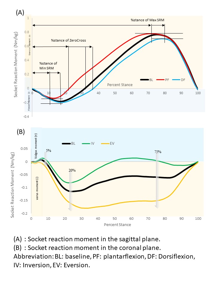
Ten users of transtibial prostheses were recruited in this study. Temporal-spatial parameters (Cadence, walking speed, step time, single support time, and step length) and sagittal and coronal SRM were measured during walking under five alignment conditions (3-degree plantarflexion and dorsiflexion, 6-degree inversion and eversion, and baseline alignment. Minimum and Maximum SRM, % stance (timing) of Minimum and Maximum SRM and zero cross in the sagittal plane, SRM of 5% stance, 20% stance, and 75% stance in the coronal plane were extracted and analyzed. Repeated measures of ANOVA or Friedman tests were carried out for statistical analyses (p < 0.05).
In the coronal plane, SRM of 5% stance, 20% stance, and 75% stance showed significant differences under both inversion and eversion (p=0.018, <0.001, <0.001, respectively). Minimum SRM, % stance of Minimum and Maximum SRM, and zero cross showed significant differences under sagittal alignment changes (plantarflexion/dorsiflexion) (p=0.028, 0.017, <0.001, respectively). There were also significant differences among single support times of the intact side in sagittal alignment changes, step time of the prosthetic side and single support times of the intact side with relation to coronal alignment changes.
Our findings suggest that inversion and eversion of transtibial prosthetic feet would affect the magnitude of SRM systematically in the coronal plane, while plantarflexion and dorsiflexion would affect the timing of SRM and Minimum SRM in the sagittal plane. These findings may be beneficial to evaluate and adjust alignment of feet in transtibial prostheses.
In the UK, the majority of amputations are transtibial, a result of diabetes and/or vascular disease, and in patients over 50 years.(1) Many older patients are prescribed a standard prosthesis with an ankle-foot that is not self-aligning. These types of prostheses are designed for level walking, and do not adjust to sloped surfaces. A self-aligning prosthesis, designed for everyday environments and for patients categorised as having ‘limited community mobility’ (K2), is available on the NHS, but seldom prescribed.
This multi-centre, randomised controlled trial assessed the feasibility of conducting a full-scale trial of the effectiveness and cost-effectiveness of a self-aligning prosthetic ankle-foot for older patients compared with a standard prosthetic ankle-foot.
We aimed to recruit 90 participants aged ≥ 50 years with a vascular-related or non-traumatic unilateral transtibial amputation for ≥1 year, categorised as having ‘limited community mobility’, and using a non-self-aligning prosthetic ankle-foot. Participants completed a baseline assessment including four clinical tests and questionnaires and wore an activity monitor on their normal prosthesis for one week. They were then randomised into one of two groups for 12 weeks: self-aligning prosthetic ankle-foot or existing standard prosthesis. Participants completed the same questionnaires at the interim follow-up and the full set of baseline assessments at final follow-up. Feasibility measures included recruitment, consent and retention rates; and completeness of clinical tests and questionnaire datasets.
Fifty-five participants were randomised (61% of the target 90 participants): n=27 self-aligning ankle-foot group, n=28 standard prosthesis group. Their mean (SD) age was 68.8 (9.6) years. Fifty-one participants were included in the final analysis (71% of the target number of participants). The consent rate and retention at final follow-up were 86% and 93%, respectively. The average recruitment rate was 1.25 participants/site/month (95% CI 0.39 to 2.1). Completeness of questionnaires ranged from 89-94%, and clinical tests were 92-95%, including the activity monitor data. The average completion rates for the data ranged from 63% to 93%. Of the clinical tests, only the 2-minute walk test appeared to show a change between baseline and final follow-up. Participants with the self-aligning prosthetic ankle-foot walked on average 6.2 (16.2) metres further while participants in the standard prosthesis group walked 9.0 (29.8) metres less.
This feasibility trial recruited and retained participants who were categorised as having ‘limited community mobility’ following a transtibial amputation. The high retention rate of 93% indicated the trial was acceptable to participants and feasible to deliver as a full-scale RCT. The findings support a future, fully-powered evaluation of the effectiveness and cost-effectiveness of a self-aligning prosthetic ankle-foot compared to a standard non-self-aligning version with some adjustments to the trial design and delivery.
(1) University of Salford. LIMBLESS STATISTICS: Repository for quantitative information on the UK limbless population REFERRED for prosthetics treatment, Annual report 2011-2012. University of Salford; 2013.
This paper presents independent research funded by the National Institute for Health Research Research for Patient Benefit Programme (ref:PB-PG-0816-20029).
Modular prosthetic ankle components have been shown to improve walking in transtibial prosthesis users, but they may reduce stability during standing. Therefore, the stiffness of the prosthetic foot-ankle components may need to be appropriately tuned to provide optimal balance between mobility for walking and stability for standing. The present research investigates both concepts to improve our knowledge about how the prosthetic ankle stiffness influences standing and walking performance.
Two aims were addressed:
Subjects: A diverse group of ten individuals with unilateral transtibial amputation and categorized as K3 level of ambulation participated.
Apparatus: Kinematic and kinetic data were collected using a digital motion capture system (Motion Analysis Corp, CA) and force platforms in the JBVAMC Motion Analysis Research Laboratory.
Procedures: Gait and balance analysis were performed at three different levels of ankle dorsiflexion stiffness (Firm, Medium and Soft) using the College Park Venture foot (College Park, MI).
Data Analysis: Data were grouped and analysed accordingly. Repeated measurements ANOVA were performed for the temporal-spatial, quiet standing and Roll-Over Shape (ROS) values. Kinetic and kinematic data were analysed using a one-dimension statistical parametric analysis.
The results suggest an influence of foot-ankle stiffness on gait and balance, specially on ROS radii and movement of center of pressure (COP).
The analysis of ROS radii indicates significant differences due to stiffness condition [F(2,98)=38.622, p<0.001] and walking speed [F(2,98)=24.522, p<0.001]. Pairwise comparisons indicated significant differences between all three levels of stiffness (p<0.001) and between slow and fast walking speeds (p<0.001).
Figure 1. Radius of ROS (Normalized) at different stiffness conditions and self-selected walking speeds
On quiet standing, at the eyes closed condition, movement of COP demonstrated a significant difference [F(2,58)=8.044, p=0.003] based upon bumper durometer. Moreover, the pairwise comparison indicated a difference between Firm and Soft bumpers (p<0.001).
Figure 2. Root Means Square Distance of Sway during quiet standing at different stiffness conditions.
Anatomical ankle stiffness has been reported to adapt to different walking speeds to maintain a uniform ROS radius (Hansen et al., 2010), contrary to our data that indicate prosthetic ankle components with constant stiffness produce different ROS radii at different speeds.
The Firm stiffness was able to best replicate the able-bodied individuals’ ROS radius and appeared to provide the most advantages during walking and quiet standing.
This study was supported by Veterans Health Administration Rehabilitation Research and Development Service. (grant no. RX002107).