The anterior cingulate cortex (ACC) shows preferential activation to anxiety-related and social stimuli, but it is unknown how microcircuits within the ACC encode these distinct stimuli. One type of inhibitory interneuron, which is positive for vasoactive intestinal peptide (VIP), is known to modulate the activity of pyramidal cells in local microcircuits, but it is unknown whether VIP cells in the ACC (VIPACC) are engaged by particular contexts or stimuli. Using in vivo Ca2+ imaging and miniscopes in freely behaving mice to monitor VIPACC activity, we identified individual VIPACC that preferentially activated to different stimuli across diverse tasks. Next, animals underwent multiple trials in behavioral paradigms that assessed anxiety-like behavior or interactions with other mice or objects. Although the population-level activity of the VIPACC remained stable across trials, the stimulus-selectivity of individual interneurons changed rapidly. These findings demonstrate marked functional heterogeneity and instability within interneuron populations in the ACC.
Studies from the psychiatric genomics consortium have concluded that common schizophrenia risk variants are regulatory and have low effect sizes. Thus, one could either inherit many of these variants or have a dysfunction in an upstream mechanism that controls their expression; however, demonstration of the latter has remained elusive.
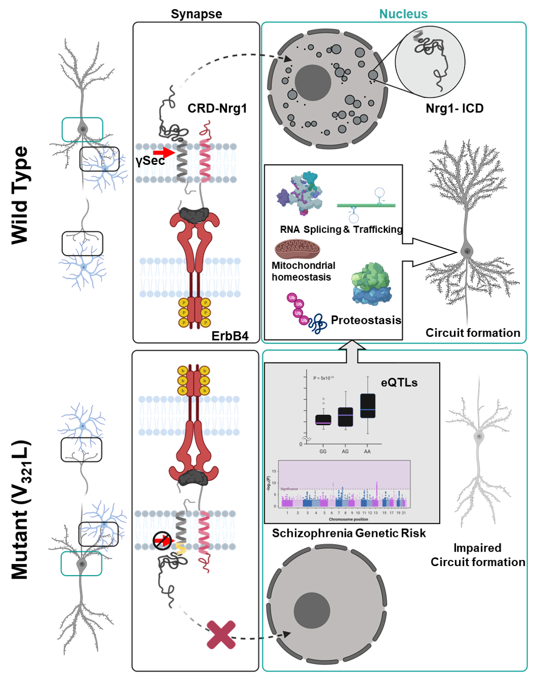
Neuregulin1 is a schizophrenia-susceptibility gene. Type III Neuregulin1 is cleaved by gamma secretase to produce a nucleus-targeted cytosolic fragment (ICD). A psychosis-associated mutation in Nrg1 (V321L) disrupts this cleavage implicating nuclear signaling in underlying pathology.
Using transcriptomic and network analyses, we uncovered a common gene regulatory network providing insights into the molecular pathology underlying genetic susceptibility to psychosis. Our data show that nuclear ICD regulates the expression of schizophrenia risk genes. The nuclear ICD signal originates at the synapse, hinting that expression of these risk genes is normally regulated during synaptogenesis; a process dysregulated in schizophrenia.
Heterozygous loss-of-function (LoF) mutations in SETD1A, which encodes a subunit of histone H3 lysine 4 methyltransferase, were shown to cause a novel neurodevelopmental syndrome and increase the risk for schizophrenia. We generated excitatory/inhibitory neuronal networks from human iPSCs with a SETD1A heterozygous LoF mutation (SETD1A+/-) using CRISPR/Cas9. Our data show that SETD1A haploinsufficiency resulted in morphologically increased dendritic complexity and functionally increased bursting activity. This network phenotype was primarily driven by SETD1A haploinsufficiency in glutamatergic neurons. In accordance with the functional changes, transcriptomic profiling revealed perturbations in gene sets associated with glutamatergic synaptic function. At the molecular level, we identified specific changes in the cAMP/PKA pathway pointing toward a hyperactive cAMP pathway in SETD1A+/- neurons. Finally, by pharmacologically targeting the cAMP pathway we were able to rescue the network deficits in SETD1A+/- cultures. Our results demonstrate a link between SETD1A and the cAMP-dependent pathway in human neurons.
Recent research has shown opposite-sex molecular mechanisms of memory formation regulated by the Tac2 pathway in the Central Amygdala (CeA). In this study, we aim to discover sex-differences in the macrocircuitry of CeA-Tac2 pathway, and how this differential macrocircuitry eventually evokes different neuronal activation ensembles upon fear recall in the extended amygdala. First, our results manifest much denser innervation of CeA-Tac2 fibers to the posteromedial-BNST (pmBNST) in males compared to females. Moreover, temporal silencing of CeA-Tac2 neurons projecting from the CeA to the pmBNST decreased consolidation in male mice, but not in females. Calcium transients during fear expression showed increased excited cells upon tone presentation in males with CeA-Tac2 silencing during consolidation, whereas activity in females remained unaltered. Altogether, these findings support the existence of sex-dimorphic macrocircuitry of the CeA-Tac2 pathway involved in fear memory consolidation.
Fragile X syndrome (FXS) is a leading genetic cause of autism-like symptoms that include sensory hypersensitivity and cortical hyperexcitability. Recent observations in humans and Fmr1 knockout (KO) animal models of FXS suggest abnormal GABAergic signaling. As most studies focused on neuron-centered mechanisms, astrocytes’ contribution to abnormal inhibition is largely unknown. Here we propose a non-neuronal mechanism of abnormal inhibitory circuit development in FXS. Astrocyte-specific deletion of Fmr1 during postnatal period leads to increased astrocytic GABA levels, but negatively impacts synaptic GABAA receptors and parvalbumin (PV) cell development. Developmental deletion of Fmr1 from astrocytes also affects communications between excitatory neurons and PV cells, impairing sound-evoked gamma synchronization in the cortex, while enhancing baseline and on-going sound-evoked EEG power, and leading to increased locomotor activity and altered social behaviors in adult mice. These results demonstrate a profound role of astrocytic FMRP in the development of inhibitory circuits and shaping normal inhibitory responses.
Schizophrenia is a psychiatric disorder, with a prevalence rate of 1-2 % worldwide. With advances in GWAS studies, hundreds of loci have been linked to an increased risk for schizophrenia. A major challenge is to understand the potential functional link between genes encoded across these many loci as well as the genomic mechanisms underlying the onset of disease. Recent analyses have pointed towards upper layer cortical excitatory neurons and inhibitory interneurons as the key biological cell types involved in pathogenesis. In this study, we applied co-IP proteomic analysis of schizophrenia risk gene networks in cultures of human stem cell-derived cortical excitatory and inhibitory neurons. From this, we have identified HCN1 as central to a protein network of synaptic proteins that is enriched for common variant risk for the disease. To study the functional impact of HCN1 loss-of-function in human neurons, we generated human embryonic stem cells (hESCs) with HCN1-knockout.
NMDA receptor (NMDAR) and GABA neuronal dysfunctions are observed in animal models of autism spectrum disorders, but how these dysfunctions impair social cognition and behavior remains unclear. We report here that NMDARs in cortical parvalbumin (Pv)-positive interneurons cooperate with gap junctions to promote high-frequency (>80 Hz) Pv neuronal burst firing and social cognition. Shank2–/– mice, displaying improved sociability upon NMDAR activation, show impaired cortical social representation and inhibitory neuronal burst firing. Cortical Shank2–/– Pv neurons show decreased NMDAR activity, which suppresses the cooperation between NMDARs and gap junctions (GJs) for normal burst firing. Shank2–/– Pv neurons show compensatory increases in GJ activity that are not sufficient for social rescue. However, optogenetic boosting of Pv neuronal bursts, requiring GJs, rescues cortical social cognition in Shank2–/– mice, similar to the NMDAR-dependent social rescue. Therefore, NMDARs and gap junctions cooperate to promote cortical Pv neuronal bursts and social cognition.
There is a great demand for non-invasive methods to prevent or ameliorate disease. We investigated a previous suggestion that 40Hz flickering light leads to brain-wide gamma oscillation and suppresses amyloid-β in an Alzheimer’s mouse model. We used multisite silicon probe recordings in the visual cortex, entorhinal cortex, and hippocampus. 40Hz stimulation did not engage native gamma oscillations, although a sizeable fraction of V1 neurons were entrained to the stimuli. Spike responses in the entorhinal cortex and hippocampus were weak suggesting 40Hz flickering light does not effectively modulate deep structures. 40Hz stimulation appeared aversive to mice and was accompanied by elevated cholinergic activity in the hippocampus. Using immunohistochemistry and in-vivo two-photon imaging, we found large variability in plaque load among young animals. Alterations in microglia morphology and plaque count were not seen following stimulation. To use 40Hz light stimulation for Alzheimer’s Disease therapy will require further extensive control experiments.
Results of clinical studies investigating the effect of oxytocin on social behavior are inconsistent. Therefore, the present study aimed to investigate the effect of chronic intranasal oxytocin treatment on the autism-like phenotype in Shank3-/- mice (KO). KO and wild-type
mice of both sexes received either intranasal oxytocin or vehicle daily for one month. Following 2 weeks of daily oxytocin treatment, KO mice spent twice as much time interacting with the social partner in comparison to vehicle-treated controls. The explorative behavior of
oxytocin-treated KO mice was 50% lower in comparison to controls. The oxytocin-induced higher sociability and lower exploration lasted even 4 weeks following the treatment termination. Oxytocin did not affect the repetitive behavior of mice. In conclusion, our results show potential therapeutic effect of oxytocin in autism, but also point towards likely behavioral side effects. These outcomes should be proved in other autistic models.
Impairments found in neurodevelopmental disorders (NDDs) are thought to be associated with disrupted homeostatic plasticity, a process known to stabilize neuronal network activity by balancing neuronal excitation and inhibition. Several studies have shown that homeostatic plasticity is regulated by epigenetic mechanisms, such as DNA-methylation. However, an epigenetic profile involved in homeostatic plasticity in NDDs is currently insufficiently defined. We established a model of homeostatic plasticity in human induced pluripotent stem cell (hiPSC)-derived neurons, grown on micro-electrode arrays (MEAs). Our data show that chronic deprivation of neuronal activity through the inhibition of sodium channels with TTX elicits a time-dependent increase in the neuronal network activity and the changes in network poperies are caused by an upregulation of postsynaptic AMPA receptors expression and changes in intrinsic properties. This model is now being used to test whether hiPSC-derived neurons with mutations in NDDs genes linked to epigenetic mechanisms display altered homeostatic plasticity.
Higher order feedback projections to sensory cortical areas converge upon layer 1 (L1) of the cerebral cortex, the primary site for integration of top-down information via the apical dendrites of pyramidal neurons and L1 GABAergic interneurons. We investigated the contribution of early thalamic inputs onto L1 interneurons for the establishment of top-down inputs in the primary visual cortex. We found that bottom-up thalamic inputs predominate during early L1 development and preferentially target neurogliaform cells. These projections were critical for the subsequent strengthening of feedback inputs from the anterior cingulate cortex. Enucleation or selective removal of thalamic afferents blocked this phenomenon. Notably, while early activation of anterior cingulate afferents resulted in a premature strengthening of these top-down inputs to neurogliaform cells, this was also dependent on thalamic inputs. These results demonstrate that the proper establishment of top-down feedback inputs critically depends on bottom-up inputs from the thalamus during early postnatal development
Many transcription regulators are linked to the biology of neuropsychiatric disorders. We found that the neuron-specific transcription repressor MYT1L enhances neurogenesis by repressing progenitor and non-neuronal programs, acting as novel “many-but-one” repressor. Myt1l is expressed throughout life and haploinsufficiency is linked to mental disorders, such as autism, suggesting that faulty repression could contribute to disease.
Using stem cell-derived human neurons and mice, we show that MYT1L-deficiency caused upregulation of non-neuronal programs and is sufficient to induce autism-associated phenotypes ranging from altered neurodevelopment to behavior. Unexpectedly, we found that loss of MYT1L affected synaptic transmission that could be rescued by MYT1L overexpression in postmitotic primary neurons. Strikingly, acute application of approved drugs rescued the electrical phenotypes in vitro and behavior effects in mice. Hence, failure to repress unwanted genes upon MYT1L-deficiency is sufficient to cause mental disease but pharmacological intervention can normalise electrophysiologic and behavior phenotypes even after development is complete.
Symptoms of neuropsychiatric disorders emerge from erroneous communication in the brain. To avoid unwanted interferences and maximize combinatorial power, neuronal circuits use dynamic input selection that coordinates afferent synaptic activity in converging pathways. The lack of understanding of such regulatory mechanisms represent a major obstacle in developing new treatment options for neuropsychiatric disorders.
We pursued cellular and network mechanisms of input selection in hippocampal circuits, where pathways carrying sensory and mnemonic information converge. We discovered a small population of GABAergic inhibitory interneurons with spike timing selectively locked to oscillatory dynamics of cortical afferents. Analysis of neurobiotin-labelled cells identified these as neurogliaform cells inhibiting pyramidal cell dendrites where cortico-hippocampal axons terminate. After neurogliaform cell spikes, spike timing of pyramidal cells decoupled from cortical oscillations, with no detectable decrease in firing rate.
Neurogliaform cells regulate information transfer by pathway specific decoupling, and may represent a novel intervention target in treating neuropsychiatric disorders.
Serotonin is considered to be the major neurotransmitter involved in depression and its treatment. Thus, 5-HT1A receptors have attracted interest as targets for therapeutic intervention. Notably, the activation of presynaptic 5-HT1A autoreceptors slows down the antidepressant effects of serotonin reuptake inhibitors whereas the stimulation of postsynaptic 5-HT1A heteroreceptors is needed for an antidepressant action. NLX-101 exhibits preferential activation of cortical and brain stem 5-HT1A receptors. Here we used behavioral, neurochemical and molecular methods to examine its antidepressant effects. NLX-101 (0.16 mg/kg) reduced immobility in the forced swim test when measured 30 min (albeit not 24 h and 7 days) after drug administration. NLX-101 increased the dialysate dopamine and glutamate in the medial prefrontal cortex, but no changes were observed in the prefrontal output of noradrenaline and serotonin. NLX-101 also produced a rapid increase in the synthesis of pmTOR, pERK1/2 and PSD95, which may contribute to its rapid antidepressant action.
Tyrosinemia type 1 is caused by mutations in fumarylacetoacetate hydrolase, resulting in the accumulation of toxic intermediates of tyrosine catabolism. Treatment with nitisinone limits cellular damage but further increases tyrosine concentrations. It has been speculated that extreme levels of tyrosine could inhibit tyrosine hydroxylase (TH), the rate-limiting enzyme in dopamine biosynthesis. We exposed rat pheochromocytoma (PC12) cells to different levels of tyrosine and measured its effects on cellular viability, TH protein levels, site-specific TH phosphorylation and rates of dopamine synthesis. At extracellular tyrosine levels of 240-800 µM, dopamine synthesis was significantly reduced, with corresponding changes in TH phosphorylation and protein levels as well. In addition to decreased TH levels, PC12 cell proteome analyses showed changes in multiple proteins and cellular pathways upon exposure to high extracellular tyrosine concentrations. Hence, neuropsychological symptoms observed in tyrosinemia patients treated with nitisinone could be related to decreased dopamine synthesis in the central nervous system.
Auditory evoked-potential (AEP) abnormalities are common in people with genetic risk for schizophrenia, including carriers of the 22q11.2 deletion. We examined AEP abnormalities in the Df1/+ mouse model of human 22q11.2 deletion syndrome (22q11.2DS). Like 22q11.2DS patients, Df1/+ mice exhibit high inter-individual variability in peripheral hearing sensitivity. We measured both peripheral hearing sensitivity and cortical AEPs in 30 Df1/+ mice and 21 WT littermates. We then analysed the influence of genotype and hearing sensitivity on loudness-dependent AEP (LDAEP) and inter-tone interval time-dependent AEP (TDAEP) growth functions. Both LDAEP and TDAEP growth functions were abnormal in Df1/+ relative to WT mice. Moreover, the nature of the AEP abnormalities in Df1/+ mice correlated with inter-individual variability in peripheral hearing sensitivity. Results suggest that AEP abnormalities depend not only on genetic risk factors for schizophrenia but also on hearing impairment, which has been associated with schizophrenia in both cross-sectional and longitudinal studies.
Restricted behaviors and impaired social interaction are hallmarks of autism spectrum disorder (ASD), and reflect a compromised reward system. Key to reward circuitry is the integration of dopaminergic and glutamatergic inputs to the striatum. The glycine receptor α2 (GlyRα2) has been linked to ASD, and is ideally positioned to modulate striatal signal integration.
Here, we report excessive appetitive responding in GlyRα2 KO and show an excessive dopamine-induced increase in striatal projection neuron activity in GlyRα2 KO, but no changes in dopamine neuron activity. We report an increased locomotor response to d-amphetamine in GlyRα2 KO, which correlates to c-fos expression in the dorsal striatum. 3D modeling revealed activation of cell ensembles in response to d-amphetamine, an increase in number of cells but no shift in intercell distance histograms.
We conclude that GlyRα2 acts as a gatekeeper of signal integration within the dorsal striatum, and crucially affects reward-motivated behavior.
Microglia are the phagocytes of the central nervous system and are involved in various processes that regulate brain homeostasis. Disruption of microglial movement and synaptic elimination gives rise to different pathologies including schizophrenia. We hypothesize that the psychiatric risk gene Disrupted-In-Schizophrenia 1 (DISC1) controls microglial movement and phagocytosis. Our results show that DISC1 is highly expressed in mouse and human microglia. DISC1 locus impairment (LI) microglia phagocytose slower but their final synaptosome uptake is increased compared to wildtype (WT) microglia. DISC1 LI microglia migrate slower compared to WT microglia in vitro and in embryonic brain slices. In contrast, the surveyed brain area of DISC1 LI microglia in adolescent brain slices is increased compared to WT microglia, whereas their morphological complexity is impaired. We are currently validating our findings using a DISC1 LI bone marrow transplantation in WT mice to study the cell-autonomous effects of DISC1 locus impairment on microglial movement.
Psychedelics, since time immemorial, have been known to alter sensory perceptions, and have been thought to have positive effects on mood. Over the last few decades, experimental findings have shown that serotonergic psychedelics modulate brain regions that are involved in mood regulation, and can be possibly used as therapeutics against mood disorders. However, it is not well known how these drugs influence different regions of the brain to drive positive effects on mood-behaviours. We found that acute administration of a serotonergic psychedelic DOI (2,5-dimethoxy-4-iodoamphetamine) exhibits anxiolytic response in rodents, which is driven via the CA1/subiculum field of the ventral hippocampus. Furthermore, we see that 5-HT2A receptors on the Parvalbumin-positive interneurons in this region may be sufficient to drive DOI-evoked anxiolysis. This work not only explores the therapeutic potential of serotonergic psychedelics, but also tries to understand how information is relayed in the brain to harness their potential against anxiety-like behaviour.
The functional maturation of PFC circuits is characterized by critical developmental time windows, which shape functional connectivity among neurons. Indeed, early life insults, such as perinatal exposure to fluoxetine (FLX), can alter PFC function, increasing the risk for depression and anxiety in the adult. Yet, the underlying cellular and circuit mechanisms are poorly understood. We found that early postnatal FLX-treatment results in a profound in vivo network hypoexcitability and reduced firing rate of PFC neurons of adult mice. This was accompanied by alterations of intrinsic excitability of a specific subtype of pyramidal neurons, which transiently expresses the serotonin transporter SERT. Surprisingly, local glutamatergic and GABAergic neurotransmission were unaffected by perinatal FLX treatment. Genetic and pharmacological experiments indicate that FLX-dependent alterations of PFC neurons mainly depend on 5HT-7 receptor. Our results provide a potentially novel neurobiological mechanism underlying the development of adult psychiatric conditions induced by early-life environmental insult.
Circadian rhythm synchronizes each body function with the environment and regulates physiology. Disruption of normal circadian rhythm alters organismal physiology and increases disease risk. Recent epidemiological data and studies in model organisms have shown that maternal circadian disruption is important for offspring health and adult phenotypes. Less is known about the role of paternal circadian rhythm for offspring health. Recently, we showed that paternal circadian disruption at conception is important for offspring feeding behavior, metabolic health, and oscillatory transcription. Mechanistically, our data suggest that the effect of paternal circadian disruption is initiated by corticosterone-based parental communication at conception and programmed during in utero development through a state of fetal growth restriction. These findings indicate paternal circadian health at conception as a newly identified determinant of offspring phenotypes. Now, we are examining whether the metabolic health impairment has a neurobehavioral origin, performing an in-depth neurobehavioral phenotyping and transcriptomics studies.
PPM1F is regulated in PTSD patients and in mPFC and amygdala of male/female mice exposed to IMO. Chronic unpredictable mild stress (CUMS) could impact on PPM1F expression participating as a marker of chronic stress. We evaluated if PPM1F mediate the response to CUMS. PPM1F expression resulted upregulated in the amygdala and blood after 3 day CUMS. Our results support a role of these markers in the response to chronic stress. Insights into the regulation of CaMKIIg-PPM1F pathway may contribute to the development of treatments for stress-related disorders. Furthermore, IMO impairs fear extinction but sex differences have not been studied. We have explored sex differences in calcium activity of mPFC neurons during fear conditioning after exposure to IMO. We found sex differences on calcium activity during fear extinction. Sex differences observed in calcium activity patterns could partially explain the different impact of chronic and acute stress depending on the sex.
Impaired inhibition and parvalbumin (PV) interneuron dysfunction are thought to underlie the development of hyperactive neuronal networks in neurodevelopmental disorders. Previous work implicated trans-synaptic ephrin/EphB signaling in excitatory synapse development. We recently made a new discovery linking astrocytic ephrin-B1 to the development of connections between inhibitory PV interneurons and CA1 pyramidal cells (PCs) in the hippocampus. Deletion of ephrin-B1 from astrocytes during the postnatal day (P)14-P28 developmental period impaired PV->PC connectivity, resulting in reduced inhibition of hippocampal PCs, increased seizure susceptibility, and reduced sociability in mice. Conversely, overexpression of astrocytic ephrin-B1 increased PV->PC connectivity, and increased inhibition of PCs, possibly through the removal of EphB from PV boutons. EphB signaling in PV cells may negatively regulate inhibitory synapse formation by influencing ErbB4 signaling. Our findings suggest that direct interactions between astrocytic ephrin-B1 and EphB receptor in PV boutons regulate the establishment of PV perisomatic inhibitory synapses.
Bipolar disorders are defined by recurrences of depressive and manic episodes. The pathophysiology is still unknown, and translating clinical symptoms into behaviors explorable in animal models is challenging. Beyond mood, emotional biases differentiate bipolar states in humans. Mania is associated with positive biases, e.g. emotional stimuli become more rewarding and less aversive, and the opposite for depression. We propose to assess behavioral hedonic responses to innately appetitive and aversive olfactory and gustatory cues in mice as proxies for the assigned emotional valence. A mania model is therefore supposed to display positive hedonic bias. Using the GBR-12909 mania model, we observed classical hyperactivity. Unexpectedly, these mice also exhibited strong negative hedonic biases. Consequently, the GBR-12909 model might not be appropriate for studying emotional disturbances associated with mania. We propose olfactory and gustatory preference tests as crucial assessment of positive and negative valence biases, necessary for characterizing animal models of bipolar disorders.
Manganese-enhanced magnetic resonance imaging (MEMRI) holds exceptional promise for preclinical studies of brain-wide physiology in awake-behaving animals. Mn(II) is a powerful contrast agent because: 1) High signal intensity at low doses without adverse effects; 2) Projection tracing and neural activity mapping via its entry into electrically active neurons and transport along axons. We pioneered longitudinal MEMRI for gene-x-environment effects on brain-wide forebrain projections and on neural activity as imaged at 100 µm resolution. We use mage analyses (statistical parametric mapping and graph theory) to define network dynamics over time. We reported that genetic disruption of monoaminergic systems alters prefrontal cortical (PFC) projections, and that ethological fear leading to anxiety-like behavior alters brain-wide neural activity. We are now defining biological bases for changes in PFC projections, and are using DREADD chemogenetics to drive activity experimentally in the locus coeruleus followed by longitudinal MEMRI to investigate biology of stress responses.
During corticogenesis, neural stem cells (NSCs) undergo rapid proliferation to produce carefully titrated populations of neurons. Expediated NSC divisions rely on efficient maintenance of genome integrity. Polycomb repressive complex 1 (PRC1) plays an increasingly recognized role in DNA damage repair. RING1 is an E3-ubiquitin ligase within PRC1 that catalyzes monoubiquitination of histone 2A (H2AUb1). A de novo pathogenic RING1 missense variant was detected in an individual with early-onset schizophrenia and developmental delay. NSCs differentiated from RING1G284A/G284A human embryonic stem cells exhibit reduced genome wide H2AUb1, increased baseline DNA damage, and delayed DNA repair following chemotoxic stress. Additionally, an accumulation of NSCs in the G2/M phase were observed in forebrain organoids characterized by single-cell RNA sequencing. Cell cycle defects translated into altered timing of neural differentiation and increased apoptosis. These results indicate that PRC1-mediated H2AUb1 is necessary for efficient DNA damage repair and cell cycle progression in developing human NSCs.
Intracranial local field potentials (LFP's) involved in cognitive processes are affected by antipsychotic drugs (APD's); additionally voltage gated potassium channels (Kv's) are inhibited by APD's and have been implicated in the pathophsyiology of schizophrenia. In this study we a) extended observations of APD effects on LFP's and b) performed the same LFP recordings and analysis in response to Kv inhibitors (KVI's).
LFP recordings were made in CA1 hippocampal region (HIP) and pre pre-frontal cortex (PFC) regions of 8 mice. Power spectra, coherence and cross-frequency coupling were measured.
Gamma power was reduced by all APD's and KVI's except 4-AP, which increased power. HIP/PFC coherence was reduced by clozapine and retigabine but increased by 4AP. Theta/gamma cross-frequency coupling was reduced by haloperidol and retigabine.
This study reveals complex effects of APD's on LFP cognition-related spectra and demonstrates novel modulation of LFP properties by KVIs intriguingly overlaping with APD's.
Our study aims to create a human in vitro model for DiGeorge syndrome (DGS) using induced pluripotent stem cells (iPSCs). DGS is a complex disease caused by the monoallelic 3 Mb long deletion of 22q11.2 chromosome region and affecting the cardiovascular, neural and immune systems. A family with three DGS patients from three generations manifesting the disease with different severity, and two healthy relatives were involved in this study. We generated iPSCs from blood mononuclear cells by Sendai virus transduction and then differentiated them into neural progenitors. Hippocampal progenitors of the child lost their proliferation capacity and showed more mature phenotype than the other samples. Similar results were found in case of cortical differentiation. Further comparative analyses are warranted for better understanding the development of the disease. Supported by; National Brain Research Program of Hungary (NAP 2017-1.2.1-NKP-2017-00002, KTIA_NAP_13-2014-0011), and National Research, Development and Innovation Office (NVKP_16-1-2016-0017, OTKA- K128369).
The clustered protocadherins (PCDHs) provide individual neurons with unique cell surface identity codes required for neurite self-avoidance and neuronal tiling. Recent studies show that DNA sequence variants across the protocadherin gene cluster associate with neuropsychiatric disorders in large cohort studies. However, the mechanisms by which these variants might contribute to disease etiology are unknown. To better understand protocadherin function, we performed PCDH-BioID studies to identify specific protein-protein interactions at the cell surface, and PCDH-DamID studies to understand membrane to nucleus signaling events. Preliminary BioID studies identified specific interactions between PCDHs and actin remodeling components in the cytoplasm, and with specific DNA binding proteins. The DamID studies revealed that the PCDH intracellular domain (ICD) interacts with specific DNA sequences, and transcriptionally activates a specific set of genes. Our goal is to understand how disease associated PCDH sequence variants might disrupt normal PCDH functions, and thus contribute to neurological disease risk.
After chronic stress, a fraction of individuals shows stress resilience, preventing long-term mental dysfunction. The underlying molecular mechanisms are complex and have not yet been fully understood. Here, in a mouse model of chronic social defeat stress, a data-driven behavioural stratification identified three groups of stressed animals. A single-cell transcriptomic of their hippocampal sub-regions revealed that, in a sub-group of mice exhibiting adaptive behavioural and molecular responses upon chronic stress, the dorsal hippocampus is particularly involved in neuroimmune responses, angiogenesis, myelination, and neurogenesis; thereby promoting brain restoration and homeostasis after stress. Based on these molecular insights, a resilience drug candidate, rapamycin, applied after the stress, was able to promote stress resilience. Altogether, our in-depth analysis of behavioral and molecular stress resilience mechanisms opens new avenues for the treatment of mental disorders.
Disease variants in the TRIO gene are enriched in individuals with neurodevelopmental disorders (NDDs). TRIO encodes a cytoskeletal regulatory protein with three catalytic domains – two guanine exchange factor (GEF) domains, GEF1 and GEF2, and a kinase domain, as well as several accessory domains. Variants in the GEF1 domain or the nine adjacent spectrin repeats (SRs) are enriched in NDDs, suggesting that dysregulated GEF1 activity is linked to these disorders. We provide evidence that the Trio SRs interact intramolecularly with the GEF1 domain to inhibit its activity. We demonstrate SRs 6-9 decrease GEF1 activity in vitro and in cells and show NDD-associated variants in the SR8 and GEF1 domains relieve this autoinhibitory constraint. Our results from chemical cross-linking and BioLayer Interferometry indicate that the SRs primarily contact the PH region of the GEF1 domain, reducing GEF1 binding to Rac1. Together, our findings reveal a key regulatory mechanism that is disrupted in multiple NDDs.
Chloride-mediated GABA signaling shifts from depolarizing to hyperpolarizing during development, due to an increase in activity of chloride exporter KCC2. In various neurodevelopmental disorders, the GABA shift is delayed. It is unclear whether a delayed GABA shift affects synaptic development.
We assessed the consequences of mistimed GABA shift in hippocampal slice cultures. We set out to delay the GABA shift by blocking KCC2 with VU0463271 or furosemide from between day in vitro (DIV)2-8. VU0463271 treatment resulted in depolarizing GABA at DIV8. At this timepoint, inhibitory and excitatory postsynaptic currents (IPSCs and EPSCs) were not affected. Yet, when chloride levels restored at DIV22, the frequency of spontaneous IPSCs was increased.
Unexpectedly, furosemide treatment resulted in hyperpolarizing GABA and an activity-dependent decrease in the frequency of spontaneous IPSCs at DIV8. At DIV22, the IPSC frequency was increased. Our results indicate that the timing of the GABA shift affects inhibitory synapse development.
Deep visual proteomics (DVP) is a newly developed method for spatially-resolved sub-cellular proteomics that is applicable to cell culture systems and sectioned tissues (frozen or paraffin-embedded). DVP combines microscopy, image segmentation algorithms, laser micro-dissection, and mass spectrometry to provide unprecedented spatial resolution and sensitivity to proteomic studies. I will present the application of DVP to neurons of various sources: iPSC-derived neurons in culture, neurons in maturing organoids, and neurons in the developing mouse brain.
Human genetics has identified numerous genetic and chromosomal mutations such as copy number variations (CNVs) associated with autism spectrum disorders (ASD). However, the lack of standardized biological resources impedes understanding of the common pathophysiology of ASD. Here, we established a biological resource including 65 genetically modified mouse embryonic stem cell (mESC) lines which mimic human SNVs and CNVs associated with ASD. To illustrate cell-type and CNV specific molecular features of ASD, we performed single-cell RNA sequencing, morphological, and physiological analyses using 12 representative cell lines. Through these analyses, we also found that reduced expression of Upf3b, a core member of the nonsense-mediated decay (NMD) pathway, as a common phenotype in glutamatergic and GABAergic neurons. This finding emphasizes the dysfunction of translational machinery in the developing neurons. Our ES cell bank of CNVs developed in this study offers opportunities to identify cell-type specific abnormalities and novel drug target for ASD.
Neuronal dendritic arbors are essential elements of neuronal networks. Once established during development, mature dendritic arbors remain relatively stable. Yet, the loss of dendrite stability has been associated with mental disorders, including major depressive disorder. The molecular mechanisms accountable for the dendritic arbor stability remain largely unknown, and they are the subject of our research. To reveal them, we firstly developed in vitro models of mature dendritic arbors’ destabilization using cultured neurons and stimuli often linked with depression (e.g., changed neuronal activity or inflammatory cytokines). Next, we compared transcriptomes of control mature neurons, mature neurons with unstable dendrites and young neurons with naturally dynamic dendritic arbor. Finally, the 77 differentially expressed genes were selected for further functional analysis to verify their role in dendrite dynamics. As a result, we identified several proteins previously unknown to have a role in mature dendritic arbor dynamics.
Excitatory neurons in the claustrum mediate anxiety responses to acute psychological stressors. However, it is unclear whether these neurons represent information related to basal anxiety and anxiety responses to a stressor. Here, we performed freely moving calcium imaging of the claustrum on a battery of behavioral tests that are used to assess anxiety. We found that a subset of claustral neurons display a marked change in calcium levels during behaviors associated with anxiety. Interestingly, the calcium responses in the claustrum were altered after an exposure to a psychological stressor. This change in calcium response suggests that stress related information that controls anxiety-related behaviors is represented in the claustrum.
Autism Spectrum Disorder (ASD) is a neuropsychiatric disorder that often co-occurs with Intellectual Disability (ID). Monogenic ID-ASD disorders are currently untreatable, at least in part due to limited information on adult reversibility of the characteristic sensory and cognitive deficits. To overcome this bottleneck, we employ the powerful genetic toolbox of the fruit fly Drosophila to study habituation: the learning process which causes the reduction in response to a repeated irrelevant stimulus. Habituation deficits are thought to underlie the sensory and cognitive deficits in ID-ASD. We determine which of our previously identified >150 habituation-deficient ID-ASD Drosophila models can be reversed by genetic rescue specifically in adulthood. Supported by mechanistic research (see abstract Van Reijmersdal et al.) and candidate drug testing, we aim to identify acute regulators of habituation and thereby identify which ID-ASD disorders are most likely responsive to treatment. This will contribute to catapulting ID-ASD disorders into the therapeutic era.
Onset of many psychiatric disorders occurs during adolescence, yet molecular mechanisms underlying adolescent brain development remain poorly understood. Using a multiomics approach, we examined protein, mRNA, and noncoding RNA dynamics in mouse cortex during early, middle, and late adolescence and adulthood. Protein and mRNA profiles revealed pathways critical for mRNA processing, protein synthesis, and mitochondrial activity are highly dynamic during the transition from early to mid-adolescence. Integration of mRNA and microRNA sequencing data revealed frequent downregulation of mRNAs targeted by upregulated miRNAs, but rarely the converse. Surprisingly, average miRNA length decreases with age due to increased 3’ trimming, reduced 3’ tailing, and arm switching from longer -5p to shorter -3p miRNAs. The greatest changes were observed in miR-338-3p, a miRNA linked to behavioral and circuit deficits in 22q11 deletion syndrome mouse models. Together, our data reveal widespread changes in core molecular pathways that might contribute to disease onset during adolescence.
Mutations in CNTNAP2 are a risk factor for development of autism spectrum disorder (ASD). Our recent work showed that Cntnap2 KO animals exhibit a loss of parvalbumin (PV)+ interneurons and decreased perisomatic inhibition in hippocampal area CA1 (Paterno et al. Cell Rep. 2021). We also reported reduced spontaneous inhibitory postsynaptic current (IPSCs) frequency in CA1. Here, we used 32-channel silicon probes across CA1 to simultaneously record in vivo local field potentials and single units while Cntnap2 KO mice navigated a novel/familiar paradigm task or a delayed spatial alternation task. During navigation, Cntnap2 KOs showed a complex disruption of theta-gamma oscillations along CA1 and reduced sharp waves ripple (SWRs) power. Firing patterns of pyramidal cells and PV+ interneurons correlated with pathological information processing in CA1. These data suggest altered hippocampal circuit dynamics play a role in the autism phenotype.
Understanding glutamate-related hippocampal dys-function in schizophrenia (SCZ) can tailor pharmacologic intervention. Here, we provide first known evidence of such dysfunction gleaned using in vivo ¹H functional MRS. Continuous ¹H fMRS spectra were acquired (42 SCZ, 36 healthy adults) during an associative learning task (learning nine object-location associations). Eight epochs of Encoding and Retrieval were used to maximize learning proficiency. Both groups showed negative accelerated learning, though proficiency in SCZ was lower. We observed significantly increased Glu modulation during both conditions indicating sensitivity of ¹H fMRS for detecting brain activity. However, only during Encoding did we observe a significant group-by-time interaction (time reflects 4 groups of pairs of contiguous epochs); In healthy controls, Glu modulation was strongest during the early stages of learning, with an opposite trend observed in SCZ. These results provide novel and direct evidence of neurochemical dysfunction related to glutamatergic neurotransmission in SCZ, evoked during learning and memory.
Habituation, a fundamental form of learning, is the decrement of a response to repeated irrelevant stimuli. Habituation is defective in >150 Drosophila models of monogenic intellectual disability/autism (ID/ASD) disorders and provides a promising biomarker of cognitive (dys)function across evolution.
Strikingly, numerous habituation-deficient genes act in a few related signaling pathways, including PI3K-Akt, Ras/MAPK, and cAMP signaling. A balanced activity profile of these pathways appears crucial for habituation, and we indeed find that restoring this balance pharmacologically can rescue cognitive deficits. We hypothesize that the same pathways may be out of balance in other ID/ASD models, and currently profile these systematically.
This approach, and parallel efforts to systematically map adult reversibility of habituation deficits (see abstract M. Boon et al.) aim to identify - genetically heterogeneous but molecularly converging - groups of adult reversible ID/ASD disorders that can benefit from common treatment.
Hoxa5 is a member of the Hox gene family that plays critical roles in successive steps of the central nervous system formation. In mouse, we showed expression of Hoxa5 in the brainstem from fetal stages to adulthood, notably in pre-cerebellar neurons. Using a conditional postnatal Hoxa5 loss-of-function mouse model (Hoxa5 cKO) associated to a comparative transcriptomic analysis, we observed the downregulation of genes essential for synaptic function in Hoxa5 mutant specimens, including genes previously related to the autism spectrum disorder (ASD) in mice and/or humans. Based on these data, we analyzed the behavior of Hoxa5 cKO mice, using tests aimed at the evaluation of cerebellar functions and autistic-like behaviors. While the Hoxa5 cKO mice show no defects in cerebellar functions, they exhibit deficits in autism-related behaviors, such as impaired social interactions and increased repetitive and stereotyped behaviors, supporting the hypothesis of a correlation between HOXA5 and ASD.
Cytokines are the primary mediators of systemic inflammation and neuroinflammation; amongst these interleukin (IL)-1β is the most potent pro-inflammatory cytokine. It is produced as response to tissue damage or infections by cells of the immune system. The cytokine is synthesized as inactive form (proIL-1β) which is cleaved by activated caspase-1 to its mature form (mIL-1β) which is rapidly secreted to the extracellular space without entering the classical endoplasmic reticulum-Golgi pathway. Generating an IL-1β reporter cell line is challenging due to the complex process of generating the biologically active form of the protein and any overexpression system would induce a constitutive inflammatory response which does not maintain the physiological conditions. Therefore, we generated a reporter cell line by endogenously tagging IL-1β using CRISPR/Cas9 technology, which recapitulates the physiological response to classical inflammatory stimuli and can be used for quantitative assessment of IL-1β secretion associated with neuroinflammation.
Shank2 is an excitatory postsynaptic scaffolding protein strongly implicated in autism spectrum disorders (ASD). Shank2-KO mice show NMDAR hypofunction and autistic-like behaviors at juvenile and adult stages that are rescued by NMDAR activation. However, these mice at P14 show NMDAR hyperfunction, and NMDAR activity suppression prevents NMDAR hypofunction and autistic-like behaviors at later stages. To better understand the mechanisms underlying this rescue, we analyzed the forebrain transcriptomes from WT and Shank2-KO juvenile mice after early chronic memantine treatment. Vehicle-treated KO mice showed upregulated synaptic genes, downregulated ribosome and mitochondria genes, and a reverse-ASD pattern. Upregulated chromatin genes, and downregulated mitochondria, extracellular matrix (ECM), and actin genes, and weakened reverse-ASD pattern were observed in memantine-treated KO mice. Memantine-treated WT mice exhibited upregulated synaptic genes and downregulated ECM genes, and the reverse-ASD pattern. Therefore, early chronic treatment of Shank2-KO mice with memantine alters expression of chromatin, mitochondria, ECM, actin, and ASD-related genes.
GRIN2B is a gene encoding the GluN2B subunit of NMDA receptors associated with autism spectrum disorders (ASD). Sensory dysfunction is observed in about 90% of individuals with ASD. Previous studies reported that ASD patients with GRIN2B mutations show sensory hypersensitivity. To explore whether and how GRIN2B mutations lead to sensory hypersensitivity, we characterized mice carrying the patient-derived Grin2b-C456Y knock-in mutation.
The mutant mice showed sensory hypersensitivity in several behavioral tests. In functional MRI combined with electric whisker pad stimulation, the anterior cingulate cortex (ACC) was a key brain region with significant increases in BOLD signals in the mutant mice. In c-fos staining combined with electric foot shock, the ACC showed increased signals in the mutant mice. Electrophysiologic recordings indicated increased baseline and evoked excitatory synaptic transmission in mutant ACC pyramidal neurons. These results collectively suggest the Grin2b-C456Y mutation in mice leads to sensory hypersensitivity involving excessive ACC neuronal activity.
Fig1.
FOXG1 serves pleiotropic functions in brain development and has been linked to the neurodevelopmental FOXG1 syndrome and Autism Spectrum Disorders (ASD). Increased Foxg1 expression was detected in iPSCs from individuals with sporadic ASD and linked to overproduction of GABAergic neurons. We leverage the fact that the two principal neurogenic niches of the adult brain harbor differentially committed progenitors: while hippocampal precursors give exclusively rise to glutamatergic neurons, precursors from the subependymal zone mainly give rise to GABAergic interneurons. We demonstrate that these niches display differential vulnerability to increased FOXG1 dosage: while high FOXG1 levels severely compromised the acquisition of a dentate granule neuron fate/survival in the hippocampus, neurogenesis in the subependymal zone/olfactory bulb was unimpeded. Furthermore, we found that vulnerability of hippocampal precursors was at least in part caused by a FOXG1/NR4A1/apoptosis pathway. Future studies will address whether the FOXG1/NR4A1-axis contributes to overproduction of GABAergic neurons in ASD.
We have developed a robust functional ultrasound (fUS) imaging pipeline to measure drug-induced changes in brain activation and functional connectivity (FC) patterns, through the intact skull, in awake and behaving mice. We report that major opioid and cannabinoid drugs lead to highly-reproducible, dose- and time-dependent reorganization of brain activation and FC patterns. While displaying individual spatio-temporal profiles, which parallel the development of behavioral parameters such as analgesia, all these drugs lead to drug-specific dysconnectivity fingerprints between cortical, hippacampal and subcortical regions. These effects are sensitive to pharmacological and genetic receptor inactivation, but are independent from animal movement and from global changes in brain perfusion. Our results both suggest the reorganization of inter-regional connectivity as an important brain effect of opioids and cannabinoids and validate a new approach in the development of neuropsychiatric drugs, with the potential of identifying compounds with improved pharmacological profiles.
ADNP and POGZ are chromatin remodelers involved in gene repression. Both are top-ranking risk factors for autism spectrum disorder (ASD). To understand their convergent functional roles, we used RNA interference to knock down Adnp or Pogz in mouse PFC. Adnp- or Pogz-deficient mice exhibit impaired cognitive performance and glutamatergic signaling. RNA-sequencing analysis reveals predominant transcriptional upregulation induced by either Adnp- or Pogz-deficiency, with significant overlap in upregulated genes. Strikingly, 48% of commonly upregulated genes are involved in inflammation. Furthermore, immunohistochemistry displays significant microgliosis in each condition, comparable to human findings of gliosis and pro-microglial signaling in ASD. These data suggest that ADNP or POGZ deficiency elevates pro-inflammatory gene expression in the PFC through insufficient transcriptional repression. Inflammatory signaling causes increased microglial activation and impaired synaptic transmission, leading to cognitive behavioral deficits. This study represents a novel insight into the convergent pathophysiology of two top ASD risk factors.
Psychiatric diseases such as depression and anxiety are multifactorial conditions, highly prevalent in western societies. Human studies have identified a number of high-risk genetic variants for these diseases. Among them, polymorphisms in the promoter region of the serotonin transporter gene (SLC6A4) have attracted much attention. Using FACS-sorted cells from different brain regions, we revealed that marmosets bearing different SLC6A4 variants exhibit distinct microRNAs signatures in a region of the prefrontal cortex whose activity has been consistently altered in patients with depression/anxiety. We also identified DCC, a gene previously linked to these diseases, as a downstream target of the dysregulated microRNAs. Significantly, we showed that levels of both microRNAs and DCC in this region were highly correlated to anxiety-like behaviors Our findings establish links between genetic variants, molecular modifications in specific cortical regions and complex behavioral/pharmacological responses, providing new insights into gene-behavior relationships underlying human psychopathology.