To save this page as a PDF, click this button and choose the PDF destination.

Poster Session 3

17:00 - 18:00 Wednesday, 28th July, 2021

Presentation type Poster


P3.001 Bloch surface wave biosensors for real-time characterization of bioactive coatings for biomedical applications

A. Sinibaldi1, V. Montano-Machado2,3, N. Danz4, P. Munzert4, F. Chiavaioli5, D. Mantovani2, F. Michelotti1
1University of Rome La Sapienza, Italy. 2Laboratory for Biomaterials and Bioengineering (CRC-I) & CHU de Quebec Research Center, Canada. 3Illumina, USA. 4Fraunhofer Institute for Applied Optics and Precision Engineering IOF, Germany. 5Institute of Applied Physics “Nello Carrara” (IFAC)-CNR, Italy

Abstract

Real-time characterization of the biomolecular interactions with surfaces is of high relevance to understand the biological performance of biomaterials. Recently, there has been growing interest about a new generation of biomaterials with the aim to improve the interactions with the biological environment. In this study, it is presented a case where the interacting molecules have a high difference in molecular weight as fibronectin (FN, 450 kDa) and phosphorylcholine (PRC, 181 Da). Thanks to their intrinsic capability to improve endothelialization and hemocompatibility, these biomolecules are largely studied for medical applications as bioactive coatings. Here, a combined label-free and fluorescence optical technique is used to quantify adsorbed/grafted FN and PRC. To assess such bioactive coatings, one-dimensional photonic crystals (1DPCs) supporting Bloch surface waves (BSW) has been used. To excite a BSW, a prism coupler is used to produce a dip in the reflectance; by monitoring such a minimum, it allows to detect refractive index changes related to FN and PRC interactions at the interface. In the case of FN adsorbed (FNa) and grafted (FNg), similar mass densities (160  185 ng/cm2) are recorded. The addition of PRC to FNa produces a significant decrease in the total mass deposited, implying desorption of proteins. Conversely, for FNg, an increase of 10  12 ng/cm2 is detected after either adsorption or grafting of PRC, showing a higher stability of the coating. Furthermore, the proposed optical approach enables to work in a fluorescence configuration, exploiting resonant excitation conditions to enhance emission features. This aspect permits to improve the resolution of the system and further confirm label-free results. To summarize, this approach could give an experimental evidence of the growth of bioactive films in real-time offering a new tool for the further understanding of the biological performance of such biomolecular coatings for biomedical applications.


P3.002 Microacoustic biosensor for label-free assessment of von Willebrand factor function in primary haemostasis

A. Oseev1, T. Lecompte2, F. Remy-Martin1, C. Élie-Caille1, G. Mourey3,4, E. de Maistre5, F. Chollet1, J-F. Manceau1, W. Boireau1, T. Leblois1
et al
1Université de Bourgogne Franche-Comté, France. 2Geneva University, and University Hospital HUG, Switzerland. 3Université Bourgogne Franche-Comté, France. 4Laboratoire de Biologie Médicale et de Greffe, France. 5Centre Hospitalier Universitaire de Dijon-Bourgogne, France

Abstract

Under shear rate conditions prevailing in the microcirculation, vWF- platelet GpIb interaction is a prerequisite of haemostatic plug formation (primary haemostasis: platelet adhesion followed by aggregation). In order to better investigate von Willebrand disease (vWD) and platelet disorders associated with bleeding, we propose to perform real-time evaluation under several shear rates at which the contribution of vWF in platelet plug formation differs.

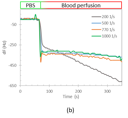
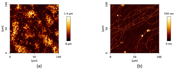
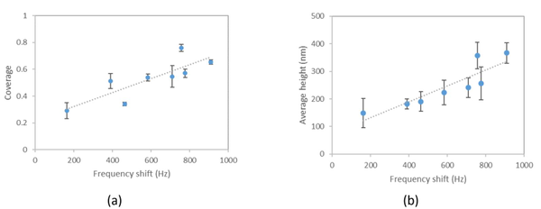
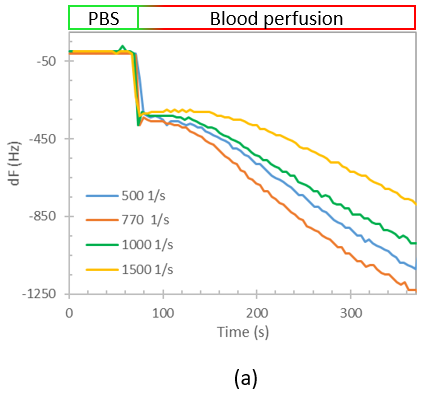
We designed a label-free assessment with a microacoustic biosensor based on quartz crystal microbalance (QCM), Figure 1. The biosensor frequency shift was prior compared with atomic force microscopy (AFM) images of the biointerface after perfusion and found to be in agreement with both coverage and average height of platelet deposits, Figure 2. The test results for 5 minutes perfusion of anticoagulated normal whole blood are shown in Figures 3(a)-4(a). Defective adhesion was induced by inhibition of GpIb platelet receptor with a monoclonal antibody at 10ug/mL. Perfusion results are shown in Figures 3(b)-4(b). Inhibition of GpIb affected initial platelet plug formation in all cases but less at 200 1/s when the contribution of vWF is expected to be the lowest.

The approach we developed is intended to improve evaluation of vWD in a label-free manner by integrative shear dependent phenotyping that is not realised by currently existing devices.

Uncaptioned visual

Figure 1. Parallel plate flow perfusion chamber with assembled biosensor (a); AFM image of collagen type I biointerface (b); experimental setup (c).

Uncaptioned visual

Figure 2. Frequency shift versus AFM measured surface platelet coverage (a) and average height (b) for two blood samples.

Uncaptioned visualUncaptioned visual

Figure 3. Test results of perfusion of whole blood without (a) and with (b) GpIb inhibition.

Uncaptioned visual

Figure 4. Representative AFM images of platelet deposits after perfusion (5 minutes) of normal (a) and GpIb inhibited (b) whole blood.

P3.003 An electrochemical sensor for on-line monitoring of ammonium in a fermentation process

A. Hasanzadeh1, M. Dimaki1, D. Semenova1, W.E. Svendsen1, M. Kilstrup1, K.V. Gernaey1, Babak Rezaei2
1Technical University of Denmark, Denmark. 22 National Centre for Nano Fabrication and Characterization, DTU Nanolab, Technical University of Denmark, Denmark

Abstract

Fermentation processes are widely used in the production of food, pharmaceuticals, enzymes, and a number of bulk chemicals. Efficient real-time monitoring of bioprocesses generates data that allows for improved process modeling and control, which can lead to increased process yield, productivity and reproducibility [1]. Therefore, instrumentation capable of real-time monitoring is essential for bioprocess optimization [2]. Recently, there has been a great effort in developing methods for real-time monitoring of fermentation processes using various advanced sensors such as electrochemical sensors / biosensors [3]. Ammonium is one of the central nutrients in media for most fermentation processes and needs to be present in relevant levels to promote growth and enzyme production. In this study, an electrochemical sensor based on polyaniline (PANi) and Nafion in combination with copper oxide (CuO) nanoparticles has been developed for the on-line determination of ammonium in the fermentation broth.

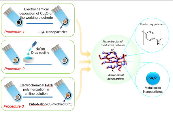
An amperometric ammonium sensor is fabricated by immobilization of three different layers on top of a graphene working electrode in a screen-printed electrode (SPE), including electrodeposited CuO, a Nafion membrane and electropolymerized PANi (Fig. 1). The presence of ammonium causes complex formation with copper, and this causes electroreduction of oxygen to result in an increased current.

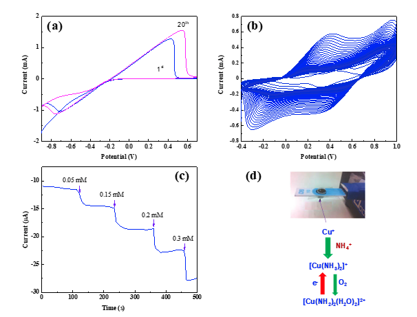
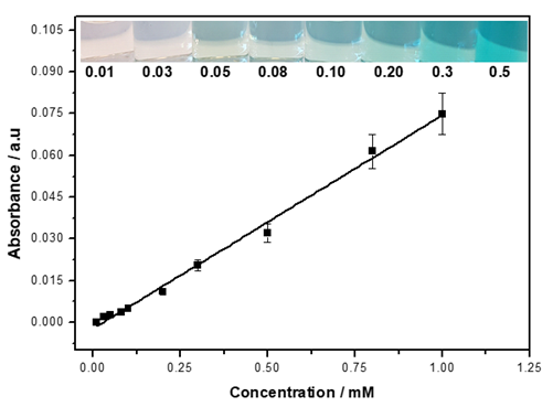
Figures 2a-b show the voltammogram of electrochemical synthesis of CuO and PANi on the surface of graphene, respectively. Amperometry was used for measurement of ammonium at the developed electrode (Fig. 2c). A proportional increase in the cathodic current was observed as a result of the increased ammonium concentration in the range from 0.05 to 1.0 mM. The whole process is realized on ammonia-associated oxygen reduction reaction electrocatalysis, as illustrated in Fig. 2d.

The here presented results show a promising path for quick detection of ammonium in the fermentation broth. Besides, the sensor can be further miniaturized and mounted on free-floating sensor particles.

Uncaptioned visual

Figure 1. Schematic representation of the fabrication of the PANi/Nafion/CuO/SPE electrochemical sensor

Uncaptioned visual

Figure 2. (a) modification of SPE by CuO (0.1 M HNO3, 50 mM Cu(NO3)2, scan rate 50 mV/s); (b) electrochemical polymerization of PANi on a Nafion/CuO-modified SPE (0.2 M aniline, 0.5 M HCl, scan rate 50 mV/s); (c) an example of amperometric response of electrocatalyst to successive additions of NH4Cl (−0.45 V, phosphate buffer); and (d) The oxygen reduction reaction at the Cu-based PANi composite in the presence of ammonium.

References:

[1] L. Mears, R. Nørregård, G. Sin, K.V. Gernaey, S.M. Stocks, M.O. Albaek, K. Villez, AIChE J. 62 (2016) 1986‐1994.

[2] D.W. Kimmel, G. LeBlanc, M.E. Meschievitz, D.E. Cliffel, (2019) Anal. Chem. 84 (2019) 685‐707.

[3] D. Semenova, A. Zubov, Y. Silina, L. Micheli, M. Koch, A.C.C. Fernandes, K.V. Gernaey, Sensors Actuators B Chem. 259 (2018) 945–955.


P3.004 Gold-seeded lithium niobate nanoparticles as NanoZyme biosensors

A.M. Pablo-Sainz-Ezquerra1,2, R. Taitt1, M. Urbain3, S. Beauquis3, Y. Mugnier3, P. R. D. Mariathomas2, R. Ramanathan2, V. Bansal2, Y. Chevolot1, V. Monnier1
1University of Lyon, France. 2RMIT University, Australia. 3Univ. Savoie Mont Blanc, France

Abstract

The need for a versatile, simple, fast and sensitive biosensor is crucial for environmental and health applications. Nowadays, enzyme-based assays in biosensing are one of the most used methods due to the high sensitivity and selectivity they present. Yet, there are some drawbacks such as instability and high cost. One strategy to overcome them is to employ the capability of metal nanoparticles deposited onto oxide nanoparticles to mimic the catalytic performance of enzymes. The immobilization of the metal nanoparticles on the oxide reduces the loss of performance related to aggregation. Besides, it produces a synergetic effect motivated by two complementary processes: a charge transfer between the materials and the creation of defects. This generates a hotspot for catalysis. We synthesized gold-coated lithium niobate nanoparticles with branched-polyethyleneimine as an intermediate linker between these two components. We showed that the intrinsic peroxidase-mimic NanoZyme activity of gold nanoparticles is upgraded when they are deposited onto lithium niobate nanoparticles. We also probed that the presence of a link between the two nanomaterials was necessary to ensure maximum efficiency. In addition, we optimized the conditions to maximize the colourimetric response (pH, temperature, quantity and surface coverage). The subsequent addition of aptamers, which bind to the gold nanoparticles, quenches the enzyme-mimics which is restored in the presence of the target. We managed to successfully attach the aptamer and the optimization of the biosensing of antibiotics is being carried out.


P3.005 Rapid nucleic acid lateral flow immunoassay for the detection of Pseudomonas aeruginosa

A. Brunauer1, R. Verboket2, D.M. Kainz1,3, F. von Stetten1,3, S.M. Früh1,3
1University of Freiburg, Germany. 2Hospital of the Goethe University, Germany. 3Hahn-Schickard, Germany

Abstract

Introduction:

Paper-based immunosensors are highly attractive tools for performing point-of-care (POC) diagnostics. Due to their simplicity and relative low costs they are promising for the detection of pathogens [1,2]. In comparison to the time-consuming conventional bacteriological culture, methods like the recombinase polymerase amplification (RPA) focus on the amplification of bacterial DNA [3,4]. Here, we present for the first time a RPA assay combined with a lateral flow immunoassay (LFA) for the detection of Pseudomonas aeruginosa (P. aeruginosa) – one of the most common bacteria that causes nosocomial infections [5]. Furthermore, the LFA includes an internal amplification control (IAC) and a flow control (FC).

Methods:

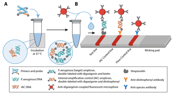
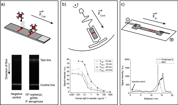
Our approach includes the generation of a double-labelled amplicon via RPA (Figure 1A) and detection of it via LFA (Figure 1B). Therefore, primers and probe were designed to target the lasB gene of P. aeruginosa. The amplification was performed at 37 °C within 15 minutes. Subsequently, we detected the amplicon using (1) lateral flow dip-sticks functionalized with streptavidin to capture the amplicon at the test line and (2) anti-digoxigenin-coupled fluorescent microspheres for signal generation. Furthermore, the validity of the test result was assessed by a FC and an IAC, which was co-amplified simultaneously with the target sequence.

Results and Discussion:

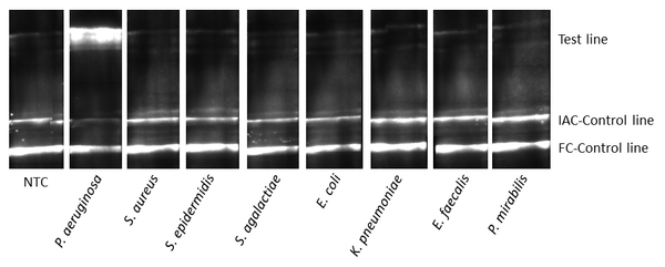
We successfully developed a rapid nucleic acid LFA for the detection of P. aeruginosa including a FC and an IAC (Figure 2). The approach is capable of detecting P. aeruginosa DNA within 30 minutes including both amplification and LFA. Specificity tests showed no cross-reaction with other microorganisms. Therefore, it provides a rapid method for pathogen identification. In future, our focus will be on integrating all three fundamental operation steps - sample preparation, amplification, and detection – into one single lateral flow strip. This technology paves the way towards reliable pathogen detection for a target-oriented therapy at the POC.

Figures:

Uncaptioned visual

Figure 1: RPA assay for the detection of P. aeruginosa combined with a LFA including a FC and an IAC. (A) Amplification of P. aeruginosa DNA and IAC DNA via RPA at 37 °C. (B) Design of the nucleic acid LFA including a FC and an IAC.

Uncaptioned visual

Figure 2: Specificity of the nucleic acid LFA was determined by using the DNA of P. aeruginosa and seven other microorganisms (S. aureus, S. epidermidis, S. agalactiae, E. coli, K. pneumoniae, E. faecalis, P. mirabilis). NTC, no template control.

References:

[1] Z. Guanyang, Trends in Analytical Chemistry. 2019, 111,100–117

[2] K. Koczula, Essays in biochemistry. 2016, 60,111–120

[3] J. Li, The Analyst. 2018, 144,31–67.

[4] O. Piepenburg, PLoS biology. 2006, 4,e204

[5] S. Wagner, Journal of medicinal chemistry. 2016, 59,5929–5969.


P3.006 A nature inspired bioluminescent paper nanosensor for affordable cancer diagnostics

A. Lopreside1,2, L. Montali1,2, M.M. Calabretta1,2, E. Michelini3,2
1University of Bologna, Italy. 2Azienda Ospedaliero-Universitaria Policlinico S. Orsola-Malpighi, Italy. 3Department of Chemistry “G. Ciamician”, University of Bologna, Italy

Abstract

Most of cancer effects on the organism are due to metastases, which often grow much faster than the primary cancer and show drugs resistance. Among others, extracellular expression of metalloproteases has been proven to be involved in tumor progression, metastasis formation and extravasation. 

Here, we present a nature inspired 3D-printed bioluminescent biosensor for rapid, cost-effective non-invasive and user-friendly cancer screening via protease activity detection.

The target sequence recognized by Matrix Metalloproteinase-2 (MMP2) was genetically fused to the Nanoluc complementation reporter system NanoBiT to create a fast and sensitive biosensor based on protein complementation technique. In absence of MMP2 protease activity, the sensor structure remains functional and it produces a bioluminescent signal by addition of furimazine substrate. Conversely, in the presence of MMP2 protease activity, the BL signals decreases in a quantitative way. 

A ready-to-use reagent-free disposable cartridge was developed lyophilizing both the bioluminescent nanosensor and the substrate into a cheap and eco-friendly paper-based device. 

3D printing technology was used to design a tunable and reusable device for smartphone interfacing and acquisition. Smartphone camera was implemented as detector for easy signal acquisition to reduce equipment cost and to allow easy data storage and labelling. 

In optimized conditions we were able to obtain as limit of detection for MMP2 in the nM range with only 30 minutes of incubation with the sample. The high stability of the sensor and the preliminary results obtained with clinical samples corroborate its potential use as point-of-care cancer diagnostic device. 


P3.007 Impact of silane monolayers on the selective adsorption of sepsis metabolites

A. Lavigne, S. Lecot, T. Géhin, M. Phaner-Goutorbe, C. Yeromonahos
University of Lyon, France

Abstract

Sepsis, blood bacteremic infection, is one of the first mortality causes in hospitalized patients. Survival chance declines by 7% each hour, while diagnosis required 5 days. There is an urgent need for rapid diagnostic strategies. Patterns of metabolites recently identified (amino acids…), might allow sepsis diagnosis and the identification of the most common pathogens. Nanoporous Silicon matrices (pSi) allow metabolites trapping from serum and are compatible with Matrix-Assisted-Laser-Desorption-Ionization Mass Spectrometry (MALDI-MS) analysis. However, for sensitive and rapid sepsis diagnosis, there is a clear need for selective trapping of sepsis metabolites among other metabolites from serum.


Silane molecules are widely used for the functionalization of silica surfaces for biomolecule adsorption applications. Here, we decipher by Molecular Dynamics (MD) simulations the efficiency of different silane monolayers (different charges and alkyl chain lengths) to selectively adsorb the sepsis metabolites among the most common blood metabolites. The results are validated by Fourier-Transform-Infrared-Attenuated-Total-Reflectance (FTIR-ATR) analysis.


Firstly, a simulation system was developed (GROMACS) to study the interactions between 14 metabolites dissolved in water (sepsis and common blood metabolites) and 4 different silane monolayers on a silica substrate. The calculated adsorption energies were correlated to the metabolite FTIR-ATR detection. The results suggest that metabolite adsorption can be tuned thanks to the nature of the silane monolayers and the physico-chemical properties of the metabolites (charge, hydrophobicity, flexibility). Moreover, we suggest that water molecules in the hydration layer are highly ordered when silica is modified with uncharged silane monolayers with short alkyl chains, whereas no order is observed with long alkyl chains. This order might be at the origin of the more favorable entropically driven adsorption of metabolites obtained with short alkyl chains. Finally, we evaluate the possibility of sepsis metabolite detection from serum, by MALDI-MS analysis, using pSi and the most suitable silane monolayers identified from simulations. 


P3.008 Double-resonant branch patterns of honeycomb-arranged gold nanoparticles for multiplexed analyte detection

Antonio Minopoli1, Bartolomeo Della Ventura1, Julian A. Tanner2, Dirk Mayer3, Raffaele Velotta1
1University of Naples "Federico II", Italy. 2School of Biomedical Sciences, University of Hong Kong, Hong Kong. 3Institute of Biological Information Processing (IBI-3), Forschungszentrum Jülich, Germany

Abstract

Development of fast and reliable biosensors able to detect disease biomarkers at very low concentrations in a complex matrix such as human whole blood is still a challenge. In this regard, optical biosensors relying on plasmon-enhanced fluorescence (PEF) provide an effective strategy to push down the detection limits while holding low-cost production and easy-to-use. In particular, honeycomb arrays of gold nanoparticle (AuNPs) realized through block copolymer micelle nanolithography (BCMN) stand out for their scalability and tunable plasmonic properties making them ideal substrates for fluorescence enhancement.1

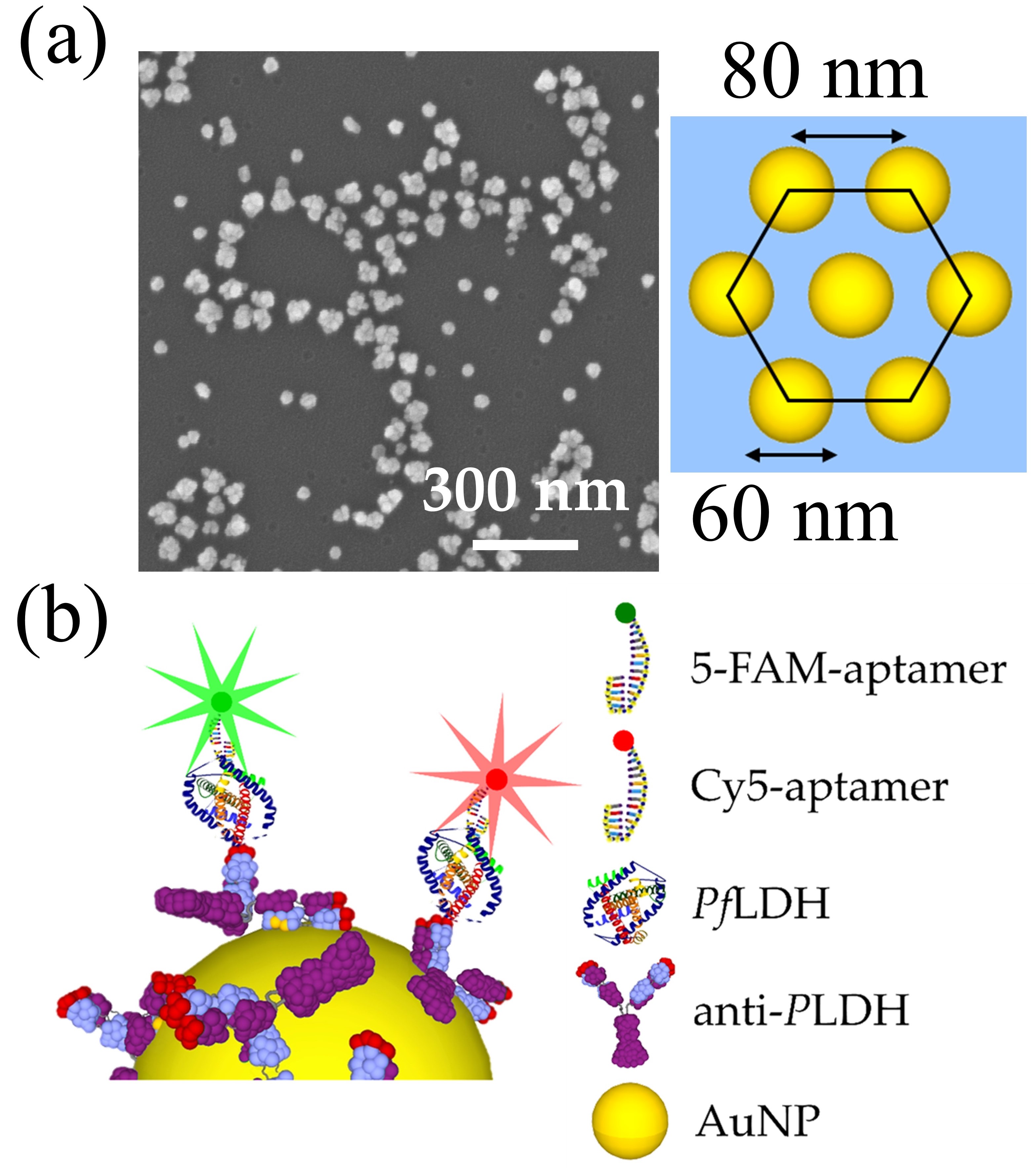
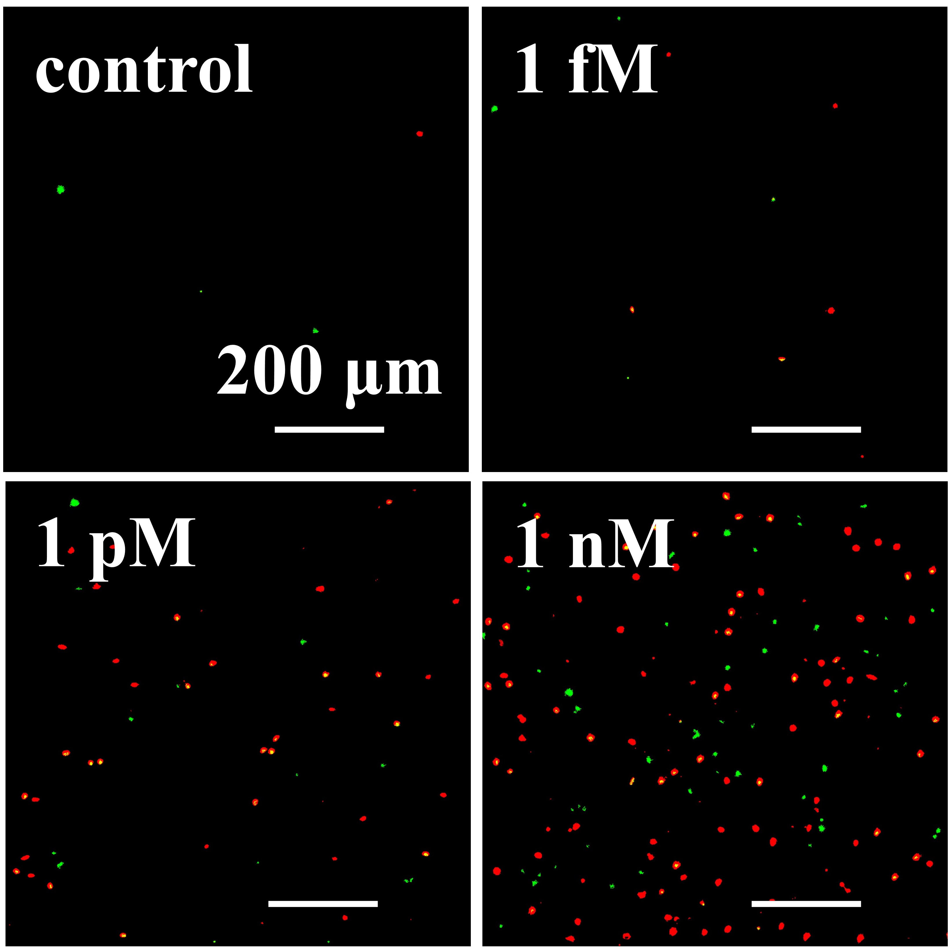
Here, we describe a novel double-resonant plasmonic nanostructure for effective signal amplification in fluorescence-based apta-immunoassay. The substrate consists of a branch pattern made of plasmon-coupled honeycomb-arranged AuNPs, which gives rise to a collective mode whose resonance lies in the far-red region, and sprinkled plasmon-uncoupled AuNPs that exhibit a narrow resonance at 524 nm (Figure 1a). The plasmon resonances are tailored to couple with the emission peak of 5-carboxyfluorescein (5-FAM) fluorescent dye and with either the excitation and emission peaks of cyanine 5 (Cy5) thereby promoting a 160-fold fluorescence amplification through emission rate enhancement and a 5200-fold signal amplification via dual-mechanism (excitation and emission rates) enhancement, respectively. Analyte capture is realized by oriented antibodies immobilized in a close-packed configuration onto the substrate via the photochemical immobilization technique (PIT), while the fluorescent labelling by a top bioreceptor layer of nucleic acid aptamers recognizing a different surface of the analyte in a sandwich conformation (Figure 1b). When implemented in a malaria fluorescence-based apta-immunoassay for detecting Plasmodium falciparum lactate dehydrogenase (PfLDH), such a plasmonic nanostructure allowed us to detect analyte concentrations over five decades down to 50 pM (1.6 ng/mL) – if explored with the green probe – and 260 fM (8.6 pg/mL) – by using the red probe – in whole blood without any sample pretreatment. Figure 2 shows some fluorescence images recorded at different PfLDH concentrations spiked in human whole blood. The proposed approach is very attractive for simultaneous monitoring different proteins in complex matrices such as human blood and may be easily implemented in automated multi-well plate readers paving the way to high-throughput analysis.

Uncaptioned visual

Figure 1: (a) SEM micrograph (left panel) and sketch (right panel) of the branch pattern of honeycomb-arranged AuNPs (b) antibody-analyte-fluorescently labelled aptamer configuration.

Uncaptioned visual

Figure 2: Example of fluorescence pictures acquired at different PfLDH concentrations spiked in whole blood.


P3.009 Novel selective and sensitive amperometric biosensors based on hybrid nanoplatforms

Artur Jędrzak, Maria Kuznowicz, Teofil Jesionowski
Poznan University of Technology, Poland

Abstract

The development of technology has enabled the advancement of research into the design of more important hybrid materials. Various inorganic-organic materials that were previously commonly used in many fields of science and a variety of industries can meet such demands [1].

Hybrid architectures can be used to develop and build new biosensor systems that are more selective, sensitive, stable (3S). Due to the need for efficient and cost-effective biodetectors, novel advanced materials for biosensors have gotten a lot of attention [2]. 

Glucose determination is a common and important assay in a variety of media, including blood plasma, medications, juices, and dietary supplements. Despite the availability of commercial glucose biodetectors, there are still some limitations in accuracy and errors due to aging, manufacturing variability, storage, temperature, coding, or incorrect data measurement.

Herein, we introduced a novel glucose-model biosensors based on a hybrid nanoplatform. We used techniques like photometric assay, commercially available glucometers, and our proposed biosensors, which include hybrid carriers and dedicated electrodes, to arrange real samples.

Magnetite, silica, lignin, polydopamine, and glucose oxidase are examples of natural or nature-inspired materials used in biosensor platforms. Biosensor systems with higher materials sensitivity, stability, selectivity, and durability are appealing alternatives among biosensors for determining the level of glucose in various media such as commercial samples (drinks, glucose-based supplements juices, fruits) or body fluids (blood plasma, whole blood).

Our proposed biosensors, which are based on a hybrid micro and nanoplatform, show promise as a biodetector in the food industry and diabetes care.

This work was financed and prepared as part of a research project supported by the National Science Center Poland, no. 2017/27/B/ST8/01506.

[1] L. Qian, S. Durairaj, S. Prins, A. Chen, Biosens. Bioelectron. 2021, 175, 112836.
[2] A. Jędrzak, T. Rębiś, M. Kuznowicz, T. Jesionowski, Int. J. Biol. Macromol. 2019, 127, 677-68.


P3.011 The evaluation the efficiency of photodynamic therapy with meso-tetraphenylporphirin as a photosensitizer and modified graphene oxide as a drug carrier using microfluidic device

B. Dabrowski, A. Zuchowska, A. Kasprzak, K. Kaminska, M. Poplawska, Z. Brzozka
Warsaw University of Technology, Poland

Abstract

Despite advances in medicine cancer diseases are still an urging problem. Due to its high surface area and affinity towards cancer cells, graphene oxide (GO) has a great potential to find an application as a novel drug carrier in photodynamic therapy (PDT).

In this study we examined the efficiency of photodynamic therapy using meso-tetraphenylporphirin (TPP) as a photosensitizer and modified GO as a drug carrier. Graphene oxide was functionalized with ethylenediamine (GO1) and with tetraethylenepentamine (GO2). The were conducted using coculture of normal and cancer breast cell lines. Cells were cultured in a microfluidic Lab-on-a-chip device as monolayer (2D) and spheroids (3D).

The design of microsystem was presented in Figure 1. The layout of microchambers corresponded to layout of wells in 384-well plate. Therefore, spectrofluorimetric measurements could be performed in standard multi-well plate reader using dedicated chip holder.

In order to evaluate efficiency of photodynamic therapy using GO1-TPP and GO2-TPP cell viability, cellular uptake and reactive oxygen species generation (ROS) were examined. Photocytotoxicity was evaluated using AlamarBlue® test. TPP accumulation in cells were evaluated by measuring fluorescence intensity of TPP and confocal microscopy. ROS generation was measured using dichloro-dihydro-fluorescein diacetate (DCFH-DA) assay. All measurements were performed for irradiated and non-irradiated cells.

Efficiency of PDT using GO1-TPP and GO2-TPP was greater comparing to free PTT. Cellular uptake and ROS generation studies are consistent with these results. Only TPP bound to the drug carrier was able to penetrate cell membrane. The results show that examined graphene oxide derivatives can be efficient drug carriers in PDT.


P3.012 Plasmonic biosensor-based detection of misfolded proteins and protein-protein interactions for diagnosis of Myelodysplastic syndromes

L. Chrastinová1, O. Pastva1, M. Bocková2, N.S. Lynn2, J. Homola2
et al
1Institute of Hematology and Blood Transfusion, Czech Republic. 2Institute of Photonics and Electronics of the Czech Academy of Sciences, Czech Republic

Abstract

Myelodysplastic syndromes (MDS) are a heterogeneous group of hematological malignancies with a high risk of transformation to acute myeloid leukemia (AML). MDS are associated with posttranslational modifications of proteins, variations in the protein expression and protein misfolding. In this contribution, we describe two novel applications of surface plasmon resonance (SPR) biosensors related to the diagnosis of Myelodysplastic syndromes (MDS) – the investigation of protein-protein interaction and detection of misfolded proteins in blood plasma of patients at different stages of MDS or AML.

In the first application, we used an SPR imaging sensor to quantify interactions between an array of six proteins (whose concentrations vary during MDS) and blood plasma of MDS patients and healthy donors. Four proteins (ICAM, VCAM, fetuin and LRG) were selected based on literature data; the other two proteins (clusterin and S100A8) were selected using the SPR/LC-MS/MS method. We applied this approach to analysis of interactions among the immobilized proteins and blood plasma samples from a total of 80 MDS patients and healthy donors and demonstrated that the used protein array allows for discrimination among different MDS and AML subgroups and healthy controls.

In the second application, we used Heat shock protein 70 (Hsp70), that is known to recognize and capture misfolded proteins, as a recognition element in a high-performance spectroscopic SPR biosensor. We applied this biosensor to the detection of misfolded proteins in blood plasma from 60 MDS patients and healthy controls. Our results demonstrated that this approach enables rapid and sensitive detection of misfolded proteins in blood plasma and revealed significantly elevated levels of misfolded proteins in plasma of patients in the two stages of MDS that are most affected by oxidative stress (RARS and RCMD).


P3.013 Paper-based electrochemical wearable sensor toward non-invasive continuous monitoring of glucose in exhaled breath

M.A. Schüler1,2, A. Altay1,2, E. Laubender1,2, S. Schumann3, F. Güder4, G.A. Urban1,5, C. Dincer1,2
1University of Freiburg, Germany. 2Freiburg Center for Interactive Materials and Bioinspired Technologies – FIT, Germany. 3MUniversity of Freiburg, Germany. 4Imperial College London, UK. 5Freiburg Materials Research Center – FMF, Germany

Abstract

Introduction

Monitoring blood glucose levels is a crucial part in the daily life of diabetes patients. Non-invasive glucose monitoring as a more comfortable alternative has recently become increasingly important1. While most studies focus on exhaled breath condensate (EBC) samples, this work deals, for the first time, with easy, low-cost and simultaneous method for sampling and measurement of glucose in exhaled breath using a paper-based electrochemical sensor.

Methods

Paper-based substrates offer a simple and cheap solution to sample and analyse respiratory fluid from exhaled breath. Figure 1 illustrates the differential chip design and fabrication. Glucose oxidase (GOx) is adsorbed into the compartment surrounding the sensing electrode. It catalyses the oxidation of glucose into hydrogen peroxide (H2O2) which can be reduced at the screen-printed Prussian Blue (PB)-mediated carbon electrode. The measured current relates directly to the sample glucose concentration. A second, identical cell, but without GOx, enables to subtract background signals and periodic variations caused by the respiratory movement.

Uncaptioned visual

Figure 1: Chip design and fabrication showing the main elements of the paper-based glucose sensor: wax isolation (lime green), electrodes from left to right: sensing, reference, blank and counter electrodes.

Results

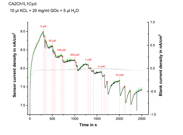
To demonstrate its feasibility, the sensor is exposed to aerosols of different glucose concentrations, using a deodorant nebulizer. Within the physiological range, a stepwise increase of the differential current signals (see figure 2) at consecutive aerosol administrations is observed. At higher concentrations, a peak followed by a current decay is noticed, possibly due to limited oxygen supply.

Glucose entrapped in aerosols can be cumulatively sampled and directly measured with paper-based sensors at concentrations of less than 5 µM. Compared to EBC analysis, our approach minimizes the risk of analyte degradation, while considerably reducing acquisition time and system’s complexity.

Uncaptioned visual

Figure 2: Proof-of-principle study of non-invasive glucose monitoring using a paper-based electrochemical sensor. Sensor (black), blank (grey) and differential (green) current density signals (at –0.2 V vs. 1 M Ag/AgCl) during a series of applications of glucose containing aerosol puffs. For each given concentration, three consecutive puffs were applied (red triangles and dotted lines).

Conclusion

We have successfully demonstrated the proof-of-principle of a facile, inexpensive and non-invasive approach for the simultaneous sampling and measurement of exhaled glucose, for the first time. Our future work will include the characterization and optimization of the developed system in simulated breath2, followed by its clinical validation.

References

  1. D. Tankasala, J. C. Linnes, Transl. Res. 2019, 213, 1.
  2. D. Maier, E. Laubender, A. Basavanna, S. Schumann, F. Güder, G. A. Urban, C. Dincer, ACS Sensors 2019, acssensors.9b01403.

P3.014 Non-disruptive electrical impedance measurement for cell analysis and classification: Application in the identification of two populations of T Lymphocytes

C.D.M. Campos1,2, P. Barmuta2,3, T. Markovic1,2, R. Yadav1,2, I. Ocket1, W. van Roy1, T. Stakenborg1, L. Lagae1,2, J. Genoe1,2, C. Liu1
1IMEC, Belgium. 2KU Leuven, Belgium. 3Warsaw University of Technology, Poland

Abstract

We developed a label-free method to characterize changes in cell physiology. Detecting membrane and cytosol alterations non-disruptively is crucial for cell engineering, personalized medicine, etc. Yet, conventionally employed destructive, end-point assays, or the requirement of fixing, staining or labelling hamper downstream uses of the cells [1,2]. Non-invasive characterization can be done with powerful but expensive techniques that demand expert users (e.g. Raman or infrared spectroscopy) [3–6]. Electrical cell impedance spectroscopy is a promising approach but requires tight cell-electrode contact, which is difficult to control in practical applications and might affect cell behaviour and signal interpretation.

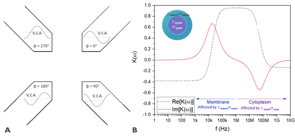
To overcome these shortcomings, we characterize cell impedance by measuring cell spinning in rotating AC electric field [7,8], generated by a set of electrodes (Fig. 1A). The dipole induced in the cell interacts with the rotating field creating a torque, leading to cell spinning. Its angular velocity, measured by imaging system, is determined by cell impedance (Fig. 1B).

Reported electrorotation devices operate under 10 MHz [9], limiting the response obtained to changes associated with the membrane and unable to resolve the physiological properties in the cytoplasm. We used microwave-based technology to extend the frequency operation to 1 GHz. At high frequencies, microwave signal passes through the membrane and allows probing the cytoplasm, bringing additional information. This enables several applications, e.g. cell classification, etc. The use of conventional microfabrication techniques reduces the cost and complexity of analysis, compared to other non-invasive methods.

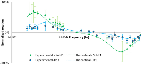
With this technique, we identified, using electrical properties, two different populations of T lymphocytes, not distinguishable through visual assessment (Fig. 2B). More studies are necessary to pinpoint the specific biological changes in membrane and cytoplasm content that entail the differences in the electric properties and the perceived rotation, but these results make clearer the application of high frequency electrorotation in non-disruptive cell analysis.

Uncaptioned visual

Figure 1. (A) Schematic of the electrode positioning for electrorotation. (B) Plot of the real (black) and imaginary (red) parts of the Clausius-Mossotti factor for a cell. Clausius-Mossotti is the factor that relates the electrical conductivity and electric permittivity of the cell and the medium.

Uncaptioned visual

Figure 1. Rotation spectrum of T lymphocytes D11 and SubT1. It is possible to see that SubT1 cells rotate at higher speed than observed for D11. Also, around 10 Mhz the rotation direction of SubT1 is opposite to the one observed for D11. Those features in the spectrum will allow us to correctly identify the two populations.

References
[1] J. Lin, Cell Analysis on Microfluidics, Springer Singapore, Singapore, 2018.
[2] D. B. Papkovsky, Live Cell Imaging, Humana Press, Totowa, NJ, 2010.
[3] C. Stringari, J. Biomed. Opt. 2012, 17, 046012.
[4] A. Downes, R. Mouras, P. Bagnaninchi, A. Elfick, J. Raman Spectrosc. 2011, 42, 1864–1870.
[5] D. Ami, P. Mereghetti, A. Natalello, S. M. Doglia, M. Zanoni, C. A. Redi, M. Monti, Biochim. Biophys. Acta - Mol. Cell Res. 2011, 1813, 1220–1229.
[6] M. E. Gosnell, A. G. Anwer, S. B. Mahbub, S. Menon Perinchery, D. W. Inglis, P. P. Adhikary, J. A. Jazayeri, M. A. Cahill, S. Saad, C. A. Pollock, et al., Sci. Rep. 2016, 6, 1–11.
[7] A. Valero, T. Braschler, P. Renaud, Lab Chip 2010, 10, 2216–2225.
[8] Y. Zhao, D. Jia, X. Sha, G. Zhang, W. Li, Micromachines 2018, 9, 118.
[9] O. J. Pletjushkina, O. J. Ivanova, I. N. Kaverina, J. M. Vasiliev, Exp. Cell Res. 1994, 212, 201–208.


P3.015 Toward "finger prick" blood tests for personalized antibiotherapy of beta-lactams

H.C. Ates1,2, N. Schneider3, Hasti Mohsenin3, W. Weber3, G.A. Urban1,4, C. Dincer1,2
1University of Freiburg, Germany. 2Freiburg Center for Interactive Materials and Bioinspired Technologies, Germany. 3BIOSS Centre for Biological Signalling Studies, Germany. 4Freiburg Materials Research Center, Germany

Abstract

Introduction

Personalized antibiotherapy is a promising remedy to maximize the therapy effectiveness while minimizing the toxic side effects and antimicrobial resistance. Hence, there is an urgent need for rapid, low-cost and simple solutions toward on-site monitoring of antibiotics. Biosensors inherently meet all these requirements, however, until now there are only attempts to measure ß-lactam concentrations in processed human body fluids, which requires additional steps[1,2]. In this study, for the first time, we present a low-cost electrochemical biosensor for on-site monitoring of ß‑lactam antibiotics in whole blood. This is the first step of developing “finger prick” blood tests for antibiotic surveillance.

Methods

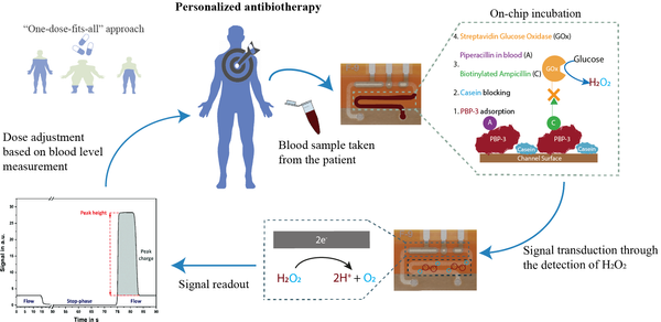
The microfluidic biosensor consists of two zones; an immobilization area and an electrochemical cell, separated by a hydrophobic stopping barrier to prevent electrode fouling[3]. Sample containing analyte and competitor goes through the immobilization area, in which competitive binding between the analyte and ampicillin-biotin conjugate to penicillin-binding-protein-3 (PBP‑3) takes place. Streptavidin-glucose oxidase is utilized for the signal generation by converting glucose to H2O2, which is then detected at the working electrode (Figure 1). On-chip ß-lactam calibration is performed by using diluted blood samples spiked with different concentrations of piperacillin.

Uncaptioned visual

Figure 1: Methodology for “one-dose-fits-all” approach to personalized antibiotherapy. On-chip incubation starts with PBP-3 adsorption by 1-h incubation of the channel surface with 250 µg ml‑1 PBP-3. Then, active surface sites are blocked with biotin-free casein for 30 minutes. The third step of the assay relies on the competitive binding of piperacillin in the sample solutions with different concentrations and 10 µg ml-1 biotinylated ampicillin to the PBP-3. Finally, 10 µg ml-1 Streptavidin-glucose oxidase is utilized with a 15-minute incubation for the signal generation. Conversion of glucose to hydrogen peroxide by streptavidin-glucose oxidase results in an electrical current through the measurement of H2O2 in the electrochemical cell, followed by the signal readout and dosage adjustment.

Results

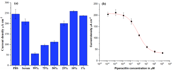
First, matrix effect was investigated by comparing whole blood signals at various dilutions with those of buffer and serum (Figure 2a). Results show that whole blood can be employed with low dilution rates (< 25%) without significant signal decrease. Consequently, on-chip calibration of piperacillin is performed in 10% whole blood (Figure 2b), resulting in a limit-of-detection of 0.058 µM and an inter-assay coefficient-of-variation below 12%.

Uncaptioned visual

Figure 2: (a) Evaluation of matrix effect on assay performance with whole blood samples at different dilution factors. No significant matrix effect is observed with untreated serum samples and whole blood at dilution factor as low as 25%. (b) On-chip piperacillin calibration in 10% whole blood (N=4). Using 4-parameter logistic fit, LOD of 0.058 µM is achieved.

Discussion

We have successfully demonstrated the proof-of-principle of on-site piperacillin monitoring in unprocessed blood samples, for the first time. Our future work will be further optimization and extension of this platform to monitor other ß-lactams and sepsis biomarkers simultaneously, which would pave the way for the personalized therapy.

References

[1]G. Merola, E. Martini, M. Tomassetti, L. Campanella, J. Pharm. Biomed. Anal. 2015, 106, 186.

[2]S. A. N. Gowers, D. M. E. Freeman, T. M. Rawson, M. L. Rogers, R. C. Wilson, A. H. Holmes, A. E. Cass, D. O’Hare, ACS Sensors 2019, 4, 1072.

[3]R. Bruch, A. Kling, G. A. Urban, C. Dincer, J. Vis. Exp. 2017, 1.


P3.016 Binding energy increase between the spike protein of the SARS-CoV-2 and the host cell receptor ACE2, through ACE2 adsorption on a silane monolayer: A molecular dynamic study

S. Lecot, M. Phaner-Goutorbe, C. Yeromonahos
University of Lyon, France

Abstract

Currently, antigenic tests are widely used for COVID diagnosis. They are based on molecular recognition by highly potent neutralizing antibodies. However, the Spike proteins of the SARS-CoV-2 have been shown to accumulate rapidly escape mutations, limiting the antigenic test efficiency. The entry of the virus into the host cell involves the interaction between the angiotensin-converting-enzyme-2 (ACE2) from the host cell and the ACE2 binding domain of the Spike protein (RBD). As such, this domain needs to be highly conserved to preserve viral virulence and consequently could be a target with limited mutation escaping possibilities. However, because ACE2 has not evolved to recognize RDB, the ACE2/RBD affinity needs to be improved for clinical diagnosis use. In particular, a deep mutagenesis study suggests that mutations in the residues N90/T92 of ACE2 would lead to an exceptional affinity with RBD.1

Silane monolayers are widely used for the chemical modification of silica surfaces and are commercially available. Different silane monolayers were shown able to tune the interactions between Biotin and adsorbed Streptavidin.2 Herein, we use Molecular Dynamic (MD) simulations to decipher the effects of different silane monolayers (bearing positively and negatively charged head-groups, without head-groups, with different alkyl chain lengths) on the adsorption of ACE2 and on the ACE2/RBD interactions.

Our results suggest that the monolayers made up of a mixture of silane molecules bearing positively charged head-groups and of silane molecules without head-groups, allow to adsorb ACE2 while keeping its bioactivity (favorable orientation and interaction energy with the silane monolayer and low deformations of the RBD binding site) and increasing the ACE2/RBD binding energy through local deformations on the residues N90/T92. Thus, silane monolayers could be promising candidates for reliable COVID diagnosis platform.

[1] K.K. Chan et al. Science 2020, 1261-1265

[2] S. Lecot et al. J. Phys. Chem. B 2020, 6786-6796 


P3.018 Metallic mass-produced pins as electrodes in (Bio)electroanalytical devices for clinical applications

A. González-López, A. García-Miranda Ferrari, O. Amor-Gutiérrez, M.T. Fernández-Abedul, E. Costa-Rama
University of Oviedo, Spain

Abstract

Nowadays, there is a widespread interest in developing simple analytical devices that provide fast and reliable responses. Electrochemical techniques fit perfectly with these purposes especially in combination with low-cost materials. Thus, in this work, we present innovative electroanalytical devices based on stainless-steel pins, commonly used for sewing, for the analysis of small molecules and proteins of clinical interest.


Herein, the electrochemical three-electrode cell configuration consists of a carbon-coated pin acting as working electrode (WE) and two bare pins as reference (RE) and counter (CE), respectively. Two different systems were developed for glucose analysis: i) an enzymatic biosensor using a transparency sheet as support, which was easily drilled with the pins creating an integrated device; and ii) a flow injection electroanalytical (FIA) system in which the pins used as electrodes were directly inserted in the tube of the flow system. On the other hand, a batch injection (BIA) electroanalytical system for epinephrine determination in pharmaceuticals was also developed. Taking advantage of the versatility of pins as electrodes, a multiplex BIA system was also designed based on 8 carbon-coated pins as WEs with shared RE and CE.


Lastly, pins were integrated in the cap of a microcentrifuge tube as detection system for a sandwich-type immunoassay performed after immobilization of the capture antibody at the bottom of the tube. The analytical signal is recorded in a simple way by turning the tube upside down. The reliability of this device was proved for the determination of Glial Fibrillary Acidic Protein (GFAP), which is studied as a biomarker for differential stroke diagnosis, distinguishing between hemorrhagic and ischemic strokes. This innovative strategy simplifies immunoelectroanalysis integrating the biological assay and the detection into an all-in-one device.


This work has been supported by projects CTQ2014-58826-R (Spanish Government) and FC15-GRUPIN-021 (Asturias Regional Government). 


P3.019 Facile biosensor-based system for on-site quantification of total viable counts of bacteria in food and environmental swabs

D. Papkovsky, E. Santovito, S. Elisseeva, J. Kerry
University College Cork, Ireland

Abstract

Microbial swab testing is a fundamental component of surface sampling and process monitoring, especially in the food, pharmaceutical and biotech industries. Environmental and microbiological surveillance is currently conducted through routine sampling of contact surfaces with dedicated swabs: brush swabs, sponge swabs or integrated swabbing devices.  Swab testing, aimed at tracing and controlling baseline levels of contamination and cross-contamination, is key for environmental, hygiene and safety monitoring. We have developed a new biosensor platform for rapid and simple quantification of total aerobic viable counts (TAVC) of bacteria in surface swabs by oxygen respirometry, which uses sterile disposable swab vials with culture medium and phosphorescent oxygen sensor coatings in the bottom part. Swab samples, prepared using the standard method (ISO 18593:2018), are transferred into these sensor vials and incubated at 30oC while periodically reading sensor signals with an autonomous handheld reader. The real-time contactless monitoring of the microbial growth via oxygen respiration, gives rapid and quantitative readout of sample TAVC values. The sensing measurements reveal time profiles of dissolved O2 in each sample vial, from which Threshold Time of sensor signal was determined and then TAVC values were calculated using a pre-determined calibration equation. The method covers the range of 0.65–7.87 Log (CFU/cm2) and produces results in 1-8 hours. The test was validated with swab samples from different contaminated surfaces and showed no statistically significant difference with the reference method which takes 48-72 hours to result. The sensor-based swab testing platform addresses the demand of industry, particularly the food sector, consumers, and society, in rapid, simple, and de-centralised swab testing. Unlike other methods that rely on manual plate counting, this system provides medium levels of automation, sample throughput and integration, along with modular flexibility, portability, and on-site deployment capability. 


P3.020 A bi-enzymatic paper-based sensor for glucosinolate detection in food waste

D. Moscone, N. Colozza, A. Mucaria, F. Arduini
University of Rome Tor Vergata, Italy

Abstract

Glucosinolates are characteristic phytochemicals present in the Brassicaceae family, including mustard, cabbage and horseradish. They play an important role in the plant’s defences against pests and diseases and confer typical organoleptic characteristics, such as a bitter flavour. When a plant is damaged (e.g. by a herbivorous), glucosinolates are hydrolysed by myrosinase enzyme (thioglucoside glycohydrolase), resulting in toxic effects for the predator or in biological responses in the physiology of the plant. Despite of their toxicity for some organisms, glucosinolates show antioxidant properties in humans and can reduce the risk of cardiovascular diseases. Thus, the monitoring of glucosinolate levels into Brassicaceae is a matter of interest in the agri-food field, also involving biomedical aspects. The reference methods for glucosinolate measurement include their extraction from plant samples by boiling in protic solvents and then their analysis through chromatographic techniques coupled with a mass spectrometer or a UV-visible spectrophotometer. Smarter innovative approaches have been proposed, such as the development of biosensors. 

Here we present a bi-enzymatic paper-based sensor for glucosinolate detection based on the measurement of glucose resulting from the glucosinolate hydrolysis carried out by myrosinase. Glucose oxidase and myrosinase were immobilised on two separate filter paper layers. After evaporating the solvent, the layers were overlapped onto a paper-based screen-printed sensor, which graphite conductive ink was previously modified with a Carbon Black/Prussian Blue nanocomposite. After having verified the enzymatic behaviour of both the enzymes absorbed on the paper support (overall KM = 5.2 ± 0.9 mM), the amount of the measured hydrogen peroxide was successfully correlated with standard amounts of glucosinolates (i.e. sinigrin), obtaining a LOD of 0.03 mM for sinigrin in amperometric mode. Moreover, the sensor was applied for the analysis of extracted samples from several species of Brassicaceae, observing a satisfactory comparison with the spectrophotometric analysis. 

Acknowledgement to PRIN2015FFY97L for funding.


P3.021 SuperPests LabDisk: a microfluidic-based molecular diagnostic platform for detection of biotypes, resistance mutations and plant pathogens

D. Baumgartner1, B. Johannsen2, N. Paust1,2, F. von Stetten1,2, R. Zengerle1,2, K. Mavridis3, J. Vontas3,4, K. Mitsakakis1,2
1University of Freiburg, Germany. 2Hahn-Schickard, Germany. 3Foundation for Research and Technology-Hellas, Greece. 4Agricultural University of Athens, Greece

Abstract

Arthropod pests pose an increasing threat to agriculture [1]. Intensive use of pesticides leads to the evolution of widespread resistance. This has caused the emergence of a subset of arthropod pests that are now extremely difficult to control. To ensure sustainable management of these so-called “super pests”, monitoring with molecular approaches as complement to traditional bioassays is urgently required, as part of insect resistance management (IRM) programs [2].

In this work, we demonstrate the integration of molecular assays in an automated diagnostic platform. As a model pest, we used the two-spotted spider mite, Tetranynchus urticae, which feeds on more than 1100 plant species.


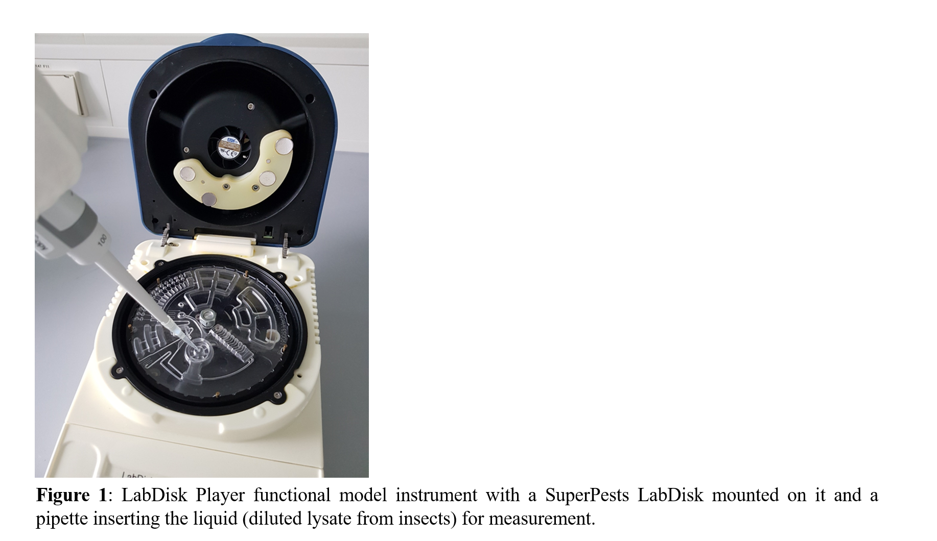
Multiplex TaqMan real-time PCR assays for Species ID (ITS region), target site mutations (G314D, G326E, L1024V, I1071F, G126S, S141F) and detoxification gene expression (GSTd05, using RP49 for normalization) [3] were transferred from a benchtop PCR device to a portable and point-of-need-compatible device, which processes a disk-shaped microfluidic cartridge (Figure 1) containing all necessary reagents and test components [4]. Crude lysis of a pool of 50 insects followed by direct amplification yielded results within 140 min.


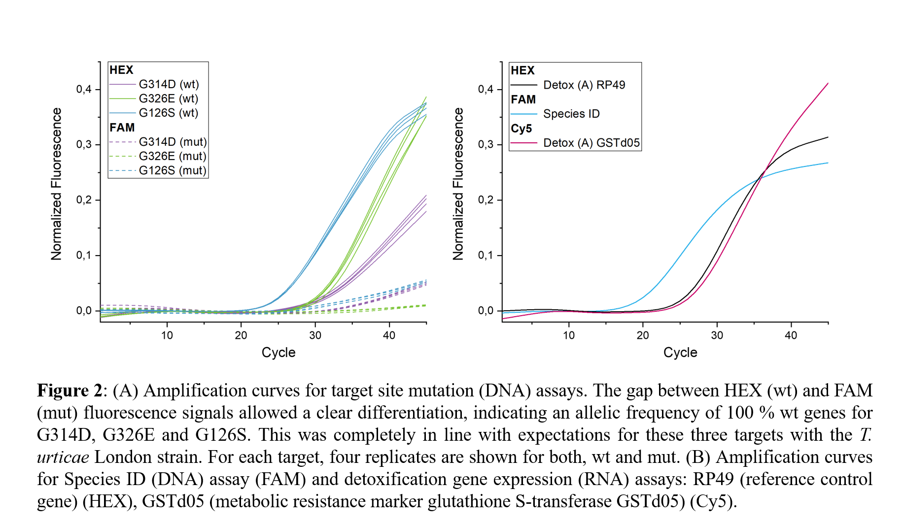
In all six target site (DNA) assays it was possible to distinguish clearly between wild type (wt) and resistance-conferring mutant (mut) genes (three of the six assays are indicatively shown in Figure 2 A). In addition to the Species ID we could also specifically detect RNA targets from T. urticae crude lysate, namely GSTd05 as a metabolic resistance marker and RP49 as a reference control gene (Figure 2 B).

This is the first time that this platform extends its utility into the field of pest control. It can comprise a valuable tool for insecticide and crop management, especially in the light of global needs for green society and sustainability. 


Uncaptioned visual

Uncaptioned visual

References:

[1] http://www.fao.org/fileadmin/templates/wsfs/docs/expert_paper/How_to_Feed_the_World_in_2050.pdf (accessed on 2021-03-23)

[2] https://superpests.eu/ (accessed on 2021-03-21)

[3] N. Pavlidi et al., Insect Biochemistry and Molecular Biology. 80, 101-115 (2017).

[4] S. Hin et al., Processes. 8, 1677 (2020). 

[?] T. Van Leeuwen et al., Current Opinion in Insect Science. 39, 69-76 (2020). 


P3.022 Novel CO2 microsensors/urease-based biosensors for urea analysis and monitoring

D. Fapyane, D. Berillo, N.P. Revsbech
Aarhus University, Denmark

Abstract

Urea has a great significance as an end product of protein metabolism and an important clinical indicator of liver and kidney functions. Urea is also an important indicator in agricultural investigations of nitrogen-cycle in nature. Electrochemical biosensors have attracted great attention due to its low detection limit, high selectivity and its possibility to be used for onsite analysis. Urease has been employed for biosensors due to its ability in catalyzing urea evolution to carbon dioxide (CO2) and ammonium (NH3). Hitherto, the analysis of urease-based biosensors is always targeted to ammonium owing to its ability to be oxidized on the electrode. However, this has drawbacks as other analytes are also able to be oxidized at ammonium-oxidizing working potential windows.

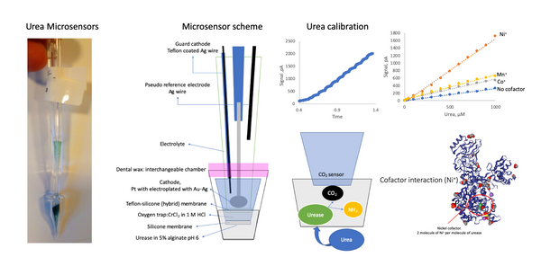
A novel amperometric CO2 microsensor-based urea sensor has been constructed and used for urea concentration analysis in water. The urease enzyme is entrapped in alginate polymer, buffered with citrate-phosphate at pH 6, placed in front of CO2-microsensor in a glass casing and fixed with dental wax to make it interchangeable. The CO2 as urea hydrolysis product, diffuse into the microsensor through the gas-selective membrane where it is detected by its reduction reaction in EMIM-DCA ionic-liquid/aprotic solvent electrolyte-based microsensor with a Ag cathode as catalyst and 1-Ethyl-3-methylimidazolium (EMIM) as co-catalyst. Our urea microsensor response time is one minute with linear response-urea concentration lies between 0-1000 M urea. Urease is metalloenzyme which has moiety interacting with two molecules of Ni2+, Co2+ or Mn2+ for one molecule of urea. The addition of metal cofactor to urease-alginate mixture was shown to boost the microsensor performance. The further method about immobilization of urease (entrapment, covalent-bond with alginate, cross-linking with BSA as stabilizer) in the microsensor and its effect on the microsensor stability is also elaborated.

Uncaptioned visual

Figure. Photo, schematic illustration and performance of CO2-based urea microsensor.


P3.023 Graphene based electrical biosensors for label-free monitoring of adalimumab towards personalized management of rheumatoid arthritis

D. Khan1, N Samane1, A. Roy1, A. Dipta Das1, C. Zhu1, G. Vasudevan Rajeswari1, A. Malisetty1, T. Wahlbrink2, M. Lemme1,2, S. Ingebrandt1, V. Pachauri1
1RWTH Aachen University, Germany. 2AMO GmbH, Germany

Abstract

Rheumatoid Arthritis (RA) is an autoimmune chronic inflammatory disorder, affecting 1% patients worldwide. RA involves inflammation and debilitation of the synovium, leading to pain and stiffness in joints. Adalimumab (Am) as a biological drug, relieves RA symptoms in patients and is generally tolerated well by the patients; however, around 10–30% of patients do not respond to the initial treatment and 23–46% of patients lose response over time due to non-optimal doses. Rapid, point-of-care monitoring of Am, is therefore, a critical requirement towards personalizing the management of RA for better treatment.

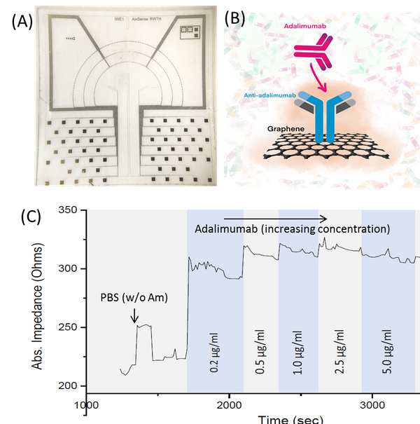
In this work, we realized a new sensor platform for fast and accurate measurement of Am concentrations in physiological conditions. The sensor platform is developed using graphene-based transducers with gold microelectrode arrays. Graphene was biofunctionalized with anti-Am Type I capture antibody, having specific binding to Am. The transducers are operated as biological ion-sensitive field-effect transistor (BioFETs) and electrochemical impedance spectroscopy (EIS) against different concentrations of Am covering clinical ranges (0.3 μg/ml to 10 μg/ml) for RA. The newly established system-integrated, miniaturized biosensor platform, as presented in this work, are expected to strengthen personalized management of diseases like RA by enabling rapid and high-accuracy monitoring of analytes by the end users themselves, away from centralized testing laboratories.

Uncaptioned visual

Figure 1: Electrical label-free monitoring of Adalimumab for Rheumatoid Arthritis management, (A) Biosensor chip, (B) Schematics showing the biofunctionalization and analyte detection strategy where graphene transducer is functionalized with Am-specific anti-adalimumab, and (C) graph showing typical changes in the electrical characteristics of the biosensors while administering different concentrations of Am – here shown as change in the impedance values in real-time.


P3.025 A DNA-based biosensor for the fast and sensitive detection of ochratoxin A in urine with isothermal rolling circle DNA amplification

E. Santovito1, D. Greco1, V. D'Ascanio1, S.M. Sanzani2, Giuseppina Avantaggiato1
1Istituto di Scienze delle Produzioni Alimentari (ISPA-CNR), Italy. 2CIHEAM Bari, Italy

Abstract

Ochratoxin A (OTA) is a toxic and teratogenic metabolite produced by fungal species of the genera Penicillium and Aspergillus. OTA analysis in food and its biomonitoring in biological samples are recommended to assess individual exposure to the mycotoxin. The main technique used for OTA detection in biological samples is LC-MS/MS. Unlike chromatographic techniques, biosensors represent a valid alternative for mycotoxin detection due to their high portability potential and ease of use. For OTA detection, different types of biosensors have been developed, based on the specific recognition of OTA by DNA aptamers. The engineering versatility of DNA makes it a powerful and programmable element for the construction of micro-scale systems that find many applications in biosensing. In our study, we describe a DNA-based biosensor for the detection of OTA in urine. The sensor is composed with a DNA-based capture-system and detection-system. We assembled paramagnetic microbeads carrying a capture-aptamer for OTA that allows its specific capture in liquid samples. A detection complex triggering an isothermal rolling circle amplification (RCA) was assembled using the same aptamer, annealed to a circularized probe, and used to detect the toxin capture event. We designed the RCA to generate autocatalytic units with peroxidase activity (DNAzyme). In the presence of OTA, the circular DNA triggers its isothermal amplification at 30°C, producing a single-stranded and tandem repeated long homologous copy of its sequence. In the amplified DNA strand, a peroxidase self-catalytic structure induces the development of colour reaction that is visible at naked eye. The resulting biosensor showed high sensitivity and selectivity for the detection of OTA as low as 1.09×10⁻¹² ng/mL. Moreover, the biosensor was used for the detection of OTA in naturally contaminated urine. Accuracy and repeatability data obtained in recovery experiments showed recoveries >95%, and relative standard deviations in the range 3.6–15%.


P3.026 Immobilization of Rab7 protein on nanoporous silicon photonic crystals combined with deposition of thin polypyrrole films: toward surface imprinting for high-sensitive optical sensing

T. Di Giulio1, C. Malitesta1, M. De Luca1, C. Bucci1, S. Mariani2, L. Strambini3, G. Barillaro2, E. Mazzotta1
1Università del Salento, Italy. 2Università di Pisa, Italy. 3Istituto di Elettronica e di Ingegneria dell’Informazione e delle Telecomunicazioni, CNR, Italy

Abstract

In this work we report preliminary results on the development of an optical sensor for Rab7 protein based on the integration of imprinted polypyrrole (PPy) with nanoporous silicon photonic crystals (nanoSi-PhCs). Molecular imprinting allows generating materials with “molecular memory” by performing a polymerization of suitable monomers in the presence of a target molecule acting as a template. Subsequent template removal creates recognition sites in the polymer that can selectively rebind the target [1]. Some specific problems can arise if the target is a protein [2] due to possible conformational changes and to difficulties in removing protein from, or rebind to a highly cross-linked polymer. Among the approaches introduced to overcome such issues, surface imprinting emerged as successful strategy [2] restricting the formation of imprinted sites to the polymer surface. The immobilization of the protein offers additional advantages allows generating uniformly accessible binding sites.

Herein, we propose a new method to prepare Rab7 surface-imprinted PPy on nanoSi-PhCs [3]. A his-tag is used to immobilize protein on Ni2+ bearing PhC surface. Rab7 is used as target molecule as it has a key role on different cellular pathways [4]. PPy is selected due to the simplicity in achieving thin polymeric layers along with its largely reported use in imprinting procedures [5]. PPy is deposited on PhCs surface by vapor- and liquid-phase chemical polymerization for different time intervals.

Each step of PhC functionalization is monitored by visible reflectance spectroscopy analysing the Fourier transform of each spectrum, which affords a peak proportional to effective optical thickness of the porous layer [3].

Obtained results evidence a stable Rab7 immobilization along with a highly uniform PPy deposition on PhCs and represent the first attempt of combining the selectivity of imprinted polymers with the optical properties of PhCs, as well as the first example of Rab7 protein imprinting.

[1] C. Malitesta, E. Mazzotta, R.A. Picca, A. Poma, I. Chianella, S.A. Piletsky, Anal Bioanal Chem 402 (2012) 1827

[2] J. Erdossy, V. Horváth, A. Yarman, F.W. Scheller, R.E. Gyurcsányi, Trend Anal Chem 79 (2016) 179

[3] S. Mariani, L.M. Strambini, G. Barillaro, Anal Chem 88 (2016) 8502

[4] F. Guerra, C. Bucci, Cells 5 (2016) 34

[5] A. Turco, S. Corvaglia, E. Mazzotta, Biosens Bioelectron 63 (2015) 240.


P3.027 Composite amperometric glucose sensors based on nano and micro structured copper and copper oxide grown on laser-induced carbon

E. R. Mamleyev, P.G. Weidler, D. Vinga Szabó, D. Mager, M. Islam, J. G. Korvink
Karlsruhe Institute of Technology, Germany

Abstract

Glucose detection is the essential information needed for people suffering from diabetes mellitus, raising the risk of cardiac arrests, renal failure, blindness, cerebral and neuronal damage. New generation flexible and wearable sensors allow constant monitoring of glucose level from blood and sweat towards timely estimation of health risks and their prevention. Transitional metal and metal oxide based amperometric biosensors have emerged as a suitable electrode material for non-enzymatic biosensors, featuring high sensitivity, selectivity and longevity. Among these electrode materials, CuO and Cu nanoparticle based substrates serve as a suitable candidate due to their abundance and ease in the fabrication.

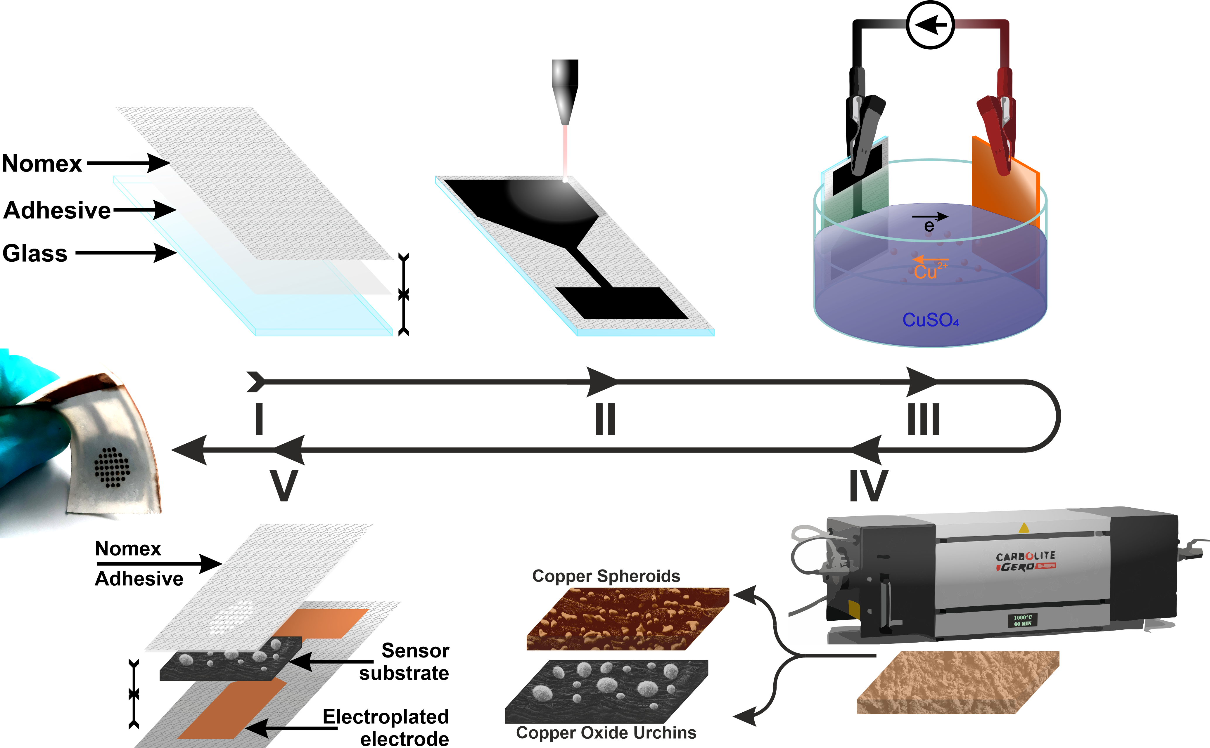
Here, we report about the fabrication and analysis of new composite films with Cu microspheroids (Cu-MS) and CuO urchins (CuO-U, spheroids with nanowires) dispersed on laser-induced carbon (LIC) substrates. Flexible meta-polyaramid (Nomex) sheets were rendered into conductive carbon- rich film using IR laser radiation, providing a seed layer for the copper electroplating. Upon electroplating, the free standing copper/LIC films were annealed in nitrogen and nitrogen/air working environments, leading to the formation of Cu-MS and CuO-U respectively (Figure 1). The aggregation mechanism, crystallographic properties and surface chemistry of the films were studied with SEM, XRD and XPS.

Both Cu-MS and CuO-U films attained activity for glucose detection, and showed a high amperometric sensitivity of 2.61 and 3.38 mA/(cm2 mM) and a limit of detection of 104 and 140 nM respectively. CuO-U film retained facile chemical composition after amperometric testing, which allowed multiple repetitions with reproducible results, and after consecutive bending.

Uncaptioned visual

Figure 1. The process flow: (I) Nomex paper attached to the glass slide, (II) the initial electrode patterned with IR laser, (III) the electroplating with Cu, (IV) the electrode annealed in nitrogen (Cu-MS) and nitrogen/air (CuO-U) environments, (V) CuO-U film attached to contact pad then tested multiple times before and after bending.


P3.028 Real-time inflammatory biomarker detection by long period grating-based fiber optic biosensing

F. Chiavaioli1, F. Esposito2, L. Sansone3, A. Srivastava2, F. Baldini1, S. Campopiano2, M. Giordano3, A. Giannetti1, A. Iadicicco2
1Institute of Applied Physics “Nello Carrara”, National Research Council of Italy (CNR), Italy. 2University of Naples “Parthenope”, Italy. 3Institute for Polymers, Composites and Biomaterials, National Research Council of Italy (CNR), Italy

Abstract

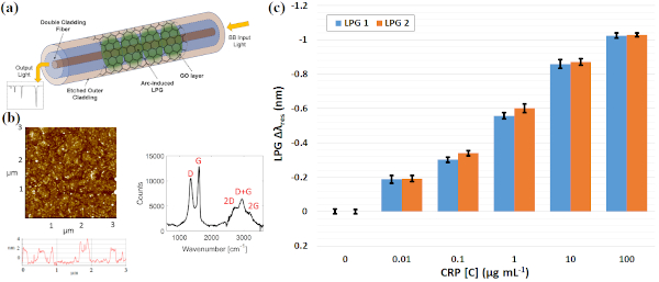
The development of biosensors able to guarantee rapid, reliable and in-situ monitoring of molecular interaction is quite an actual topic, as occurring for SARS-CoV-2 infection. Commercially-available optical systems for the detection of inflammatory biomarkers, such as C-reactive protein (CRP), cytokines or procalcitonin, are based on the popular ELISA method, with limit of detections (LOD) of 0.01 ng/mL. Different techniques, such as immunoturbidimetric or immunonephelometry assay, underpin other approaches attaining a LOD of 30 ng/mL and 200 ng/mL, respectively. The peculiarities of fiber optic technology, especially in light control, combined with nanotechnology can ensure superior performances over other sensing platforms in measuring refractive index (RI) changes. Several biosensors based on surface plasmon resonance (SPR), interferometric configurations or fiber gratings, have been proposed for the detection of inflammatory biomarkers. Among them, those based on SPR are so widespread, with an attained LOD of 9 ng/mL using plastic fibers. Here, the design, development and assessment of a label-free fiber optic biosensing platform based on long period grating (LPG) for real-time detection of CRP in serum are discussed. The device consists of a novel LPG inscribed into a double cladding fiber (DCF), with a special W-shaped RI profile (Fig. 1a). A slight chemical etching of fiber outer cladding permits to improve the device sensitivity by means of the mode transition phenomenon. A thin layer of graphene oxide (GO) is deposited around the LPG portion (Fig. 1b), to directly provide functional groups for the covalent immobilization of the receptor. A large working range of CRP concentrations of clinical relevance (1 ng/mL – 100 μg/mL) is covered, with a LOD of 0.15 ng/mL attained in repeated experiments with CRP spiked in human serum (Fig. 1c).

Uncaptioned visual

Fig. 1 (a) Sensor sketch. (b) AFM analysis and Raman spectrum of GO. (c) Biosensor calibration curve as histogram using two LPGs.


P3.029 Improving referencing in optical affinity biosensors for analysis of complex biological samples

G. Finocchiaro, M. Bocková, J. Homola
Institute of Photonics and Electronics, Czech Republic

Abstract

In optical affinity biosensors, referencing techniques are used to improve robustness of the measurements by reducing the interfering effects (e.g. non-specific binding from complex media). The single surface referencing (SSR) approach uses a surface functionalized with receptors of a single type split into the sensing and reference channel, to which complex sample (sensing channel) and complex sample mixed with the receptor, thus inhibiting the binding of the analyte to the surface (reference channel), are injected. While the SSR approach has been demonstrated to provide more accurate results than the conventional referencing approaches, usually involving surfaces functionalized with different antibodies, its potential is also limited by the cross-reactivity of interfering matrix components with the receptor. In this paper, we investigated these interferences to overcome the cross-reactivity limitation. We combined the SSR approach with the SPR biosensor for the detection of two cancer biomarkers (human chorionic gonadotropin and carcinoembryonic antigen) in blood plasma. We investigated the effect of blood plasma dilution factor on the biosensor response to different pooled blood plasmas (BP) and observed increasing false positive response (not associated with the presence of analyte) with decreasing BP dilution. We showed that this false positive response is in large part due to the cross-reactivity of the used receptors with BP components. Further study has revealed the large contribution of the so-called interfering antibodies and showed that their effect can be reduced by means of a special blocking agent by up to a factor of 6. We used this knowledge to demonstrate the possibility to detect low levels of biomarkers in blood plasma samples, reducing the risk of false positive responses.


P3.030 Strategies for voltammetric signal discrimination of dopamine derivatives as basis for activity measurements of a metabolic enzyme

G. Göbel1, A. Talke2, U. Ahnert2, F. Lisdat1
1Technical University Wildau, Germany. 2BioTeZ GmbH, Germany

Abstract

Dopamine and its derivatives are involved in numerous signal transduction processes in the central and peripheral nervous system. As representatives of the group of catecholamines they are very suited for electrochemical sensing because of their redox properties.

Carbon-derived materials are frequently used as sensor electrodes for analytical detection. However, several problems are connected with these electrodes: Dopamine metabolites and interfering substances often superimpose the dopamine signal. Furthermore, the formation of polymerization products of the oxidized dopamine causes a successive reduction of the sensor response.

Here we show that fluorine doped tin oxide (FTO) electrodes can be advantageously applied for a stable dopamine detection [1]. As transduction method differential pulse voltammetry (DPV) is used.

The measurements reveal a well-pronounced current peak for dopamine at +320 mV vs. Ag/AgCl. For the dopamine precursor L-dopa a significantly lower oxidation current is obtained, and even more importantly the degradation product methoxytyramine causes nearly no current signal [2]. Further investigations demonstrate that interferences such as ascorbic or uric acid, generate nearly no oxidation signal at the FTO electrode, even at concentrations of 1 mM.

These properties have been used as basis for the sensorial activity determination of one enzyme of the dopamine metabolism – catechol-O-methyltransferase (COMT). The enzyme and its inhibition plays an important role in the treatment of Parkinson disease and thus, its activity determination is of medical interest. By following the dopamine concentration during the action of captured enzyme, we have been able to quantify the COMT activity without having electrochemical signals from commonly used inhibitors.

[1] G. Göbel, A. Talke, F. Lisdat, Electroanalysis, 2018,  Vol. 30 (2), p. 225-229

[2] G. Göbel, A. Talke, U. Ahnert, F. Lisdat, ChemElectroChem, 2019, Vol. 6 (17)


P3.031 Selective laser etching fabricated micro cavity arrays for high-resolution screening of 3D cell cultures

F.D. Zitzmann, S. Schmidt, M. Naumann, H-G. Jahnke, A.A. Robitzki
Leipzig University, Germany

Abstract

Three-dimensional cell culture models represent the native in vivo situation more closely than 2D cultures and are therefore preferred today for in vitro studies. In this context, there is a great demand for fast, non-invasive, real-time and label-free methods that are capable for detailed analyses of three-dimensional cultures. To characterize heterogenous 3D cultures or detect locally restricted effects of drugs in 3D cultures, measurement method that offers spatial resolution capabilities are desired. A highly suitable technique is impedance spectroscopy in combination with micro cavity arrays (MCA). Already described MCA are based of opaque silicon substrates with four measurement electrodes located in one level for the analysis of 3D cultures. Due to the crystal structure of silicon, the geometry of etched microcavities is limited to an initial square shape and therefore, an increase in the number of measuring electrodes is not feasible.

To overcome this limitation, we investigated alternative substrates as well as structuring techniques and identified the selective laser etching (SLE) process as highly suitable for fabrication of microcavities with various geometries in fused silica and borosilicate glass without geometric restrictions. To increase the number of possible electrode combinations for an improved spatial resolution, we successfully developed MCA with a hexagonal or octagonal shape including six or eight measuring electrodes respectively in one cavity. In addition, we integrated a central pin electrode structure at the bottom of the cavity to extend the spatial resolution on the z-axis. To demonstrate the capabilities of our novel MCA, we analyzed and compared various human tissue models.

In conclusion, our SLE-fabricated MCA clearly improve the bioelectronic analyses of cellular changes in heterogeneous three-dimensional tissue models. Thus, bioelectronic analysis of e.g. tumor biopsy samples itself as well as chemosensitivity analytics of therapeutics could be benefit from our development.


P3.032 Inkjet-printed customizable electrodes matrix for gesture recognition

G. Rosati1, G. Cisotto1, D. Sili2, L. Compagnucci2,3, C. De Giorgi2,3, E.F. Pavone2, A. Paccagnella1, V. Betti3,4
1University of Padova, Italy. 2BrainTrends Ltd., Italy. 3Sapienza University of Rome, Italy. 4Fondazione Santa Lucia, Italy

Abstract

Gesture recognition is constantly raising interest in the bioengineering and clinical communities for purposes ranging from attention monitoring (e.g. in driving) to active prosthetics and rehabilitation haptics controlled by the residual muscular activities [1-3].

In this context, the ultra-short design-to-prototype time and the customization offered by inkjet printing by nanostructured ink is filling the gap between lab experiments and real-life applications. Indeed, innovative nanoparticle-based inks allow to print sensors’ matrices sensitive to muscular activity on the skin surface, implementing surface electromyography (sEMG) measurements [4-6].

In our work we have developed an easy to use and low-cost platform based on office printers for the production of such matrices, that we used to measure the electrical activity of the forearm muscle in response to different positions of the dominant hand. For this purpose, we employed two commercial multichannel sEMG amplifiers (Biopac MP35 by BIOPAC Systems, Inc., USA), acquiring data in a synchronized way, and a custom-built PCB for the connections of the matrix. We tested the system on 12 subjects, each one performing 16 simple exercises, 8 for extensor and 8 for flexor muscles, with rest periods of 5 seconds.

Our results proved the reliability of inkjet printing with consumer printers for the production of electrodes and circuits for sEMG, giving the possibility to any laboratory or clinical ambulatory to setup a cheap and stable platform for the production of customized high-density sEMG matrices.

Uncaptioned visualWe validated the acquisition system in a study of sEMG-based fatigue assessment with 6 subjects, well-matched to the current literature [4].
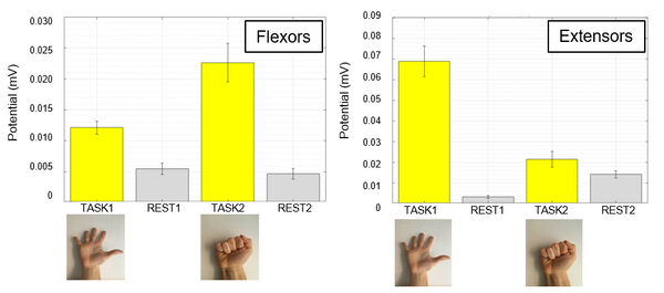
Finally, we found specific patterns of muscular activation corresponding to all the tested movements. The evaluation of the intra-subject and inter-subject recording variability showed that the system is relatively stable and reliable for the single subject sEMG acquisitions, despite the high physiology-related differences between subjects.

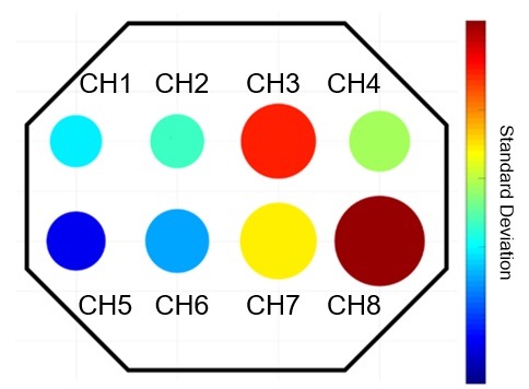
Figure 1: Bubble plot of the intensity of the signals (bubbles diameter) and of their standard deviations (bubble colour) recorded during the flexion of all the fingers

Uncaptioned visual

Figure 2: Average potentials and respective standard deviations calculated for the task and rest periods for flexors and extensors during tasks 1 and 2.

References:

[1] S. Haufe et al. (2011) Journal of neural engineering, Vol. 8 No. 056001.

[2] G. Cisotto et al. (2018) IEEE Healthcom conference, pp. 1-6

[3] T. Ono et al. (2014) Frontiers in neuroengineering, Vol. 7, pp. 19.

[4] G. Cisotto et al. (2019) IEEE EMBC conference, pp. 5765-5768

[5] R.G. Scalisi et al. (2015) Organic electronics, Vol. 18, pp. 89-94.

[6] M. Gazzoni (2015) IEEE MeMeA conference, pp. 79-83


P3.033 Surface biofunctionalization techniques and their pivotal role in the performance of AChE biosensors for pesticide detection

G. Tsekenis1, D. Soulis1, C. Chandrinou2, A. Klinakis1, I. Zergioti2
1Biomedical Research Foundation of the Academy of Athens, Greece. 2National Technical University of Athens, Greece

Abstract

The development of field-deployable biosensors for the rapid, on-site detection of pesticides in food is essential in order to alleviate the need to transport samples to a centralized laboratory. Despite the considerable amount of effort that has already been invested in the development of such biosensors, their reliability and sensitivity with regards to real sample analysis is still low, which has hampered their deployment in portable devices and commercialization. Herein, an electrochemical enzymatic biosensor based on the inhibition of acetylcholinesterase (AChE) for the detection and quantification of traces of organophosphate and carbamate pesticides is presented. The enzyme was immobilized on commercially available screen-printed carbon electrodes (SPEs) modified with a carbon black-chitosan matrix, as a means to enhance their conductivity. Most interestingly, two different methodologies for the deposition of the enzyme onto the sensor surface were followed (Figure 1), with strikingly different results obtained depending on the family of pesticides under investigation and the immobilization techniques. Furthermore, and towards the uniform application of the functionalization layer onto the SPEs’ surfaces, the laser induced forward transfer (LIFT) technique was employed, which not only permitted the sensor biofunctionalization in one step but also allowed a considerable improvement of the sensor’s performance to be achieved. Under optimized conditions, the fabricated sensors can effectively detect carbofuran over a linear range from 1.1 × 10⁻⁹ to 2.3 × 10⁻⁸ mol/L, with a LoD equal to 0.6 × 10⁻⁹ mol/L and chlorpyrifos over a dynamic range from 0.7 × 10⁻⁹ up to 1.4 × 10⁻⁸ mol/L and a LoD of 0.4 × 10⁻⁹ mol/L in buffer[i].

Uncaptioned visual

Figure 1. Graphical representation of the fabricated enzyme-based biosensor using (a) the multistepand (b) the one-step approaches


[i] Soulis, D.; Trigazi, M.; Tsekenis, G.; Chandrinou, C.; Klinakis, A.; Zergioti, I. Facile and Low-Cost SPE Modification Towards Ultra-Sensitive Organophosphorus and Carbamate Pesticide Detection in Olive Oil. Molecules 2020, 25, 4988. https://doi.org/10.3390/molecules25214988


P3.034 Substrate displacement colorimetry for the detection of diarylethylamines

J. Lowdon1, K. Eersels1, R. Rogosic1, B. Heidt1, H. Diliën1, E. Steen Redeker1, M. Peeters2, B. Van Grinsven1, T. Cleij1
1Maastricht university, The Netherlands. 2Newcastle University, UK

Abstract

Introduction:

In this study a novel molecularly imprinted polymer (MIP) based dye displacement assay is introduced for the rapid detection of the new psychoactive substance (NPS) sub-class known as diarylethylamines. The competitive nature of the assay facilitates displacement of a pre-bound dye molecule by a selected target molecule in a process coined Substrate displacement colorimetry (SDC).

Methods:

The assay was fully characterized by conducting affinity studies on the synthesized MIP, evaluating multiple dyes and expressing these affinities as binding factors (BF). The result of this study indicates the mathematical relationship between the established BF and the imprinting factor (IF) of the MIP towards the target analyte (2-MXP), allowing a prediction of efficacy towards dye displacement. Off the back of this study, dye-loaded MIPs were incubated with two common adulterants, two legal pharmacological, and the target diarylethylamine assessing the selective nature of the assay.

Results:

Of the dye’s tested malachite green was found to displace most readily when incubated with the target analyte, demonstrating easily quantifiable concentrations even when exposed to lower concentration of analyte. When incubated with the fore mentioned adulterants and pharmacological molecules, no dye displacement was observed; yet the target analyte stimulated clear dye displacement coloring the filtrate from the assay. This dye displacement was shown to be quantifiable, with a dose response being generated and the linear range of the assay determined by means of UV-visible spectroscopy.

Discussion:

The low–cost, robust and rapid nature of the proposed assay, combined with its tailorable selectivity and generic nature makes this an ideal tool for the screening of unknown seized samples. Displaying high sensitivity towards the desired analyte without sacrificing selectivity to other molecules, generating a highly reliable analytical protocol that can be both qualitative and quantitative depending upon the environment it is employed (Figure 1).

Uncaptioned visual

Figure 1. Dose response of the dye displacement assay when incubated with 2-MXP, and caffeine

Reference(s): Lowdon, J. W.; Eersels, K; Rogosic, R; Heidt, B; Diliën, H; Redeker, E. S; Peeters, M; Grinsven, B. v; Cleij, T. J. Substrate displacement colorimetry for the detection of diarylethylamine. Sensors & Actuators B: Chem. 2019, 282, p137-144


P3.035 Point-of-care therapeutic drug monitoring for adalimumab in blood using a self-powered (i)SIMPLE microfluidic chip

H. Ordutowski, F. Dal Dosso, Y. Yildizhan, S. Vermeire, D. Thomas, A. Gils, J. Lammertyn, D. Spasic
KU Leuven, Belgium

Abstract

We present here a novel “whole blood in – result out” self-powered microfluidic device for quantifying adalimumab (ADM) and enabling therapeutic drug monitoring (TDM) at the point-of-care (POC).

Chronic inflammation plays a central role in autoimmune diseases, like inflammatory bowel disease and rheumatoid arthritis. Possible treatments include TNF-α inhibitors, among which the fully human monoclonal antibody ADM.1 It has been shown already that TDM can (1) maximize clinical response, while minimizing loss-of-response2 and (2) reduce treatment cost, which amounts to €18,000/patient/year for ADM. Several assays quantifying ADM in serum are currently available, including classical ELISA and lateral flow tests.3–5 However, they either suffer from long time-to-result or require off-chip sample preparation, respectively, emphasizing that there is a need for a true POC TDM test.

The (i)SIMPLE microfluidic platform6,7, used in this study, can exceptionally both push and pull liquids without requiring any external pumps (Fig.1), while being inexpensive (i.e. made from plastics and filter paper) and easy to use/fabricate. For integrating a clinically validated ELISA assay on the (i)SIMPLE chip, we miniaturized the protocol in microtitre plates towards an on-chip POC assay (Fig.2), yielding similar sensitivity. Next, to avoid sample pre-treatment, we integrated a plasmapheresis filtration system on the (i)SIMPLE chip capable of yielding plasma in a few minutes with a purity equal to standard centrifugation (Fig.3). Lastly, we designed an (i)SIMPLE device to perform all ADM ELISA steps and incubations upon a single finger-press activation with 15 µL of ADM-spiked plasma (Fig.4).

In conclusion, we presented a one-press-activated POC microfluidic chip capable of autonomously handling plasma samples to detect ADM. Additionally, we demonstrated the compatibility of the same platform with on-chip plasmapheresis, thereby revealing flexibility of the (i)SIMPLE chip to accommodate complex bioassays (i.e. other therapeutic monoclonal antibodies besides ADM) and to utilize a wide range of biological samples.

1.Colombel, J. et al. Gastroenterology 132, 52–65 (2007).

2.Ordás, I. et al. Clin. Gastroenterol. Hepatol. Off. Clin. Pract. J. Am. Gastroenterol. Assoc. 10, 1079-1087; (2012).

3.Bian, S. et al. J. Pharm. Biomed. Anal. 125, 62–67 (2016).

4.Rocha, C. et al. Ther. Adv. Gastroenterol. 12, 1–11 (2019).

5.Verstockt, B. et al. Aliment. Pharmacol. Ther. 48, 731–739 (2018).

6.Dal Dosso, F. et al. Anal. Chim. Acta 1000, 191–198 (2018).

7.Kokalj, T. et al. Lab. Chip 14, 4329–4333 (2014).

Uncaptioned visualUncaptioned visual

Figure 1: The SIMPLE consists of a working liquid (blue) that is brought into contact with a porous material by a finger press, after which it starts wicking into the porous material. The generated underpressure pulls the sample (red) into the channel. The infusion SIMPLE (iSIMPLE) works in reverse and pushes the sample.

Uncaptioned visual

Figure 2: A) Scheme of the sandwich ELISA assay and table with incubation times/volumes for each step of the standard and downscaled ELISA. B) The on-chip ELISA was done in flow with syringe pumps, in single channels made of (i)SIMPLE materials. C) and D) POC ELISA calibration curves in plasma spiked with ADM, performed in microtitre plates and on-chip in flow, respectively. Curves were fit with Michaëlis-Menten and logistic growth, respectively. Error bars represent one standard deviation (n=3).

Uncaptioned visual

Figure 3: Plasmapheresis on the (i)SIMPLE platform. A) The chip is composed of working liquid (1), porous material (2), sample channel (3) and filter material chamber (4). B) Upon whole blood deposition on the inlet, a SIMPLE pump created sufficient underpressure to elute the plasma from the filter. C-D) RBCs were counted with bright field microscopy with phase contrast (381 ± 91 RBCs/mm³), which was comparable to standard centrifugation method.

Uncaptioned visual

Figure 4: Single-press activated, autonomous (i)SIMPLE chip for ADM detection. A) Digital design of the chip layout. B) Chip is activated with a single finger press after sample loading. C) The activation pump (AP) pulls the trigger liquid to the first trigger pump (TP1), activating the first reagent pump (RP1). D) RP1 pushes washing buffer and conjugate over the sensing zone. E) After the RP1 finishes, TP2 – RP2 pair is activated. F) The RP2 pushes washing buffer and substrate over the sensing zone. After incubation, the signal is measured with a reflectometer.


P3.036 Lab-on-chip platform for on-field analysis of grapevine leafroll-associated virus 3

I. Buja1,2, E. Sabella1, A.G. Monteduro1,2, M.S. Chiriacò2, S. Rizzato1,2, L. De Bellis1, A. Luvisi1, G. Maruccio1,2
1University of Salento, Italy. 2CNR-Institute of Nanotechnology, Italy

Abstract

Worldwide phytopathological adversities are often attributable to human activities (as a consequence of the globalization of trade or tourism mass, changes in common agricultural practices and climate change) and pathogens, with serious repercussion in quantity and quality of yield. As a consequence, “early detection” in combination with “fast, accurate and cheap” diagnostics become a categorical imperative, especially for emerging diseases or challenging pathogens that spread thanks to asymptomatic individuals, with subtle initial symptoms but are then difficult to face (Buja et al., 2021). For this, we are developing a lab-on-chip (LOC), as a diagnostic approach to phytopathological problems caused by infectious agents as the virus Grapevine leafroll-associated virus 3 (GLRaV-3). Grapevine leafroll disease (GLD) is one of the most important viral diseases, with GLRaV-3 affecting grapevines worldwide. Symptoms of GLD can vary greatly with the season, grape cultivar, and climatic conditions and some varieties can be completely symptomless (Maree et al., 2013). There is no cure for the virus, but only preventive actions (the use of certified material). These pathogens can have serious economic/environmental repercussions on two of the major cultivated woody plant of Mediterranean basin, due to the absence of therapeutic techniques and the need of rapid, in-field and low-cost detection methods. Here we present a lab-on-chip platform, coupled with microfluidic module of polydimethylsiloxane (PDMS), based on an electrochemical transduction method, able to recognize serial dilutions of GLRaV-3. LOC require small sample volumes, allowing a rapid detection of the target. Through PDMS, is possible to realize biochemistry conventional laboratories functions such as sample preparation, reaction, separation and detection (McDonald et al, 2000). This device can show competitive performances with conventional diagnostic methods in terms of reliability, with further advantages of portability, low costs and ease of use, making the difference in real time detection of the pathogens.


P3.037 Single-cell biosensing with cellular adhesion and migration studies using Field-Effect Transistor Cell-substrate Impedance Sensing - FETCIS

Sven Ingebrandt
RWTH Aachen University, Germany

Abstract

Electrical Cell-substrate Impedance Sensing (ECIS) using ion-sensitive field-effect transistors (ISFETs) is a technique, which offers a true single cell resolution for adhesion and migration studies. This novel concept of Field-Effect Transistor Cell-substrate Impedance Sensing (FETCIS) can be utilized with various types of transistors down to silicon nanowires [1]. We present the versatility of our method addressing cell lines, cardiac myocytes, neurons and individually acting cells of the immune system.

Depending on the application, we tuned the bioassays to small sensor dimensions using silicon nanowire arrays or to micro-scale, open-gate ISFET arrays. We optimized the latter devices by local oxidation of silicon followed by repetitive thermal oxidation and etching to fabricate ultra-smooth, all silicon dioxide surfaces. With these devices, migration of cardiac fibrobasts can be studied in a time-dependent manner. With similar devices, it was possible to biochemically modify the chip surfaces and to observe the tendency of T lymphocytes to form tight junctions to specific coating materials.

In recent years, we utilized transparent, low-cost sensor arrays using of poly(3,4-ethylenedioxythiophene) doped with polystyrene sulfonate (PEDOT:PSS) as transducer material in electrochemical transistors. Such organic thin film transistors are another version of FET devices, which gained a lot of attention in the field of biosensing in recent years. Their versatility, easy fabrication and their extremely high biocompatibility make those devices a new and exciting alternative for cell sensing applications. OECTs offer a completely novel gating mechanism compared to the classical, purely capacitive coupling of cells to silicon ISFETs. We realized a wafer-scale fabrication protocol for OECTs on silicon and glass substrates.

In future, OECTs with smooth surfaces could be produced such cheap that disposable sensors embedded in standard plastic cell culture dishes would be possible. This could be a very interesting alternative to the classical sensor types in ECIS.


P3.039 Human liver cell-encapsulated microgels for organ-on-a-chip toxicity sensing applications

Jonathan O'Connor1,2, Estelle Regnault1,3, Margaux Delafosse1,3, Jasmin Gebauer-Barrett1,4, Nina Apushkinskaya1,5, Seongho Jeong1,6, Heesoo Kim1,7, Eunheui Gwag1,8, Leon Abelmann1,2,9, Changseon Ryu1, Young Jun Kim1,8, Jaeho Lee1, Baeckkyoung Sung1,8, Andreas Manz1,2
1KIST Europe, Germany. 2Universität des Saarlandes, Germany. 3Ecole Supérieure de Chimie Organique et Minérale, France. 4University of British Columbia, Canada. 5Hochschule Fulda, Germany. 6Helmholtz Zentrum München, Germany. 7Hanyang University, Republic of Korea. 8UST, Republic of Korea. 9University of Twente, The Netherlands

Abstract

Introduction:

Recent developments in synthetic 3D gel microenvironments for cells have been enabling bio-engineered organ-specific cellular niches which resemble native tissue architectures. Especially, applications of the cell-laden microgels, coupled with microfluidic systems, are attracting increasing interests due to their facile adaptability to high-throughput screening processes for drug toxicity sensing. The purpose of this study is to create miniaturized liver tissues with about 100 µm in size which are compatible to microfluidic manipulation and long-term 3D cell culture in lab-on-a-chip platforms.

Methods:

Gelatin microgel spheres were fabricated using prototypic microfluidic channel setup and water-in-oil mini-emulsification techniques: The suspension of human hepatocytes was mixed with aqueous solution of gelatin type A, and the mixture was emulsified to generate gelatin droplets where the cells are seeded. After a rapid cooling step, the gelatin gel particles were covalently crosslinked by genipin at low concentrations for a short period in order to minimize cytotoxic effects. Then, the liver cell-laden microgel beads were incubated up to 2 months in a conventional cell culture condition. Fluorescence light-sheet microscopy and bright-field time-lapse imaging were applied for the morphological observation, cell viability assay, and microchannel-based single-bead manipulation experiments.

Results:

Spherical gelatin gel microbeads could act as an artificial 3D extracellular matrix which support the adhesion and spreading of human hepatocytes. In the microgel matrix, the liver cells maintained well-characterized long-term viability which decreased slowly from 95% after encapsulation down to 50% after 5 weeks. A single microgel sphere could be manipulated by controlling shear flow in a channel, showing translational and rotational motion simultaneously.

Discussion:

The genipin-crosslinked 3D network of gelatin was proven to efficiently support the prolonged viability of human liver cells in a form of microscale colloidal sphere, which showed a potential to be fully adaptable for microfluidic biosensor systems in vitro.


P3.040 Proof-of-concept of an aptasensor for detection of Bacillus cereus spores in milk and ready-to-eat food

J. Vidic, F. Rizzotto, M. Marin
Université Paris-Saclay, France

Abstract

Bacillus cereus is ubiquitously present in nature and can contaminate milk through a variety of means from the farm to the processing plant, during transport or distribution. There is a need to detect and quantify spores directly in food samples, because B. cereus might be present in food only in the sporulated form. We developed a colorimetric aptasensor based on spores enhanced peroxidase-like catalytic activity of gold nanoparticles (AuNPs) for selective and sensitive detection of B. cereus spores in food. The aptasensor developed by covalent immobilization of the 5’-thiol-BAS6R aptamer on AuNPs (20 nm) possessed a high intrinsic peroxidase-like activity as evidenced using 3,3′,5,5′-tetramethylbenzidine (TMB) as substrate. B. cereus spores (µm sizes) were able to capture and concentrate BAS6R@AuNCs and thus to greatly increase the local concentrations of the enzyme-mimetic nanoparticles. As a result, when TMB and H2O2 were added into a sample containing B. cereus spores, the aptasensor generated an intense blue coloration of solution allowing naked eye detection of spores within a few minutes. The efficiency of reaction was tested using UV-vis spectroscopy and transition electron microscopy. Under optimized conditions, the aptasensor detected B. cereus spores in the concentration range of 101–1010 CFU/mL. The limit of detection was 104 CFU/mL in infant formula suspension and 105 CFU/mL in homogenized ready-to-eat mashed potatoes, suggesting that the biosensor was not inhibited by complex food matrices. The set of tests using Bacillus subtilis spores, or B. cereus, B. subtilis, Escherichia coli and Salmonella Typhimurium vegetative cells demonstrated the specificity of this aptasensor. The designed method may become a powerful tool for a label-free screening on-site for food contamination with B. cereus spores.


P3.041 Graphene SGFET for biosensing : From transductor design to tripod-based non-covalent functionalization

Juliette Simon1, A. Hugo1, M. Kumar1, R. Othmen2, J. Renard2, V. Bouchiat1, C. Sun3, W.R. Dichtel3, T. Alava3, H.G. Craighead3
et al
1Univ. Grenoble Alpes, CEA, LETI, F-38000 Grenoble, France. 2Department of Chemistry, Northwestern University, Evanston, Illinois, USA. 3Univ. Grenoble Alpes, CNRS, France

Abstract

The accurate detection of biomarkers for medical diagnosis requires biosensors with high sensitivity and selectivity. Graphene-based Solution-Gated Field-Effect Transistors (SGFET) (Fig.1.a)) have shown superior electrical sensitivity in liquid compared to silicon and diamond-based SGFET [1], owing to the outstanding graphene electrical properties. However, selective biosensing requires the introduction of specific bioreceptors at the surface of graphene. Simple adsorption of bioreceptors onto graphene is not suitable, due to irreversible denaturation and / or to lack of orientation of the protein bioreceptor. Besides, covalent grafting of chemical moieties to graphene disrupt its honeycomb lattice, resulting in drastically reduced charge carrier mobility. Aromatic compounds such as pyrene can adsorb onto graphene by π-stacking without deteriorating its properties. Thus, several teams have reported the immobilization of bioreceptors on graphene using pyrene-based spacer molecules [3]. Nevertheless, the unambiguous demonstration that such spacers would prevent bioreceptors from denaturation by stacking on graphene, due to a rotational degree of freedom along the spacer carbon chain is still a challenge (Fig. 1.b)). In this work, we report the functionalization of graphene SGFET with a tripodal molecule including three pyrene feet (Fig. 1.c)).

Uncaptioned visual

Figure 1: a) SGFET, 3D view of b) monovalent pyrene-based spacer and the rotational degree of freedom, c) Tripod with the pyrene feet (red), backbone (blue), reactive ester group (green)

Such tripodal molecule is 103 times more kinetically stable than classical monovalent spacers, and was specifically designed to stably maintain the functional protein bioreceptors away from the graphene surface [4]. Micro-fabricated SGFET [5] functionalized with the tripod show a reproducible and significant Dirac peak shift. State-of-the-art electrical sensitivity values maintained after tripod immobilization are reported (Fig.2). Building upon these promising results, we are currently binding antibodies to the tripod-functionalized SGFET, and assessing the sensitivity of immunological sensing with our biosensors in buffer-engineered media.
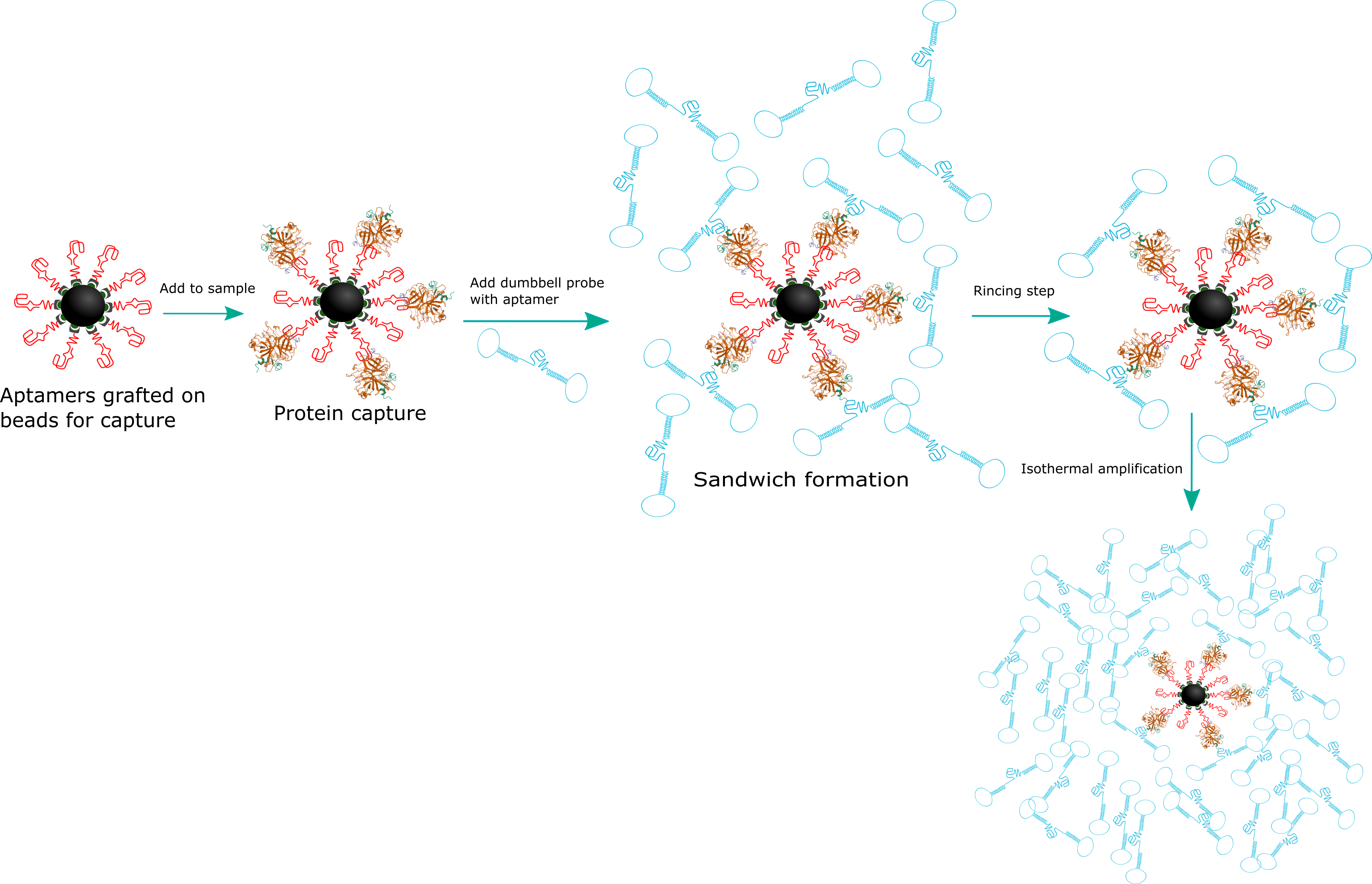
Uncaptioned visualUncaptioned visual

Figure 2: Transfer curves (left) and sensitivity (right) before (red) and after (yellow) graphene functionalization with the tripod (5 cyclic scans)

References :

[1]L. H. Hess et al., Proc. IEEE 7 (2013) 1780.

[2]T. Alava et al., Anal. Chem. 5 (2013) 2754.

[3]T. S. Sreeprasad et al., Small 3 (2013) 341.

[4]J. A. Mann et al., Angew. Chem. Int. Ed. 11 (2013) 3177.

[5] A. Hugo et al., EDM2018, Grenoble (2018)


P3.042 Development of optical dopamine neurotransmitter biosensor and its biosensor application for monitoring dopamine released from PC12 cells

W.A. El-said1,2, J.W. Choi2
1Assiut University, Egypt. 2Sogang University, Republic of Korea

Abstract

Dopamine (DA) is one of the most important catecholamine neurotransmitters that have a vital role in the transmission of nerve impulses. Several physiological processes and illnesses including Parkinsonism, Schizophrenia, and Huntington’s disease are related to the changes in the DA levels. Therefore, observing the concentrations of DA receive great attention. Here we have successfully detected different concentrations of DA, effects of pH on the UV absorbance and detection of DA in the presence of human serum solution. Although using OPA/ME enables UV detection, however, it is not stable for a long time. The challenge for us is to enhance the stability, thus we have used OPA/ME/mesoporous silica instated of ME in solution. The results indicated that using OPA/ME/mesoporous silica has long-time stability and could be used for interacting with DA after about one year of its preparation. Hence, we have successes in developing a new and stable compound that enables us to simplify the detection of DA neurotransmitters based on UV within a short time, without the need for complicated instruments and without using a large amount of solvents. Finally, the fabricated biosensor was used to monitor the dopamine neurotransmitter released from PC12 cells.


P3.043 A miniaturized device for detecting Corynebacterium diptheriae

K. Marchlewicz1,2, R. Ziolkowski1, K. Zukowski3, E. Malinowska1,3
1Warsaw University of Technology, Poland. 2University of Warsaw, Poland. 3Centre for Advanced Materials and Technology, Poland

Abstract

Fast detection of pathogens is crucial for efficient diagnosis of infectious diseases. A portable device for fast detection of Corynebacterium diptheriae was developed. It consists a miniaturized microfluidic chip, for carrying polymerase chain reaction (PCR), and an electrochemical DNA biosensor as a detector element.

The miniature PCR device was fabricated in poly(methyl methacrylate) using micromilling technique. The designed microchannel system was divided into three zones, each of which was heated by a separate heater to ensure the right temperature for each individual step of PCR. The system has been integrated with a miniaturized peristaltic pump and a valve that allows automatic introducing of reagents and circulation of the mixture in the device.

An electrochemical DNA biosensor, dedicated to detection of the asymmetric PCR product, was developed at the base of hairpin like DNA probe labeled with methylene blue. After the hybridization process the double stranded DNA was formed at the electrode surface. This resulted in the hairpin like probe structure change and the same in the change of the distance between methylene blue and electrode surface, which influence the registered current.

The developed DNA device allowed to perform any number of reaction cycles with different reagent flow rates in the range from 4 to 150 µl/min. It allowed to obtain the reaction product within 15 minutes (several times shorter than a commonly used thermocycler). For the most optimal sequence of the DNA probe the obtained signal response was proportional to DNA concentration in the range of 0.0125-0.25µmol/L with very high selectivity versus other analyzed pathogens.

The designed device consisting of microPCR and DNA biosensor allows for rapid detection of toxigenic Corynebacterium diptheriae strain based on molecular interactions. Devices of this type may in the future be a useful tool in medical diagnostics as point-of-care tests.


P3.044 Spatially resolved biomolecules immobilisation through contact printing onto chemical micropattern: step-by-step characterisation with TOF-SIMS imaging for application to interferometric biosensors on a silicon chip

Katarzyna Gajos1, Andrzej Budkowski1, Panagiota Petrou2, Konstantinos Misiakos2, Ioannis Raptis2, Kamil Awsiuk1, Jakub Rysz1, Sotirios Kakabakos2
1Jagiellonian University, Poland. 2NCSR Demokritos, Greece

Abstract

Contact pin-printing, established for microarray fabrication [1a], enables spatially controlled deposition of multiple overlapping spots on a chip [1b]. It facilities immobilisation of probe molecules on different groups of Mach Zehnder Interferometric (MZI) biosensors, each with window opened through the top cladding layer over one of the interferometer arms [2a]. Recently, MZI biosensors having both interferometer arms exposed through the window to analyte solutions have been applied in all-silicon spectroscopic chip [2b]. Such on-chip biosensors require probe molecules immobilised exclusively on sensing arm areas, and both arm areas saturated with blocking molecules. Here, we characterise with Time-of-Fight Secondary Ion Mass Spectrometry [3a] a suitable multi-step spatially resolved biofunctionalisation approach, based on contact pin-printing onto plasma treated photolithographic micropattern [3b]: Si3N4 surface with (3-AminoPropyl)TriEthoxySilane layer is examined after photolithography, oxygen plasma treatment, photoresist removal, and after printing of biotinylated Bovine Serum Albumin (probe), blocking with BSA and specific binding of streptavidin. The proposed spatially-selective biofunctionalisation meets the expectations as demonstrated using on-chip MZI biosensor detecting streptavidin.               

[1a] K. Gajos et al., Analyst 140(2015)1127,  [1b] Appl. Surf. Sci. 385(2016)529,  

[2a] K. Misiakos et al., Opt. Express 22(2014)329,   [2b] ACS Photonics 6(2019)1694

[3a] K. Gajos et al., Colloid Polym. Sci. 299(2021)385, [3b] K. Gajos et al., Appl. Surf. Sci. 506(2020)145002

Uncaptioned visual


P3.045 EGOT-based biosensor for the detection of biomarkers of multiple sclerosis

K. Solodka1, M. Berto1, F. Biscarini1,2, C.A. Bortolotti1, M. Pinti1
1Department of Life Sciences, University of Modena and Reggio Emilia, Modena, Italy, Italy. 22Center for Translational Neurophysiology of Speech and Comunication, Istituto Italiano di Tecnologia, Ferrara, Italy, Italy

Abstract

Organic electronics-based immunosensors, such as electrolyte-gated organic transistors (EGOTs), are receiving increasing attention as an alternative strategy for ultrasensitive and label-free detection of biological analytes. 

EGOTs are classified as Organic Electrochemical Transistors (OECTs) and Electrolyte-Gated Organic Field-Effect Transistors (EGOFETs), depending on the permeability of the active layer to the electrolyte ions. Both EGOFETs and OECTs are three-electrode devices, where the current flowing within the organic (semi)conductor bridging source and drain electrodes is controlled by the potential applied to the gate electrode.

Multiple sclerosis (MS) is a chronic and inflammatory disorder of the central nervous system characterized by progressive neurodegeneration. The accurate detection and quantification of MS biomarkers is an urgent need to correctly assess the diagnosis and management of the disease. Therefore, highly sensitive methods, able to detect very low concentrations of the biomarker and discriminate MS patients from healthy individuals are required. 

Here, we propose a novel EGOT-based biosensor for the detection of neurofilament light chain (NF-L), a candidate MS biomarker. In the proposed architecture, the specific recognition of the biomarker is ensured by immobilizing anti-NF-L antibodies on the gate electrode, with a controlled and uniform orientation. A concentration-dependent change in the output current was observed as a consequence of the binding events occurring at the gate surface. Additionally, different electrical parameters, including transconductance and threshold voltage, were monitored during the experiments. Finally, control experiments demonstrated the absence of a non-specific response. Our biosensor showed high selectivity for the detection of NF-L in a wide dynamic range of concentrations, even in the presence of a potentially interfering protein, indicating that it could be safely implemented at the point-of-care for real-time monitoring of the disease. 

The authors acknowledge funding from the European Union's Horizon 2020 research and innovation programme under the Marie Skłodowska-Curie grant agreement no. 813863.


P3.046 Novel diagnostic and prognostic markers of metastatic melanoma based on biophysical methods

A. Sobiepanek1, P.D. Kowalska1, T.M. Grzywa2, R. Galus2, T. Kobiela1
1Warsaw University of Technology, Poland. 2Medical University of Warsaw, Poland

Abstract

The analysis of biomolecular interactions has become essential for the development of innovative, more efficient techniques for the diagnosis and prognosis of the cancer progression. Many studies revealed that cancer development and progression, including epithelial-mesenchymal transition and invasion, is accompanied by changes in glycosylation patterns of cell surface and secreted glycoproteins [1]. 

The aim of the presented studies was establishing a protocol for distinguishing cells from different progression stages of melanoma based on the glycosylation profile of cells and biophysical methods. Interactions between specific glycans present on the surface of melanoma cells at various stages of the tumor progression with appropriate lectin were investigated [2]. For this comprehensive approach with biophysical methods such as the quartz crystal microbalance with the dissipation monitoring, surface plasmon resonance, atomic force microscopy and microscale thermophoresis were employed [3]. The first established procedure utilizes adherent cells grown directly on the given surface, like sensor or coverslip, and the second one was invented to shorten the time needed for cell spreading by using cells directly from suspension [4]. These methods were set on the model of commercially available human cell lines, and next the procedure was verified with cells isolated from patients with the confirmed melanoma metastasis. The observed changes in lectin-glycan interactions among the studied cells enabled distinction of melanoma cell types (normal, radial, vertical and metastasis). Furthermore, two prognostic markers were identified based on the lectin-glycan interaction measurements – the affinity and the viscoelastic index.

[1] Sobiepanek et al. (2021) European Biophysics Journal with Biophysics Letters.

[2] Sobiepanek et al. (2017) Biosensors and Bioelectronics, 93:274-281.

[3] Sobiepanek and Kobiela (2018) Review and Research on Cancer Treatment, 4(1):4-12.

[4] Sobiepanek and Kobiela (2021) In: Bioengineering Technologies. Methods in Molecular Biology, I.


P3.047 Orthogonal paper biosensor for mercury (II) combining bioluminescence and colorimetric smartphone detection

L. Montali1,2, A. Lopreside1,2, B. Wang3, M. Salinitro1, A. Tassoni1, M. Ferri1, M.M. Calabretta1,2, E. Michelini1,2
1University of Bologna, Italy. 2Universitaria Policlinico S. Orsola-Malpighi, Italy. 3University of Edinburg, UK

Abstract

Mercury contamination in the environment has reached alarming levels. Due to its persistence and bioaccumulation, mercury is one of the most widespread toxic heavy metals found in air, water and food. Thus, it is mandatory to monitor mercury and its compounds. The availability of sensitive and rapid biosensors is highly valuable.  

We developed a low-cost biosensor for orthogonal detection of mercury(II) integrating three different biorecognition principles on a three-leaf paper: i) a mercury-specific bioluminescent E. coli bioreporter strain expressing the mercury receptor MerR under the regulation of a constitutive promoter and NanoLuc luciferase under the regulation of the mercury-responsive promoter PmerT, ii) a purified β-galactosidase enzyme which is irreversibly inhibited by mercury and other metal ions reacting on the sulfhydryl group of cysteine, and iii) a V. fischeri bioluminescent strain which is used to quantitatively assess sample toxicity and correct the analytical signal accordingly.

Both sensory elements and substrates, furimazine for the reporter strain and chlorophenol red-β-D-galactopyranoside for colorimetric detection of β-galactosidase, were integrated in a paper sensor to provide a stable all-in-one disposable cartridge which can be easily snapped into a smartphone with a 3D printed housing. This is the first integration of bioluminescent and colorimetric detection on a smartphone-paper sensor, providing a readout within 15 min for colorimetric and 60 min for bioluminescent detection with a limit of detection for Hg(II) at the ppb levels. The device was also used to detect mercury in environmental samples, supporting its feasibility as a rapid and sensitive screening tool.


P3.049 Miniaturised sensor modified with carbon black for tyrosine and nitrotyrosine measurement

L. Fiore1, V. Mazzaracchio1, B. De Lellis1, D. Moscone1, F. Arduini1,2
1University of Rome &quot;Tor Vergata&quot;, Italy. 2Sense4Med, Italy

Abstract

Tyrosine is a very important amino acid because it is incorporated in the structure of several proteins, essential for the human body. Furthermore, it is a precursor of various hormones such as thyroxine and catecholamines, pigments such as melanin, and the antioxidant coenzyme Q10. Any disorders in the various metabolic pathways of this amino acid can cause its accumulation as in tyrosinemia and alkaptonuria diseases. In case of nitrotyrosine, abnormal values are ascribed to inflammatory diseases. The traditional and most-commonly used techniques to detect amino acids encompasse spectrophotometry, gas chromatography, liquid chromatography, high performance liquid chromatography, as well. However, these techniques are expensive, time-consuming, required laboratory set-up, and trained personnel. Herein, we propose a novel user-friendly and low cost miniaturised screen-printed sensor for the simultaneous detection of tyrosine and nitrotyrosine in human plasma samples, using square wave voltammetry as electrochemical technique. The screen-printed electrodes were printed on a polyester substrate in order to realize a platform that can be used for measurements directly in situ. Moreover, the working electrode was modified via drop casting with a carbon black dispersion, in order to increase the electrochemical performance of this device in terms of sensitivity and low applied potential. Tyrosine quantification has been achieved with a detection limit of 6 µM and a linearity range up to 0.5 mM, while a detection limit of 20 µM and linearity range up to 0.8 mM was obtained for nitrotyrosine detection. The preliminary analyses carried out in plasma demonstrated the capability of this sensor to work in biological fluid for a fast detection of these metabolites.


P3.050 Development of an ultra-sensitive, on-chip genosensor for detection of Shiga toxin-producing E. coli (STEC) using interdigitated microelectrodes modified with chitosan-gold nanocomposite.

L. Wasiewska1,2, G. Duffy2, C. Burgess2, A. O'Riordan1
1Tyndall National Institute, Ireland. 2Teagasc Food Research Centre, Ireland

Abstract

Shiga toxin-producing E. coli (STEC) is a food-borne pathogen of significant concern due to the severity of the disease it can cause. Herein we report the development of on-chip, label-free, electrochemical genosensor for detection of STEC using interdigitated gold microelectrodes (IDEs). Each chip comprised six gold IDEs, gold counter, and platinum reference electrodes. Each IDE comprised a working IDE, used for DNA probe immobilization, and generator IDE used for accumulation of methylene blue. First, the working IDE was modified with gold nanoparticles (Au NPs) and chitosan gold nanocomposite. Afterwards amine-modified probe DNA was immobilised on the chitosan modified electrode using glutaraldehyde as a linker. The label-free electrochemical detection was undertaken using methylene blue as a redox molecule, which intercalated into the double-strand DNA after applying an open potential circuit at the generator IDEs. Reduction of methylene blue was recorded using square wave voltammetry (SWV). Using this label-free detection, we have achieved linear response between 10-16 and 10-6 M synthetic target strand with the lowest limit of detection of 100 aM after 20 minutes hybridisation time. The chromosomal DNA from four different E. coli strains (two stx1 positives and two stx1 negatives) were used to confirm the selectivity of the presented method. This novel on-chip biosensor for the detection of VTEC has the potential to be used in point-of-use detection, for example, on the farm.

Uncaptioned visual

Figure 1. (A) SWV results of ssDNA (blue) and after hybridisation with target gene with open potential applied at generator electrode (yellow) and without (green); (B) SWV results of ssDNA (blue), non-complementary strand (green), 3 miss matches (yellow) and complementary (purple); (C) Linear calibration curve corresponding to DNA detection; (D) SWV results obtained for detection of chromosomal DNA from E. coli bacteria culture where strain O103 (blue) contained stx1 gene while strain 12900 (green) did not.


P3.051 Improving SPR limit-of-detection at gold-graphene oxide interfaces by thermal annealing and electrical bias

X. Lu1, P. Damborský2, W. Munief3,4, J. Katrlík2, X. Chen5, S. Ingebrandt1, V. Pachauri1
et al
1RWTH Aachen University, Germany. 2Slovak Academy of Sciences, Slovakia. 3University of Applied Sciences Kaiserslautern, Germany. 4RAM Group DE GmbH, Germany. 5Chongqing University, China

Abstract

Graphene based materials (GBMs) are promising candidates in the field of surface plasmon resonance (SPR) biosensing for advanced biomolecular recognition, converging versatile chemistries beyond gold with advanced plasmonics. Among GBMs, graphene oxide (GO) offers superior advantages for their upscale synthesis, possibility of surface functionalization, and more recently, for their potential for system integration for sensor applications. In this work, we investigated the sensing performances of gold-graphene oxide (Au/GO) interface as a test-bed for advanced SPR biosensor platform. The SPR chips with Au/GO interfaces were fabricated using top-down lithography approaches and deployed for the study of bio-specific interactions between surface immobilized prostate-specific antigens (PSA) and concanavalin A (ConA) (as depicted in Figure.1). The results demonstrated that the SPR signal of Au/GO interface can be tuned by thermal treatment and application of an external electrical bias via solid-liquid interface. For the given thermal reduction, gold-reduced graphene oxide (Au/rGO) interfaces showed much higher sensitivities and exhibited excellent biorecognition capabilities, whilst the average surface plasmons intensity and signal-to-noise ratio were respectively 1.7 and 3 times higher than the case of GO. More remarkably, taking advantage of the bipolar property of the rGO thin films, it was possible to further enhance the surface plasmon intensity by applying bias voltages to the rGO thin film. The limit of detection of ConA target molecules was achieved down to 10 ng/ml, which was unattainable by the conventional SPR biochip. Such tunable SPR platforms based on rGO thin films lighten up a wider road for diverse surface functionalization, adjustable sensing regimes and improved sensitivity in the field of SPR biosensing.

Uncaptioned visual

Figure 1: Schematic of the GO or rGO thin film based SPR biosensor platform, with six layers: prism, glass substrate, gold layer, GO or rGO, biomolecule layer, analyte solution.


P3.052 Bringing biological content into biochip and biosensor systems: scaling of multiplexed diagnostic test production on biochips and biosensors using ultra-low liquid volume dispensing

A. Simon, H. Eickhoff, W. Weigel, M. Jo
SCIENION, Germany

Abstract

Introduction

Most rapid diagnostic tests work by capturing molecules on a solid surface. Surfaces can contain discrete binding sites for analytes carrying a specific capture molecule.  The trend in the miniaturization of diagnostic test systems with the intention to increase throughput and decrease cost, requires precise handling of picoliter to nanoliter liquid volumes. Due to the possibility to use only picoliter amounts of reagents, test production costs can be significantly decreased. In addition, due to the smaller feature size of the analytes, introducing multiple other analytes onto the same test area can be easily achieved. A technology platform for seamless use from R&D to production, which delivers ultra-low volumes very accurately onto any surface, enables short development times. Sophisticated optical systems for online alignment, calibration and quality control of goods produced are important from the first development steps. 

Uncaptioned visual

Various Biochip and Biosensor formats can be loaded with Picoliters of Biomolecules

Miniaturized test formats for multiplexed testing can look very different. Either landing marks or complete areas have to be loaded with biomolecules and the active surfaces can vary from silicone and glass to polymers and metals. Non contact delivery of biomolecules onto these substrates keeps the surface coatings intact and is compatible with with electronic (amperometric, voltage metering, other) optical and mechanical detection (cantilevers).

Uncaptioned visual

Uncaptioned visual

Uncaptioned visual

Uncaptioned visual


P3.053 Aptamer based nano biosensors for the new anticoagulant drugs: Dabigatran etexilate as working model

M. aljohani1,2, R. Chinnappana3, S. Eissaa3, T. Owaidah3, D. Cialla-Mayc1, J. Popp1, M. Zourob3
1Friedrich-Schiller-University, Germany. 2Taibah University, Saudi Arabia. 3Alfaisal University, Saudi Arabia

Abstract

New oral anticoagulants (NOACs) are increasingly used both for prevention of stroke in non-valvular atrial fibrillation (NVAF) and the treatment of venous thromboembolism (VTE) replacing the use of the traditional Anticoagulant warfarin in clinical practice because of Many clinical benefits to patients over traditional anticoagulants.

NOACs include dabigatran, rivaroxaban, apixaban and edoxaban. Dabigatran is the first a NOACs increasingly used for number of blood thrombosis conditions, prevention of strokes and systemic emboli among patients with atrial fibrillation. It provides safe and adequate anticoagulation for prevention and treatment of thrombus in several clinical settings. However, anticoagulation therapy can be associated with an increased risk of bleeding.

There is a lack of specific laboratory tests to determine the level of this NOACs in blood. This is considered the most important obstacles of using these new medications, particularly for patients with trauma, drug toxicity, in urgent need for surgical interventions or uncontrolled bleeding.

The goal of this research project is to obtain and characterize DNA aptamers for Dabigatran Etexilate and to evaluate potential applications of these aptamers to develop low cost, sensitive, selective and user-friendly biosensors for the new NOACs dabigatran is the model here.

In this work, we develop a specific DNA aptamer for dabigatran Etexilate. We Studied the affinity and specificity of generated aptamers to the drug showing dissociation constants (Kd) ranging from 46.8- 208 nM.

A preliminary application of one of the selected aptamers in an electrochemical biosensor was successfully performed and showed high sensitivity and selectivity. This achievement and work plan can be considered as model for all the other new anticoagulants.


P3.054 Ultra- highly sensitive sensor for glucose detection in real samples

Maria Kuznowicz, Artur Jędrzak, Teofil Jesionowski
Poznan University of Technology, Poland

Abstract

Biosensors are devices of great importance to human society, including monitoring of glucose in human blood plasma. It is estimated that the growth of the value of the biosensor market is around 10-15% each year. Due to this reason, the application opportunities of these devices are also expanding and rising [1]. 

Actually, most of the research focused on biosensors is aimed at building systems with high precision, repeatability, stability, selectivity, sensitivity, and rapid response. The long-term stability of amperometric glucose biosensors is an important aspect when designing new detectors. Most biosensors presented in literature have good stability only for a relatively short work-time, up to 30 days. Long-term stability is an important property, especially due to its potential use in environmental and medical research, reducing cost of using devices [2]. 

Herein, a hybrid nanoplatform magnetite@polydopamine@β-cyclodextrins with glucose oxidase to construct a biosensor was presented. The electrocatalytic efficiency tests showed the effectiveness of the proposed matrix as a carrier for the immobilization of enzymes. Long-term reliability of the proposed system was demonstrated for up to 9 months. The excellent selectivity against crucial interferences like ascorbic acid, uric acid, L-cysteine, saccharose, maltose, and fructose was confirmed. It is worth mentioning that commercially available glucometers can last up to three months, while biosensors mentioned in the literature can last up to 30 days. Finally, the proposed system was subjected to electrochemical tests to optimize work on the model and real glucose solutions.

This work is financed and prepared as part of a research project supported by the National Science Center Poland, no. 2017/27/B/ST8/01506

[1] A. Jędrzak, T. Rębiś, M. Kuznowicz, T. Jesionowski, Int. J. Biol. Macromol. 2019, 127, 677-68.
[2] E. Sehit, Z. Altintas, Biosens. Bioelectron. 2020, 159, 112165.


P3.055 Innovative electrochemical sensors based on modified nano-structured electrodes as devices for the measurement of oxidative potential of particulate matter

M.P. Romano1, M.E. Giordano1, R. Caricato1, A.R. De Bartolomeo1, D. Contini2, M.G. Lionetto1, M.R. Guascito1
1Università del Salento, Italy. 2Istituto di Scienze dell'Atmosfera e del Clima, ISAC-CNR, Italy

Abstract

INTRODUCTION: Human exposure to particulate matter (PM) air pollution is associated with morbidity and mortality. The term “particulate” refers to the solid and liquid particles dispersed in the atmosphere and it is responsible for the generation of the biological oxidative stress. The goal is to produce an innovative electrochemical sensor (Flow Injection Analysis-FIA) for the measurement of oxidative potential, OP, of PM, as an alternative to the classic spectrophotometric methods (DTT assay).

METHODS: Our sensor is a Glassy Carbon electrode, modified with Gold nanoparticles (GC/AuNPs), useful for the dithiothreitol (DTT) detection, to evaluate the OP. The operating principle is the measurement of the loss in DTT content in the reduced form, exploiting its electro-active properties. The preparation of the CME is based on the electrochemical deposition. Chronoamperometry to promote the Au deposition was performed and the DTT detection was studied through the cyclic voltammetry and the chronoamperometry. In parallel, the sensor validation was also performed by biological assays (cytotoxicity MTT assay and intracellular oxidative stress detection on the human A549, cell line representative of type II pneumocytes).

RESULTS: The electrochemical properties of the sensor are considerably influenced by the Gold nanoparticles, which were optimized to yield a stable and reproducible response. The MTT shows an increase in cell mortality after 24h of exposure to the PM aqueous extracts. The CM-CM-H2DCFDA shows the PM cytotoxic effect which results in an increase in endogenous production of reactive oxygen species in cells exposed to extracts.

Uncaptioned visual

DISCUSSION: In the trend of sensitive detection, our innovative electrochemical sensor for PM oxidative potential promote the simplicity and the highly sensitivity, with a minimal equipment. Thanks to the encouraging results, the industrial development with the miniaturization of the sensor and its technological integration for the prototype, is being carried out. 


P3.056 Ambient electrospray deposition: An efficient technique to immobilize laccase on cheap electrodes with improved reuse and storage performances

M.C. Castrovilli1, J. Chiarinelli1, P. Bolognesi1, E. Tempesta2, P. Calandra3, A. Cartoni4, P. Plescia2, N. Cicco5, D. Centonze6, L. Gullo4, L. Avaldi1
1Institute of Structure of Matter-CNR, (ISM-CNR), Italy. 2CNR-Institute of Environmental Geology and Geoengineering (CNR-IGAG), Italy. 3CNR- Institute for the Study of Nanostructured Materials (CNR-ISMN), Italy. 4Sapienza University, Italy. 5CNR-Institute of Methodologies for Environmental Analysis, (CNR-IMAA), Italy. 6Università degli Studi di Foggia, Italy

Abstract

The Electrospray Ionisation (ESI), a well established technique widely used to produce beams of biomolecules in mass spectrometry (ESI-MS), can be used for ambient soft landing of biomolecules, as enzymes, on a specific substrate1,2. In this work we show how the ambient electrospray deposition (ESD) technique can be successfully exploited for manufacturing a new promising green friendly electrochemical amperometric Laccase based biosensor with unprecedented reuse and storage performance. 

These biosensors have been manufactured by spraying a laccase solution of 2μg/μL at 20% of methanol on a commercial carbon screen printed electrode (C-SPE) using a custom ESD set-up3.

The laccase-based ESD biosensor has been tested for the detection of catechol compound in the linear range 2.5–100 μM, with a limit of detection of 1.9 μM, without interference from lead, cadmium, chrome, arsenic and zinc, but showing a matrix effect in lake and well water. 

The ESD biosensor shows enhanced performances compared to the ones fabricated with other immobilization methods, like e.g. dropcasting. Indeed it retains a 100% activity up to one month of storage at ambient conditions without any special care and a working stability up to 60 measurements on the electrode just made and 20 on one year old electrode subjected to redeposition together with a 100% resistance to use of the same electrode in subsequent days.

The ESD method appears to be a one-step, environmentally friendly method allowing deposition of the bio-recognition layer without using any additional chemicals. The promising results in terms of storage and working stability obtained also with lactate oxidase enzyme4, suggest these improvements should be attributed to the ESD immobilization technique rather than to the peculiarities of the bioreceptor.


[1]Anal. Chem. 2011, 83, 2648–2654, DOI: 10.1021/ac102940q

[2] Phys Chem Chem Phys, 2005;7,1490-500. DOI: 10.1039/b418056a.

[4] Castrovilli et al, paper in preparation


P3.057 Checking hydrogen peroxide in milk samples and commercial cosmetic solutions using new Clark-LDH enzyme device

M. Tomassetti1, R. Pezzilli2, G. Prestopino2, F. Di Biagio1, P.G. Medaglia1, C. Di Natale1
1University of Rome ‘‘Tor Vergata’’, Italy. 2University of Rome ‘‘Tor Vergata’', Italy

Abstract

A good number of analytically efficient enzymatic biosensors have been developed in recent years, which usually have long lifetimes, as the enzyme was immobilized into nanostructured materials known as hydrotalcite-like compounds, also called Layered Double Hydroxides (LDH). In almost all cases these new materials, containing the enzyme, were adsorbed, or glued, on glassy carbon (GC) electrodes, while the measurement was carried out amperometrically, chronoamperometrically, or voltammetric, by two - or three electrode systems. Despite the good analytical performance, the main drawback of these biosensors were several interferences of redox species, which often limited their use in real systems; this is evidenced by the fact that they have several times been used as inhibition sensors, for the determination of interfering species, rather than to determine the specific substrate of the enzyme used. To overcome this drawback we therefore decided to develop a biosensor which, while using an LDH of the type [ZnII AlIII (OH)2]NO3- , for enzymatic immobilization, in our case it was not combined with a GC electrode, but with a Clark type electrode. The result was a new Clark-Layered Double Hydroxide enzyme device, which not suffer in practice any interference, even operating in turbid, or intensely colored samples, although its sensitivity was lower and detection limits higher than the corresponding GC biosensors of the same type. By immobilizing the enzyme catalase, it was therefore possible to determine the concentration of hydrogen peroxide, with good accuracy and precision in very complex matrices, such as bovine milk and viscous cosmetic solutions, containing concentrations of H2O2 of the order of about 2 mg L-1, in the first case and about 0.8 – 2 g L-1, in the second case.

Uncaptioned visual


P3.058 Silver nanowires: remote-controlled biosensors with propagating plasmons

M. Cwik1, D. Buczynska1, K. Sulowska2, E. Rozniecka1, S. Mackowski2, J. Niedziólka-Jönsson1
1Polish Academy of Sciences, Poland. 2Nicolaus Copernicus University Torun, Poland

Abstract

The ability of metallic nanowires to focus down the electric field of incident light, can be utilized in surface enhanced Raman spectroscopy (SERS) or metal-enhanced fluorescence. On the other hand, due to their elongated shape collective oscillations of electrons (plasmons) may propagate over long distances. The plasmon propagation along silver nanowires (AgNWs) is an effect, which has been actively studied over the last 15 years [1]. Although AgNWs have previously been used as in remotely controlled SERS platforms [2], there is limited knowledge about using AgNWs as remotely controlled fluorescence biosensors.

In this work we show that propagation of plasmons in AgNWs can be controlled with morphology of the nanowires, and thus by using different reducing agents in their hydrothermal synthesis [3]. Fluorescence studies employing a two-objective fluorescence microscope for independent control of excitation and detection spots, revealed that the emission intensity of molecules decays exponentially from the excitation spot over the length of nanowire. The decay lengths correlate with diameters of AgNWs measured using scanning electron microscopy, proving that thicker nanowires (150 nm) are able to propagate plasmons over longer distances as compared to the thinner ones (50 nm). Furthermore, wide-field fluorescence measurements revealed plasmonic enhancement of molecules in the vicinity of AgNWs. The largest enhancement of fluorescence (20x fold) is observed for the thinnest nanowires.

These measurements show that nanowires synthesized with specific diameters can be used as building blocks of remotely controlled biosensors.

[1] J. Niedziółka-Jönsson, S. Mackowski, Materials 12, 1418 (2019)

[2] Moskovits, Martin. (2012). Remote Sensing by Plasmonic Transport. Journal of the American Chemical Society. 134. 11384-7. 10.1021/ja3046662.

[3] M. Ćwik, et al. Materials. 2019 12: 721.

Acknowledgments:

The research has been partially financed by the National Science Centre Poland within grant no 2016/22/E/ST5/00531, 2016/21/B/ST3/02276, 2017/27/B/ST3/02457.


P3.059 Wireless epidermal electronic system to simultaneously measure electrocardiograms and seismocardiograms from human body

M. Eyvazi Hesar, N. Seyedsadrkhani, D. Khan, S. Ingebrandt
RWTH Aachen University, Germany

Abstract

Electrocardiogram (ECG), which is the state-of-the-art technique to acquire the body surface potentials induced by the heart activity, can be measured by integrated epidermal electronic systems. However, Seismocardiogram (SCG), which measures the vibration of the chest due to the mechanical activity of the heart, needs more investigation. Having an epidermal electronic system (EES) to separately measure the mechanical and electrical cardiac activity will introduce new ways of measuring continuous blood pressure.  

In this study, after stacking of a thin film of piezoelectric polyvinylidene difluoride (PVDF) on a stretchable dielectric substrate, an onboard amplifier circuit used to record the SCG signal via a near field communication (NFC) reader.

As the result, the simulation results of applying maximum principal strain show the mechanical stability of this patch while measuring the tiny vibrations. Moreover, we compared the recording results with the conventional methods and discussed the limitations. Measurements showed a good signal-to-noise ratio of the readout signal. However, the transferred power from NFC reader to the coil limited us in data transferring, which needs more improvement in future designs.


P3.060 Direct electrochemistry and electrocatalysis of hemoglobin on graphite paste/TiO2 film electrode and its biosensing for hydrogen peroxide

E. A. Economou, E. Deskoulidis, G. Papathanidis, E. Topoglidis
University of Patras, Greece

Abstract

The present work describes a novel hybrid material consisting of graphite, TiO2 nanoparticles synthesized at low temperature and cetylramethylammonium bromide (CTAB), functionalized with Hemoglobin (Hb) for the development of an electrochemical hydrogen peroxide (H2O2) biosensor.1,2 The hybrid graphite-TiO(G-TiO2) paste deposited on indium tin oxide (ITO) glass substrates as films, was investigated in terms of morphology and structural analysis using FE-SEM, EDS, XRD and FT-IR. The high surface area provided by the porous structure of TiO2 and the highly conductive graphite offer a very promising electrode material for the immobilization of redox proteins like Hb. Modification of the electrodes with CTAB facilitates Hb immobilization providing a favorable microenvironment to enhance protein binding and maintaining its activity. The direct interfacial electron transfer between the immobilized Hb and the film electrode without the use of electron transfer mediators and its electrocatalytic behavior towards the reduction of H2O2 was demonstrated using cyclic voltammetry (CV) and differential pulse voltammetry (DPV). Quasi-reversible redox peaks were obtained for the Hb/G-TiO2 electrodes with a midpoint potential of -0.08 V (vs. Ag/AgCl) and an interfacial electron transfer rate constant of 0.3 ± 0.01 s-1. DPV was used for displaying the high electrocatalytic activity of the proposed biosensor for H2O2 detection exhibiting a linear range from 1 to 100 μΜ with a correlation coefficient R2 = 0.997 and a limit of detection (LOD) of 0.7×10⁻⁶ M. Our biosensor demonstrated good repeatability, stability and selectivity and was found to be applicable for use in determining H2O2 concentration in commercial honey samples.  


References:

  1. A. Panagiotopoulos, A. Gkouma, A. Vassi, C. J. Johnson, A. E. G. Cass, E. Topoglidis, Electroanalysis 2018, 30, 1956 – 1964.
  2. G. Samourgkanidis, P. Nikolaou, A. Gkovosdis-Louvaris, E. Sakellis, I. M. Blana, E. Topoglidis, Coatings 2018, 8, 284.

P3.061 Electrochemical detection of SARS-CoV-2 using phenol red as indicator in loop mediated isothermal amplification (RT-LAMP)

A. González-López1, J.F. Beltrán-Sánchez2, M. García Suárez3, M.D. Cima Cabal3, A.J. García Sánchez2, T. Fernández-Otero2, J. García Haro2, E. Costa-Rama1, M.T. Fernández-Abedul1
1University of Oviedo, Spain. 2Technical University of Cartagena, Spain. 3International University of La Rioja, Spain

Abstract

The current pandemic has shown how important is the ready access to rapid diagnostic and cost-effective tests as well as massive community screenings for ensuring public health. Integrated and simple devices with fast on-site response offer this possibility.


The rapid diagnosis of infectious diseases is key for preventing the spread of epidemics. The gold standard for this diagnosis is the polymerase chain reaction (PCR), which amplifies genomic sequences of the microorganism under consideration. However, PCR-based methods need exhaustive temperature control to perform successive cycles, hampering easy on-site detection. To overcome this, isothermal amplification procedures are available, which can be performed at constant temperature without requiring thermocyclers. In loop-mediated isothermal amplification (LAMP, or RT-LAMP in case RNA is amplified and reverse transcription required) the addition of an indicator molecule (e.g., colored/fluorescent dye), enables to distinguish between positive and negative samples at a glance.


Nonetheless, sensitive yet simple and reliable methodologies are required for quantitative pocket analysis. Here we describe the integration of electrochemical detection with LAMP procedures, aimed at detecting specific sequences of viruses and bacteria. In this case, methodologies for COVID-19 diagnosis, using specific genes of SARS-CoV-2 are developed. Phenol red (PR), a colorimetric pH indicator commonly used in LAMPs, is employed as electrochemical indicator. As amplification takes place, pH decreases (because of the inherent production of protons by DNA polymerase) and PR allows visual detection and, for the first time, also electrochemical monitoring.


Since visual detection can hide positive cases here we propose a novel procedure combining low-volume electrochemical cells with a low-cost and portable device mainly composed by a heater and a potentiostat. Our proposal entails an important advantage, noting the increase in the sensitivity of the measures. 


This work is supported by the project LIFE of the Fondo Supera COVID-19 from Banco de Santander, CRUE and CSIC.


P3.062 Universal multiplex mediator displacement LAMP - analytical and clinical validation on the example of HIV, Haemophilus ducreyi and Treponema pallidum

T. Härpfer1, L. Becherer1,2, M. Bakheit3, S. Frischmann3, S. Stinco4, N. Borst1,2, R. Zengerle1,2, F. von Stetten1,2
1University of Freiburg, Germany. 2Hahn-Schickard, Germany. 3Mast Diagnostica GmbH, Germany. 4Max von Pettenkofer-Institute, Germany

Abstract

Introduction:

Presented is the analytical and clinical validation of a novel method for multiplex detection of DNA and RNA by isothermal nucleic acid amplification. Detection of the target is based on loop-mediated isothermal amplification (LAMP), hybridization of novel mediator displacement (MD) probes (Figure 1A) and fluorogenic universal reporter oligonucleotides. For the first time, analytical and clinical performance are validated for the detection of HIV and for simultaneous detection of Haemophilus ducreyi and Treponema pallidum, causing yaws.

Method:

During amplification a mediator is released (step 1) and interacts with a universal reporter (step 2) generating a fluorescence signal (Figure 1B). Analytical performance is determined for HIV MD LAMP and compared to state-of-the-art method based on molecular beacons. Further, data on the analytical and clinical performance are collected for multiplex MD LAMP of T. pallidum and H. ducreyi.

Results/Discussion:

MD detection showed 4 min shorter times-to-positive (16-20 min for 103-106 HIV RNA copies/reaction) as well as doubled signal-to-noise fluorescence ratio compared to molecular beacons (Figure 2). Further, MD LAMP of T. pallidum and H. ducreyi was validated with 293 clinical patient samples. Excellent agreement between MD LAMP and qPCR is demonstrated. The sensitivities of singleplex and biplex assays are in the same order of magnitude.

The presented approach benefits not only from the energy efficient isothermal reaction conditions of LAMP, a powerful tool for point-of-care tests, but also takes full advantage of its universal character allowing simplified assay design and easy transfer to various targets. We demonstrated high analytical and clinical performance and successfully showed the universal applicability of the same mediator-reporter set used for HIV and HTLV by adaption to H. ducreyi and T. pallidum.

Uncaptioned visual

Figure1: A) Mediator displacement and B) multiplex mediator displacement LAMP.

Uncaptioned visual

Figure2: Normalized fluorescence, plotted against time for mediator displacement- (MD) and molecular beacon- (MB) HIV LAMP.


P3.063 Phagomagnetic isolation of Listeria monocytogenes: a rapid and selective approach

N.F.D. Silva1, C. Maciel2, P. Teixeira2, J.M.C.S. Magalhães1
1Universidade Do Porto, Portugal. 2Universidade Católica Portuguesa, Portugal

Abstract

Listeria monocytogenes (LM) is a ubiquitous foodborne pathogen responsible for listeriosis in humans. Several successful rapid methods have been developed to fast enumeration of LM from food samples1-3. Despite, time-consuming and laborious pre-treatment of samples and enrichment for bacteria isolation are still required4. Magnetic separation protocols based on superparamagnetic particles and highly specific bioreceptors have been shown promising in rapid sample processing2,5. Among biological receptors, bacterio(phages) evidence a high ability to stay active in harsh conditions of pH, ionic concentration or temperature, enabling its application on different and complex food matrices, without implication on method stability6. However, a correct immobilization of phages (head-down) in a functional magnetic support is essential to achieve a high capture efficiency. In this work, it was developed an innovative phagomagnetic protocol, comprising the effects of phage immobilization methods (physical or chemical), concentration and the type of magnetic particle (MP) used on the phage coupling and the capture efficiency and infectivity retention of the functionalized MP in LM pure cultures2. As a model, we used the P100 phage, one of the few USDA/FDA approved phages for use as food control additive6, 7 and different types of commercial MP, with surface terminal amino groups (-NH2, polyethylene glycol or polyethyleneimine) for optimization of the phagomagnetic separation protocol. DLS and zeta potential studies of P100 phage were made. Also, the MP were characterized by potential zeta to access the optimum pH range regarding the best orientation of the phage on the MP. Later the efficiency of immobilization and LM capture was accessed by counting the number of colonies on agar plates and visually confirmed by SEM. The applicability and validation of the method were evaluated trough the study of P100-MP infective cycle kinetics along to the ability to viable cells discrimination and its appliance to different food matrices.


1. R. Rocha, J. M. Sousa, L. Cerqueira, M. J. Vieira, C. Almeida and N. F. Azevedo, Food Microbiology, 2019, 80, 1-8.

2. Y. Zhou and R. P. Ramasamy, Colloids and Surfaces B: Biointerfaces, 2019, 175, 421-427.

3. N. F. D. Silva, M. M. P. S. Neves, J. M. C. S. Magalhães, C. Freire and C. Delerue-Matos, Talanta, 2020, 216, 120976.

4. N. F. D. Silva, M. M. P. S. Neves, J. M. C. S. Magalhães, C. Freire and C. Delerue-Matos, Trends in Food Science & Technology, 2020, 99, 621-633.

5. D. Wang, Q. Chen, H. L. Huo, S. S. Bai, G. Z. Cai, W. H. Lai and J. H. Lin, Food Control, 2017, 73, 555-561.

6. N. Komora, C. Bruschi, V. Ferreira, C. Maciel, T. R. S. Brandão, R. Fernandes, J. A. Saraiva, S. M. Castro and P. Teixeira, Food Microbiology, 2018, 76, 416-425.

7. L. O. Henderson, L. A. Cabrera-Villamizar, J. Skeens, D. Kent, S. Murphy, M. Wiedmann and V. Guariglia-Oropeza, Journal of Dairy Science, 2019, 102, 9674-9688.


P3.064 IL-6 EGOT-based biosensor: a comparison between OECT and EGOFET

P.A. Manco Urbina, M. Berto, F. Biscarini, C.A. Bortolotti
University of Modena and Reggio Emilia, Italy

Abstract

Electrolyte Gated Organic Transistors (EGOTs) are rapidly emerging as one of the architectures of choice for label-free biosensing, characterized by its high sensitivity, low-voltage operation, low-cost fabrication, flexibility, and biocompatibility. The main feature of EGOTs is that they operate in an aqueous environment, using an electrolyte containing the analyte as dielectric between the gate electrode and the semiconductive organic channel. 

Here are reported two EGOT immunosensors for the detection of pro-inflammatory cytokine Interleukin-6 (IL-6): Organic Electrochemical Transistor (OECT), usually working in the faradic regime and/or in the non-faradic or capacitive regime, and Electrolyte Gated Organic Field Effect Transistors (EGOFET) biosensor, since the question of which of both configurations is to be preferred when attempting to develop a potentiometric biosensor, which just work in the capacitive regime. The question of which of both configurations is to be preferred when attempting to develop a potentiometric biosensor remains unsolved in the organic bioelectronics scientific community. 

IL-6 is a small homodimeric protein (19-26 kDa) that can be detected in circulation at very low concentrations (1 pg/mL), expanding up to a thousand-fold during infection or any other inflammatory triggering event. Consequently, it is widely considered a biomarker of diseases associated to inflammation, such as cancer, obesity, arthritis, among others. 

We successfully demonstrated detection of IL-6 within a dynamic range from 1pM to 10nM, showing a superexponential increase of the signal response in that range, and a LOD on the subpicomolar range. The selectivity of the biosensors was tested by exposing it to potentially interfering cytokines, showing significantly lower response. Therefore, the response of the immunosensor might be safely ascribed to specific recognition between immobilized biorecognition elements and the corresponding analyte.

The authors acknowledge funding from the European Union’s Horizon 2020 research and innovation programme under the Marie Sklodowska-Curie grant agreement no. 813863.


P3.065 Biomechanical drug testing by atomic force microscopy combined with human pluripotent stem cell derived cardiomyocytes

M. Pesl1,2, J. Pribyl1, S. Klimovic1, M. Scurek1, T. Urban1, S. Jelinkova1,2, K. Brat1, V. Rotrekl1,2, Z. Starek2, P. Skladal1
1Masaryk University, Czech Republic. 2St. Anna University Hospital, Czech Republic

Abstract

Atomic force microscopy (AFM) combined with human cardiomyocytes allows the dynamic follow-up of contraction dynamics (e.g. beating rate, contraction, and relaxation time), simultaneously with other biomechanical properties. Today most drugs entering clinical usage has to be tested on arrhythmic adverse effects, nevertheless, the effect on cardiomyocyte contraction has been tested in very few substances, only related to cardiac pathologies. AFM-based biosensor allows in-vitro disease modeling but also enables monitoring the effect of cardiomyocyte-contraction affecting drugs.

The ability of selected drugs to modulate contractility and spontaneous pacing was described in animal models. This work for the first time demonstrates that basic biomechanical parameters, such as the average value of contraction force and the beat rate represent valuable pharmacological indicators of different phenotypic effects on cells without genetic burden.

The presented method is robust and has low computational requirements while keeping optimal spatial sensitivity (detection limit 200 pN, respectively 20 nm displacement). The heart stimulating activities of basic beta-adrenergic stimulators such as isoproterenol and adrenalin, but also drugs utilized in pneumology as ipratropium, or salbutamol were tested.

Stimulating drugs, e.g. methylxanthines and caffeine, showed aberrant cardiomyocyte response, confirming arrhythmogenic potential, and showing force-related fluctuations. In the experiment, those could be diminished by beta-blockers as metoprolol. Spontaneous contraction irregularities quantification and related contractility changes allow precise scaling of potential negative effects adding new safety levels to clinically relevant drug testing.

AFM combined with cardiomyocytes can serve as a robust screening platform for direct drug effects, especially contractility, which is hard to describe by other screening methods.

Ref.: Pesl M, Pribyl J, Acimovic I, Vilotic A, Jelinkova S, Salykin A, Lacampagne A, Dvorak P, Meli AC, Skladal P, Rotrekl V. Atomic force microscopy combined with human pluripotent stem cell derived cardiomyocytes for biomechanical sensing. Biosens Bioelectron. 2016 Nov 15;85:751-757. doi: 10.1016/j.bios.2016.05.073

Caluori, G., J. Pribyl, M. Pesl, G. Nardone, P. Skladal and G. Forte (2018). "Advanced and rationalized atomic force microscopy analysis unveils specific properties of controlled cell mechanics. Front Physiol 9: 1121

Pribyl J, Pešl M, Caluori G, Acimovic I, Jelinkova S, Dvorak P, et al. Biomechanical Characterization of Human Pluripotent Stem Cell-Derived Cardiomyocytes by Use of Atomic Force Microscopy.  Methods Mol Biol. 2019;1886:343–53.

Caluori, G., J. Pribyl, M. Pesl, S. Jelinkova, V. Rotrekl, P. Skladal and R. Raiteri Non-invasive electromechanical cell-based biosensors for improved investigation of 3D cardiac models. Biosens Bioelectron 2019, 124: 129-135.,10.1016/j.bios.2018.10.021

Jelinkova S, Vilotic A, Pribyl J, Aimond F, Salykin A, Acimovic I, et al. DMD Pluripotent Stem Cell Derived Cardiac Cells Recapitulate in vitro Human Cardiac Pathophysiology. Front Bioeng Biotechnol 2020, DOI:10.3389/fbioe.2020.00535


P3.066 Islet-on-a-chip - Microfluidic system for a three-dimensional pancreatic islet model

P. Sokolowska1,2, K. Zukowski1,3, J. Janikiewicz2, E. Jastrzebska1, A. Dobrzyn2, Z. Brzozka1
1Warsaw University of Technology, Poland. 2Nencki Institute of Experimental Biology, Poland. 3CEZAMAT WUT, Poland

Abstract

Diabetes Mellitus is one of the most common chronic disease causing a significant reduction in the quality of patient’s life. Currently, the highest incidence of diabetes is diabetes mellitus type 2 (80%). For this reason, it is extremely important to thoroughly understand the kinetics of insulin release in response to various conditions, which can help develop effective therapy.

In this study, we present a Lab-on-a-chip system in which a pancreatic “pseudoislet” model will be developed. PDMS/GLASS microfluidic system consist an elliptical cell culture chamber in which there are 15 round microtraps. Each of the microtraps are made of 7 micropillars, which forces the aggregation of cells by limiting the growth surface. Measurements in a multi-well plate reader are possible because the geometry of the developed system is consistent with the culture wells on a standard multi-well plate. All experiments were performed using two commercially purchased pancreatic islets cell lines: β-cells (INS-1E) and α-cells (α-TC1-6).

Spherical aggregates with dimensions of about 150 µm were obtained succesfully. Due to the development of immunostaining protocol using antibody solutions and confocal microscope, the appropriate cell ratio and “pseudoislet” composition were confirmed. A high viability was confirmed after 24 and 48 hours of culture in the microchip and was 97% and 95% respectively.

The next stage of this study was to examine the insulin secretion profile from the obtained “pseudoislets” after stimulation with various concentrations of glucose solutions. For this purpose, the standard ELISA test procedure was adapted to the microfluidic conditions. The next step will be to develop a method for analyzing insulin secretion in real time (e.g. Surface Plasmon Resonance).

This study presents basic research and in the future, this model can be utilized to simulate diabetes, testing new drugs and therapy in diabetes mellitus treatment.


P3.067 Wearable electrochemical microneedle sensor for real-time continuous catecholamine detection

Cristina Tortolini1, Amir Hossein Amjad1, Riccardo Pofi1, Mary Anna Venneri1, Andrea Isidori1, Antony Cass2, Riccarda Antiochia1
1Sapienza University of Rome, Italy. 2Imperial College, UK

Abstract

Microneedle arrays for minimally invasive continuous sensing in the dermal interstitial fluid (ISF) have been demonstrated in both amperometric [1,2] and potentiometric [3] modes for detection of several biomarkers of clinical interest [4], however there are no publication where microneedle arrays have been used for direct monitoring of catecholamine in ISF. 

Dopamine, epinephrine and norepinephrine are the main catecholamine of clinical interest, as they play crucial roles in the regulation of nervous and cardiovascular systems and are involved in some brain behaviors, such as stress, panic, anxiety and depression. Changes of catecholamines concentrations in organisms have a close connection with some neurological disorders and certain diseases. Therefore, there is an urgent need for a reliable sensing device able to provide their continuous monitoring in a minimally invasive manner [4].

In this work, we present the first highly nanoporous gold (h-nPG) microneedles-based sensor for minimally invasive monitoring of catecholamine in ISF.

The highly nanoporous microneedles-based gold electrode was prepared by a simple electrochemical self-templating method that involves two steps, gold electrodeposition and hydrogen bubbling at the electrode, which were realized by sweeping the potential between +0.8 V and 0 V vs Ag/AgCl for 25 scans in a 10 mM HAuClsolution containing 2.5 M NH4Cl, and successively applying a fixed potential of -4 V vs. Ag/AgCl for 60 s [5]. The resulting microneedle-based h-nPG sensor displays an interference-free catecholamine detection without compromising its sensitivity, stability and response time.

The performance of the h-nPG microneedle array sensor for catecholamine detection was successively assessed in a hydrogel skin model at typical physiological concentrations.


[1] Cass, A. E. G., & Sharma, S. (2017). Microneedle Enzyme Sensor Arrays for Continuous In Vivo Monitoring. Meth.Enzymol., 589, 413-427. doi:10.1016/bs.mie.2017.02.002

[2]   Ventrelli, L., Strambini L.M., Barillaro, G. (2015). Microneedles for transdermal biosensing: current picture and future direction, adv. Healthcare Mater., 4, 2606-2640. doi:10.1002/adhm.201500450

[3]   Rawson, T. M., Sharma, S., Georgiou, P., Holmes, A., Cass, A., & O'Hare, D. (2017). Towards a  minimally invasive device for beta-lactam monitoring in humans. Electrochemistry Communications, 82, 1-5. doi:10.1016/j.elecom.2017.07.011

[4] Ribeiro, J.A.; Fernandez, P.M.V.;, Pereira,C. M.; Silva, F.   (2016) Electrochemical sensors and biosensors for determination of catecholamine neurotransmitters: A review. Talanta , 160, 653-679.679. doi:10.1016/j.talanta.2016.06.066

[5]  Bollella, P.; Sharma, S.; Cass, A.E.G.; Tasca, F.; Antiochia, R. (2019).  Minimally Invasive Glucose Monitoring Using a Highly Porous Gold Microneedles-Based Biosensor: Characterization and Application in Artificial Interstitial Fluid. Catalysts 9, 580. doi:10.3390/catal9070580.


P3.068 Layered double hydroxide enzyme biosensor, or catalytic sensor, both sensitive to hydrogen peroxide

R. Pezzilli1, M. Tomassetti1,2, G. Prestopino1, C. Di Natale1, P.G. Medaglia1
1University of Rome ‘‘Tor Vergata’’, Italy. 2University of Rome "La Sapienza”, Italy

Abstract

Uncaptioned visual

A sensor system based on a GC electrode modified with Layered double hydroxide (LDH) [ZnIIAlIII (OH)2]+ NO3-  (with, or without, the addition of an enzyme cross-linked in the LDH matrix), which was strongly fixed on the GC by silver paste, has been fabricated and result more than satisfactory. Its main virtue seems to be a very long duration, not less than at least two and a half months, in operative conditions. A limit of detection (LOD) to hydrogen peroxide of the enzyme catalase biosensor was about 0.20 mM. Also, the same electrode without catalase enzyme but with LDH glued on GC showed catalytic activity towards H2O2, exhibiting an amperometric response obviously much lower than that of the enzyme electrode, with a LOD of about 1.0 mM. The lifetime of this electrode too was longer than 2 months after preparation. Since, the two sensors have different but almost consecutive linearity ranges, there is therefore the possibility of determining hydrogen peroxide over a wide range of concentrations (between about 0.2 and 1200 mM). By means of XRD, thermal analysis and cyclic voltammetries it has been shown that the enzyme interacts by intercalating at least partially with LDH; this immobilization was then reinforced by cross-linking with glutaraldehyde. This confirmed once again how LDH is a suitable material for sensor and biosensor realization.


P3.069 PMMA based 96-well microelectrode arrays for bioelectronic high throughput monitoring of cells in a live mode

S. Schmidt, C. Prönnecke, H-G. Jahnke, A.A. Robitzki
Leipzig University, Germany

Abstract

Microelectrode arrays (MEA) are widely used for bioelectronic monitoring of cells and tissue, especially for detecting and quantifying e.g. drug induced cellular alterations. Microelectrode arrays are based on a substrate that is coated with electrodes and conducting paths. While cheap solutions use substrates like printed circuit board materials that offer easy production and flexible contacting, there are limitations regarding structure resolution, optical transparency and biocompatibility. In contrast, glass substrates are favoured due to its biocompatibility, chemical resistance and optical transparency. Drawbacks are high substrate costs, especially with regard to single use systems in screening and diagnostic applications as well as limited flexibility for routing of conducting paths that could become critical with several hundreds of electrodes and size restrictions according to ANSI compatible microplate formats. To overcome these limitations, we wanted to use optical transparent polymer-based substrates.

Therefore, we identified the polymer poly methyl methacrylate (PMMA) as a promising substrate material, due to its good optical and mechanical properties as well as biocompatibility. The successful optimization of the structuring process leads to comparable resolution and thus, microelectrodes with diameter of less than 100 µm. Moreover, the use of PMMA allowed the simple integration of more than 400 vias directly into the substrate for contacting of the microelectrode array from the bottom without the need of complex and error prone redirecting adapters with hundreds of additional bonding sides. In order to show that the PMMA based MEA is comparable to glass based MEA in terms of signal quality and sensitivity as well as optical and surface properties, we cultivated reference cell models like HEK-293 cells on the MEA and successfully validated our novel PMMA based 96-well microelectrode arrays by impedance spectroscopy.


P3.070 A microfluidic prototype coupling an ion exchanger microcolumn and amperometric detector for simultaneous separation and determination of heavy metals in water matrices

C. Malitesta, S. Di Masi
University of Salento, Italy

Abstract

Nowadays, heavy metals in environment represent an emergent issue, due to their toxicology and bioaccumulation through trophic chain. Therefore, monitoring of these contaminant compounds is highly required, especially for in-field purposes. Along screening purposes in environmental real time monitoring, the application of a device consisting of a preceding separation of different ionic species in line with electrochemical detection could be an attractive and alternative approach compared to that with traditional instrumentations. So that, we propose a microfluidic prototype by combining a microcolumn packed with an ion exchanger resin with an electrochemical detector to achieve the simultaneous separation and determination of heavy metals in a flow injection setup. A hand-packed microcolumn is proposed to perform the separation of metals under appropriate conditions, whereas the screen-printed graphite electrodes were the electrochemical detectors. The chronoamperometric measurements in a FIA setup were performed at different concentrations of metal ions, injected as single and mixed solutions through a flow of an appropriate carrier. The performances of the device were studied in relation to the applied potential, the flow rate and the carrier pH. A multivariate approach using a D-optimal design was employed for the optimisation of affecting variables. The working and shelf life of the microcolumn, the reproducibility and the reusability of the device were also studied and discussed. Preliminary results of the prototype performances in real water matrices are also reported.


P3.072 Streptavidin adsorption on silane monolayers and its impact on biotin recognition by molecular dynamics

S. Lecot1, A. Lavigne1, Z. Yang1, T. Géhin1, C. Botella1, V. Jousseaume2, Y. Chevolot1, M. Phaner-Goutorbe1, C. Yeromonahos1
1University of Lyon, France. 2Université Grenoble Alpes, France

Abstract

Promising clinical diagnosis tools are based on the capture of the analyte to be detected by adsorbed proteins through molecular recognition. However, protein adsorption on surfaces can induce conformational changes in the protein structure, resulting in a loss of bioactivity. Among surfaces, self-assembled monolayers of silane molecules are widely used to functionalize SiO2. Our objective is to decipher the impact of silane head-group charge and alkyl chain length, which vary the charge and hydropathy of the surface, on the conformation of the adsorbed streptavidin and on its further interactions with biotin.

Molecular Dynamics (MD) simulations are well-suited to investigate protein adsorption, as they give insight into protein-surface interactions and conformational changes at atomic scale. Furthermore, Steered Molecular Dynamics (SMD) simulations, that mimic Atomic Force Spectroscopy, provide additional information regarding forces and dynamics of ligand-receptor interactions.

Firstly, a MD simulation system was developed (Gromacs). The structure of the silane monolayers, including various alkyl chain lengths, head-group charges and surface coverages, were investigated and qualitatively validated by FTIR and XPS experiments [1].

Then, the effect of streptavidin adsorption on silane monolayers on its subsequent interactions with biotin was deciphered. It was shown that streptavidin adsorption on silane monolayers induces conformational changes which depend on the alkyl chain length and head-group charge of the silane molecules. By coupling MD and SMD simulations, it was possible to identify that silane molecules with uncharged head-group and short alkyl chain length are better suited to immobilize streptavidin while keeping its interactions with biotin [2].

This methodology could be used to decipher the effects of silane monolayers on the interactions between the receptor-binding domain of SARS-CoV-2 spike protein and cell receptor ACE2.

[1] S. Lecot et al. J. Phys. Chem. C 2020, 124, 20125–20134 

[2] S. Lecot et al. J. Phys. Chem. B 2020, 124, 6786-6796


P3.073 New optical biosensor-based assay for the detection of miRNA at attomolar levels in blood plasma

T. Špringer, J. Homola
Institute of Photonics and Electronics of the Czech Academy of Sciences, Czech Republic

Abstract

Microribonucleic acids (miRNAs) are short noncoding RNAs that play roles in various biological processes and have been linked with a multitude of human diseases (e.g. some types of cancers and heart diseases). Herein, we present a new extremely sensitive optical biosensor-based approach for the detection of miRNAs and demonstrate the detection of miRNAs related to myelodysplastic syndromes (miR125b and miR16) in human blood plasma.

The reported approach is based on a new assay that uses functional gold nanoparticles (AuNPs) as “mass tags” and can be combined with any optical affinity biosensor technology (herein, with a plasmonic sensor). Whereas in “conventional sandwich assays” the sensor output is proportional to the number of captured miRNAs and AuNPs, in the new assay, the sensor output is proportional to the number of AuNPs released from the sensor surface upon the injection of DNA competitor (see Figure).

Uncaptioned visual Uncaptioned visual

Figure. New assay format (left); calibration curve for the detection of miR125b (right).

We optimize the design of the assay and demonstrate that the optimized assay provides robust specific response to very low levels of miR125b and miR16 in blood plasma and suppresses the effect of variability of the nonspecific binding of AuNPs to sensor surface, which is a factor that limits performance of conventional sandwich assays. Furthermore, we show that the approach can be expanded through temporal and spatial multiplexing to allow for the detection of multiple miRNAs on a single chip.

We demonstrate that the proposed approach enables the detection of miRNAs in blood plasma with a limit of detection (LOD) < 400 aM which is better by two orders of magnitude when compared to the conventional sandwich assay. Moreover, this is the best LOD for miRNA achieved so far using a plasmonic biosensor and is comparable with those provided by electrochemical methods.


P3.074 Electrochemotherapy efficacy evaluation using lab-on-a-chip system

S. Skorupska, I. Grabowska-Jadach, A. Dybko, Z. Brzozka
Warsaw University of Technology, Poland

Abstract

Lab-on-a-Chip microsystems are multi-tasking tools successfully used in chemical and biological research. ECT is a cancer treatment that combines electroporation (EP) with the simultaneous administration of drugs. EP involves applying an electric field to the cells to create pores in their membranes, through which molecules can migrate. In this way, increased drug transport to cells is noted, which translates into higher treatment efficiency. Herein we present a microsystem for cell electroporation that can be used to assess the effectiveness of electrochemotherapy.

The proposed microsystem enables testing the effectiveness of both chemotherapy and electrochemotherapy. The microchip contains 4 rows of microchambers, 2 of them are parallelly surrounded by ITO electrodes. This configuration enables leading experiments for 4 different conditions: I - cells exposed neither to compound nor to electric field, II - cells exposed only to electric field, III – cells electroporated with compound, IV - cells incubated with compound. As part of the work, the optimal electroporation parameters (pulse length, their number, voltage) for two skin cell lines: normal HaCaT and tumor A375 were determined. Two sets of parameters were tested: 1 pulse 10ms and 8 pulses 0.1ms, each in three voltage variants: 150, 180 and 200V. We determined the cell viability after electroporation. Furthermore, efficiency of molecules delivery into cells was evaluated qualitatively and quantitatively.

Significantly higher efficiency of introducing molecules into both cell lines using 1 pulse of 10ms was observed. In addition, it was found that tested electroporation parameters did not affect the cell morphology. Furthermore, we confirmed the pore formation in the cell membrane during electroporation by taking SEM images. Our research has proven that the designed microsystem is characterized by high electroporation efficiency and high cell viability. The microsystem is an outstanding device that could be used to pre-testing the effectiveness of drugs delivery in electrochemotherapy.


P3.075 PDMS lenses with embedded porous silicon optical filters for fluorescence live/dead cells imaging with a smartphone

S. Mariani1, V. Robbiano1, R. Iglio1, A.A. La Mattina1, P. Nadimi1, J. Wang2, B. Kim2, T. Kumeria2, M.J. Sailor2, G. Barillaro1
1Università di Pisa, Italy. 2University of California, USA

Abstract

Last advances in a hand‐held optical/fluorescence microscopy rely on the scaling down of the lens\filter fabrication technology and in the exploitation of the computing power and networking capabilities of the last smartphones generation [1].

Contrary, in this work, both light-shaping and image magnification features are integrated into a single lens element using a moldless procedure that takes advantage of the optical properties of porous silicon (PSi) based photonic crystal.

Drop casting of a fluid poly(dimethylsiloxane) (PDMS) pre-polymer solution onto a PSi film generates a PDMS droplet with a contact angle that is readily controlled by the porous silicon nanostructure properties. The adhesion of the cured polymer to the PSi photonic crystal allows preparation of lightweight (10 mg) and freestanding lenses (4.7 mm focal length) with embedded tunable optical components (e.g., distributed Bragg reflector, rugate filter, resonant cavity). The fabrication process shows excellent reliability (yield 95%) and the lens is expected to have implications in a wide range of applications (e.g. microscopy and biosensing).

Using a single monolithic lens/filter element self-adhered to a commercial smartphone camera, we demonstrate the fluorescence imaging of live/dead isolated human cancer cells (stained with Calcein AM and Ethidium Homodimer, respectively) with 40X magnification and rejection of the blue excitation light [2].

The reported approach shows great potential for perspective applications in the field of in-cell biosensing using labelled bioreceptors (e.g. with fluorescent molecules) for the tagging of biomarkers of clinical interest.

Reference

[1] Z. S. Ballard et al., ACS Nano 2018, 12 (4), 3065–3082.

[2] S. Mariani et al., Adv. Funct. Mat., 2019, doi: 10.1002/adfm.201906836.


P3.076 Next generation Potentiostat-on-a-Disc (PoD) for electrochemical sensing on disc

S. Thoppe Rajendran1,2, H. Shamkhalichenar2, R. Slipets1, J-W. Choi2, K. Zór1, A. Boisen1
1Technical University of Denmark, Denmark. 2Louisiana State University, USA

Abstract


Reliable, cost-effective point-of-need sensing tools are required for biomedical diagnostics, food safety, and environmental monitoring. The compactness of Lab-on-a-Disc (LoD) platforms facilitates the development of portable, sample-to-answer detection units1 and the combination of electrochemical sensing with microfluidics is advantageous (e.g. miniaturization and ease of integration of sensor and detection unit)2. However, electrochemical detection in LoD is challenging due to the need for robust interfacing between electrodes and potentiostat for real-time measurement during spin3. This can be partially overcome with slip rings, but the inherent noise due to movement of contact points during rotation is inevitable4. The wirelessly powered potentiostat-on-a-disc (PoD)5 combined with LoD solved the above-mentioned noise issues, however, there is still a necessity for further simplification of the PoD for increasing robustness and ease-of-use. 

The next generation PoD (v2.0) was significantly simplified by using off-the-shelf components (Fig. 1) and consists of a core circuit (Fig. 1a) and a shield that connects the electrodes to the PoD (Fig. 2a). The data recorded with a custom-made interface was transferred via Bluetooth. A digital gain selector and Li-ion battery were also implemented to increase the compactness, portability, and operational simplicity. As a case study, the PoD v2.0 was used for the detection of ascorbic acid, an antioxidant found in various food products, and often measured from blood6. The potentiostat was connected to electrodes on disc (Fig. 2b) and the amperometric response for successive addition of ascorbic acid was recorded (Fig. 2c). The obtained calibration curve is shown in Fig. 1d, with a detection limit of 32 µM. 

Considering the user-friendliness and versatility of the PoD v2.0, as a next step, we aim for full integration of blood processing on the LoD device for sample-to-answer analysis of e.g. methotrexate and irinotecan for therapeutic drug monitoring.

Uncaptioned visual

References:

1    S. Z. Andreasen, K. Sanger, C. B. Jendresen, A. T. Nielsen, J. Emnéus, A. Boisen and K. Zór, ACS Sensors, 2018, acssensors.8b01277.

2    F. Sassa, G. C. Biswas and H. Suzuki, Lab Chip, 2020, 20, 1358–1389.

3    K. Abi-Samra, T. H. Kim, D. K. Park, N. Kim, J. Kim, H. Kim, Y. K. Cho and M. Madou, Lab Chip, 2013, 13, 3253–3260.

4    M. Bauer, J. Bartoli, S. Martinez-Chapa and M. Madou, Micromachines, 2019, 10, 31.

5    S. T. Rajendran, E. Scarano, M. H. Bergkamp, A. M. Capria, C.-H. Cheng, K. Sanger, G. Ferrari, L. H. Nielsen, E.-T. Hwu, K. Zór and A. Boisen, Anal. Chem., 2019, 91, 11620–11628.

6    A. F. Hagel, H. Albrecht, W. Dauth, W. Hagel, F. Vitali, I. Ganzleben, H. W. Schultis, P. C. Konturek, J. Stein, M. F. Neurath and M. Raithel, J. Int. Med. Res., 2018, 46, 168–174.


P3.077 Actuation principles for membrane-based bioanalytical devices - from smart membrane cartridges to highly integrated test stripes

A. Brunauer1, B. Breiner2, S. Hennig2, D. Kainz1,2, R. Verboket3, B. Johannsen2, Z. Shu1, R. Zengerle1,2, F. von Stetten1,2, S. Früh1,2
et al
1University of Freiburg, Germany. 2Hahn-Schickard, Germany. 3Hospital of the Goethe University, Germany

Abstract

Test stripes are widely applied in the analysis of biomolecules in the context of in vitro diagnostics, food safety, and many more. However, the integration of the complete bioanalytical process chain from untreated samples to a digital readout into one single test stripe, including sample preparation, separation, and a highly sensitive readout, still poses major challenges. We investigate different actuation principles for test stripes in order to optimize the development of bioanalytical devices for sample-to-answer analysis.

First, we functionalize the membranes with receptor molecules by digital printing. Second, these biofunctionalized, porous materials are integrated into the bioanalytical devices facilitating a) capillary, b) centrifugal and c) electrostatic sample actuation. Third, we investigate the usability of these actuation principles with respect to the above steps of the bioanalytical process chain (Figure 1), in order to extend the spectrum of analytes and to increase the number of integrated process steps.

a) We broaden the application of capillary-force-driven test stripes from protein biomarkers to the successful detection of genetic information from P. aeruginosa within 30 minutes. b) We control flow via centrifugal forces and show that samples of any viscosity can be processed with fine-tuned incubation times (between 4.5 to 40 minutes) without changing test stripe dimensions and chemistry, thus being compatible with various upstream sample preparation strategies. c) Finally, we show the use of electrostatic forces to drive the separation of fluorescently labelled model proteins in a novel open microfluidic approach.

We aim for the integration of crucial steps like sample preparation, amplification or digital readout into these test stripes to complete the toolbox for bioanalytical devices in many fields of application such as in vitro diagnostics, food safety, or drug development especially with respect to the growing risks associated with the spread of infectious diseases world-wide.

Uncaptioned visual

Figure 1: Overview of integration of biofunctionalized membranes into bioanalytical devices. a) Direct pathogen identification for infection monitoring on a lateral flow assay driven by capillary forces. A fluorescent signal at the test line is detected for a sample containing 106 copies/µL genomic DNA (gDNA) of P. aeruginosa versus no signal for the negative control. The flow control was verified by the signal at the control line for both samples. b) Samples are analysed on test stripes with fine-tuned incubation times driven by centrifugal forces. For a competitive human IgG assay, incubation times range from 4.5 to 40 minutes and is compared to a capillary force driven test stripe. c) Electrophoretic separation of proteins in open microfluidics driven by electrostatic forces. The separation of fluorescently labelled BSA and Proteinase K is shown.


P3.078 Capacitive sensor based on molecularly imprinted polymers for detection of the insecticide imidacloprid in water

S. El-Akaad1,2, M. Ali Mohamed3, N. Sayed Abdelwahab4, E. Abdelaleem4, S. De Saeger1, N. Beloglazova1,5
1Ghent University, Belgium. 2National Organization for Drug Control and Research, Egypt. 3National organization for Drug control and research, Egypt. 4Beni-Suef University, Egypt. 5South Ural State University, Russia

Abstract

Imidacloprid (IMD) has become the world’s largest selling pesticide for many years with registered uses for over 140 crops in 120 countries. In this work, we present a novel electrochemical biosensor based on molecularly imprinted polymers (MIPs) for the detection of IMD in water samples. To the best of our knowledge, this is the first capacitive sensor for the detection of IMD in literature.

Synthesis of polymers

An IMD selective MIP was prepared by a 1 hr emulsion polymerization using IMD was used as a template, MAA - as a monomer and EGDMA - as a cross-linker.

Electrode functionalization

A gold electrode was cleaned, MIPs or NIPs were immobilized on the electrode surface using “electropolymerization” then remaining bare sites were blocked using 10 mM dodecanethiol.

Uncaptioned visual

Capacitive determination of imidacloprid

An automated flow injection system developed by Capsenze HB (Lund, Sweden) was used to perform the measurements. A change in the capacitance is recorded as a function of different concentrations for both the MIPs- and NIPs-functionalized electrodes. The response of the MIPs- functionalized electrode showed a limit of detection of 4.61 µM for the developed platform and a working range between 5-100 µM in which the response of the electrode was linear.

Uncaptioned visual

Conclusion

In conclusion, a highly sensitive capacitive sensor based on the MIPs for the selective determination of IMD in water was successfully developed. The proposed sensor showed a linear range of 5 µM to 100 µM with an LOD of 4.61  µM. The reproducibility and the number of regeneration cycles were also checked and showed a high reproducibility and the possibility of reusing the same electrode up to 32 times due to the regeneration step. Finally, the proposed sensor was tested for environmental analysis by spiking tap and river water samples and it showed good recovery percentages


P3.079 Oriented antibody immobilization through site-specific copper-free click chemistry for design of efficient immunosensors

S.H. Hussain1,2, E. Laurenceau1, A-L. Deman3, C.S. Huertas2, A. Mitchell2, T. Gehin1, Y. Chevolot1
1Univ Lyon, ECL, France. 2RMIT University, Australia. 3Univ Lyon, UCBL, France

Abstract

Label-free plasmonic biosensors are excellent candidates for development of analytical and diagnostic devices. For immunosensors, methods employed for antibody immobilization on substrates are an important parameter for detection efficiency. One of the standard method for developing plasmonic immunosensors relies on carbodiimide/NHS coupling of antibodies onto COOH-terminated gold substrates 1. However, this method is prone to loss of bio-recognition activity due to non-specific and non-oriented antibody immobilization specifically decreasing detection efficiency at lower concentrations 2. To overcome this, we have developed a simple, efficient and oriented antibody immobilization strategy based on copper free click chemistry with dibenzocyclooctyne (DBCO) onto gold surface. Click chemistry is a recent approach for specific conjugation of biomolecules. 

In our study, gold surfaces were functionalized with various thiolated monolayers (with alkyl and PEGylated chains) having amine (-NH2), carboxyl (-COOH), hydroxyl (-OH) or DBCO- terminal group. Contact angle goniometry and polarization modulation-infrared reflection spectroscopy (PM-IRRAS) were performed for surface characterization. Stable, dense, and highly ordered monolayers were obtained at 0.5:0.5 ratio mixed with –OH terminated monolayers. Immobilization efficiency of non-modified (Ab), azide modified (Ab-N3) and F(ab’) fragmented antibodies (Ab-Fab') on mixed monolayers functionalized on gold surfaces were evaluated through SPRi analysis. Compared to standard method, better biological recognition was observed for azide-modified antibody immobilized onto mixed –OH/ DBCO-functionalized gold surfaces. Thus, for the efficient fabrication of plasmonic immunosensors, our strategy can be presented as rapid, specific and oriented immobilization alternative. Furthermore, it has the added potential of being modified for different surfaces i.e., glass and silicon substrates with silanes. 

References: 

1.        Palazon, F. et al. Carbodiimide/NHS derivatization of COOH-terminated SAMs: Activation or byproduct formation? Langmuir 30, 4545–4550 (2014).

2.        Tsekenis, G., Chatzipetrou, M., Massaouti, M. & Zergioti, I. Comparative Assessment of Affinity-Based Techniques for Oriented Antibody Immobilization towards Immunosensor Performance Optimization. J. Sensors 2019, (2019).


P3.080 Quartz crystal microbalance biosensors for studies of extracellular vesicle subpopulations

T. Liangsupree1, E. Multia1, P. Forssén2, T. Fornstedt2, M-L. Riekkola1
1University of Helsinki, Finland. 2Karlstad University, Sweden

Abstract

Extracellular vesicles (EVs) are nanosized membrane-bound biomacromolecules (30-5000 nm) released from various cell origins1. EVs actively mediate intercellular communications and physiological processes by carrying essential information, such as biomarkers for diseases, and exchanging them with recipient cells. Despite the increasing interest in EV research, interaction studies of EV subpopulations have been hindered by limitations of current isolation, separation, and characterization techniques, making it challenging to study their characteristics and functions. For instance, biological properties of exomeres, a recently discovered EV subpopulation, still remain largely unknown. A better understanding of interactions between EV subpopulations and cellular components can serve as an asset for diagnostic and therapeutic applications. 

In this study, exomere- (under 50 nm) and exosome-sized EVs (50-80 nm) isolated by our recently developed automated on-line coupled immunoaffinity chromatography-asymmetric flow field-flow fractionation method2 were studied using continuous flow quartz crystal microbalance (QCM) immunosensors. EV characterization was achieved by exploiting interactions between EV subpopulations and antibodies targeting tetraspanins on EV surfaces. To gain insights into cell-to-cell communication and EV uptake, interactions between EVs and a transmembrane protein were studied. Interaction data were analyzed using the Adaptive Interaction Distribution Algorithm (AIDA) specifically designed for complex interaction data analysis3. The findings suggested that the combination of QCM and AIDA is very useful as a quick and reproducible tool for EV characterization and interaction studies of the EV subpopulations, suggesting a potential use in diverse applications, including as diagnostic tools. As the immunosensors could be utilized simultaneously and reused multiple times, they have proven more advantageous compared to conventional characterization techniques that require manual operations, such as western blot and enzyme-linked immunosorbent assay.


References

1. Liangsupree, T. et al. J. Chromatogr. A. 1636, 461773 (2021)

2. Multia, E. et al. Anal. Chem. 9 (2020)

3. Forssén, P. et al. Anal. Chem. 90, 5366-5374 (2018)


P3.081 Organic mixed ionic and electronic conductors as on chip reference electrode for ISFET based biosensors

M. Tintelott, L. Fehlings, S. Ingebrandt, X.T. Vu
RWTH Aachen University, Germany

Abstract

Even though the ion-sensitive field-effect transistor (ISFET) is one of the promising solutions for label-free and real-time detection of certain biological species, the commercial breakthrough is still pending [1]. A microscale ISFET is normally combined with a bulky liquid-filled Ag/AgCl reference electrode, which limits the downscaling of the sensor chip and its fluidic integration [2]. Therefore, a stable alternative to standard reference electrodes is of high interest, making a commercial breakthrough possible. To overcome this issue we introduce a monolithic integrated PEDOT:PSS (Poly(3,4-ethylendioxythiophen):polystyrene sulfonate) based pseudo-reference electrode. Organic mixed ionic and electronic conductors such as PEDOT:PSS prove to be promising materials for the use as pseudo-reference electrode, as they provide electronic and ionic conduction [3]. Further developments and advances in structure and composition of such materials could prove to revolutionize electrochemical interfaces at the micro-scale.

In our work, a PEDOT:PSS layer was selectively coated by electropolymerization of its monomer EDOT (3,4-ethylene dioxythiophene) on a microscale on-chip gold electrode next to an ISFET device. The impedance and the open-circuit potential of the PEDOT:PSS coated electrodes were systematically characterized. Long-term stability of the electrodes was investigated by measuring the drain-source and gate-source currents of the ISFET using the PEDOT:PSS coated electrode in phosphate-buffered saline (PBS, pH 7.4) over 1 hour at room temperature. The results show a stable behaviour over the whole period after the monotonic drift of the drain-source current due to the gate oxide. These preliminary results indicate that a pseudo-reference electrode for a stable operation of an ISFET can be obtained using PEDOT:PSS based material. The dependence of the pH, ions concentrations and the reliability of the PEDOT:PSS coated electrodes will be presented. Furthermore we will discuss the potential usages of the on chip pseudo – reference electrode and ISFET chips for the chemical sensor and biosensor applications.

References:

  1. P. Bergveld, IEEE Transactions on Biomedical Engineering (1970), 1, 70-71.
  2. P. Bergveld, Sensors and Actuators B: Chemical (2003), 88(1), 1-20.
  3. B. D. Paulsen, K. Tybrandt, E. Stavrinidou, and J. Rivnay, Nature Materials (2019), 1-14.

P3.082 Integration of conductivity sensors for the label-free inline monitoring of chemical synthesis and purification steps in microfluidic lab-on-chip applications

T. Haensch, F.D. Zitzmann, H-G. Jahnke, A.A. Robitzki
Leipzig Universitiy, Germany

Abstract

Microfluidic lab-on-chip systems provide a broad research field with various applications. The miniaturisation of chemical reaction spaces and integration in continuous flow systems with the combination of purification or buffer exchange steps can increase synthesis efficiency while at the same time drastically reducing the amount of educts and solvents required. A critical point is the real-time monitoring of educts, emerging products or even organic solvent streams e.g. in free flow electrophoresis that is needed for regulating flow rates for achieving optimum synthesis as well as high purification rates of products. While actual approaches mostly rely on optical detection methods that need complex excitation and detection setups, we investigated the capabilities of electrochemical detection techniques. Therefore, we used electrical impedance spectroscopy for the detection of charged compounds in aqueous buffers with low ionic strength. For a proof of concept, the well-characterised chemical synthesis of propranolol was chosen with a subsequent separation from the organic solvent and the educts by free flow electrophoresis. First, we fabricated electrode structures by photolithographic lift-off techniques in microfluidic channels and optimized geometry and positioning of the electrodes with regard to sensitive detection and quantification of the organic solvent DMSO by an increased impedance magnitude. More strikingly, the synthesis product propranolol could be detected and quantified. With this knowledge, a microfluidic chip was designed with optimized interdigital electrode structures included in the outlet channels of the electrophoretic separation chamber to analyse the different outgoing fluidic streams in real-time and therefore, enabling an inline control of the several fluidic flows for the synthesis and purification step. In conclusion, we could provide a microfluidic chip that can monitor the efficiency of the separation of a compound mixture during the free flow electrophoresis without elaborate analytical techniques by the use of the electrical impedance spectroscopy.


P3.083 Printed nanoporous sensors for detection of circulating tumor DNA

V. Thoeny1, E. Melnik1, W. Pulverer1, G.C. Mutinati1, R. Hainberger1, M. Asadi2, M. Pooyan2, T. Schalkhammer2, M. Lan3, W. Huang4
et al
1AIT Austrian Institute of Technology GmbH, Austria. 2Attophotonics Biosciences GmbH, Austria. 3East China University of Science and Technology (ECUST), China. 4Shanghai Industrial Technology Institute (SITI), China

Abstract

The diagnosis and monitoring of different types of cancer is usually carried out in specialized laboratories. This can create delays between sampling and diagnosis, causing the target analyte in the sample to degrade. One viable alternative to standard methods is to set up diagnostic devices that can be implemented directly at the site of care.

Our project goal is to develop a highly integrated multiparametric electrochemical test for detection of breast cancer-associated mutations in circulating tumor DNA (ctDNA) using nanoparticle-modified screen-printed sensors. The rapid test will be able to carry out a “liquid biopsy” of the tumor by taking and examining blood directly in the doctor’s office or at the point of care. For this purpose, a hybridization-based electrochemical assay is designed to enable the specific detection of point-mutations in selected target DNA sequences. 

We first developed a microarray-based assay for detection of single-base mismatches in target DNA sequences via capture probe-target hybridization. For this purpose, we designed capture probes and reference capture probes and investigated the influence of the mismatch position on the target DNA (80‑mer) and capture probe (19‑mer) on the specificity of the hybridization. The capture probe immobilization procedures (e.g. buffer, blocking, washing solutions) and hybridization conditions (e.g. temperature) were optimized on gold-coated microarray slides. As result, we were able to define highly stringent hybridization conditions that allow the successful detection of point-mutations due to a significantly reduced signal intensity of mismatch hybridizations compared to perfect match hybridizations. 

In the further course, the optimized immobilization and hybridization conditions were used for developing an electrochemical assay for detection of single-base mismatches. Using screen-printed multi-channel sensors with gold-plated working electrodes and an HRP-based enzymatic electrochemical assay, we successfully detected three selected point-mutations relevant for breast cancer. The electrochemical test can be easily adjusted for detection of other point-mutations.


P3.085 Cost-effective way to detect L1CAM

Y. Castaño-Guerrero1, F. Moreira1, G. Sales1,2
1BioMark@ISEP, Portugal. 2BioMark@UC, Portugal

Abstract

Advances in nanoscience brought us the advantageous possibility of replacing the biorecognition elements by synthetic versions while maintaining efficiency. Molecular imprinting polymers (MIPs) in here includes, having established themselves as a well-established alternative in the area of chemical and biological biosensors. 

Herein, we present for the first time a biosensor based on MIPs for detecting L1 cell adhesion molecule (L1CAM). L1 was identified as a cell adhesion molecule (CAM) component involved in cell recognition. Originally it was described in the nervous system, but has been a cross-border process into its expression in cancer cells. 

Outside of the nervous system, L1CAM emerges as a predictor and possible target in tumor therapies. The ectodomain soluble is a marker of poor prognosis, in various cancer types: in renal cell cancer, endometrial, ovarian carcinomas, melanoma, glioblastoma, colon cancer, pancreatic ductal adenocarcinoma, and small cell lung cancer. 

Many classical techniques emerge in the literature using L1CAM as a positive marker, to name a few immunohistochemistry, for evaluated the expression of L1CAM in cancers are ELISA and RT-PCR based on gene expression. Using these complex techniques requires a lot of work and time, in addition to their high. 

As an alternative method, we present herein a novel electrochemical biosensor for the detection of L1CAM protein, using MIP technology produced in-situ by electropolymerization of thionin. This was made on the working electrode of a carbon screen-printed electrode. Results pointed out good analytical features and overall selectivity, to the presented at the conference.

Figure 1:   Scheme for building of the L1CAM electrochemical detection. Linear range 0.1 ng/ml to 100 ng/ml.

Uncaptioned visual

Acknowledgements. 

The authors gratefully acknowledge the financial support from project MindGAP (FET-Open/H2020/GA829040), supported by European Commission. Yuselis Guerrero acknowledges Fundação para a Ciência e a Tecnologia (FCT) her PhD Grant (SFRH/BD/145590/2019).


P3.086 Label-free electrochemical immunosensors based on functionalized nanomaterials for pathogen and protein marker detection

B. Mansuriya, S. Savas, Z. Altintas
Technical University of Berlin, Germany

Abstract

Considering the promptly sprouting heart disease rates and prevailing infectious diseases globally, the selective quantification of biomarkers and pathogens is crucial to efficaciously diagnose or/and monitor cardiac and pathogenic diseases. To fulfill this demand, we intend to construct a facile, rapid, economic and label-free biosensing platform, where Yersinia enterecolitica and cardiac troponin-I were selected as the model analytes for infectious diseases and acute myocardial infraction diagnosis, respectively [1,2].

Initially, the gold sensor chip was modified with graphene quantum dots (GQDs) and the optimized sensor was utilized for the detection of Y. enterecolitica in milk and human serum [1]. The GQD immunosensor was capable to catalyse H2O2 avoiding the use of peroxidase-labelled secondary antibodies; thus acting as nanozymes. We further investigated the synergistic role of GQD and gold nanoparticles (AuNP) by fabricating a nanocomposite sensor, which provided an excellent stability and a large free-room for the effective loading of bioreceptors in addition to the improved catalytic performance [2]. The stepwise construction of sensor was examined by employing voltammetric techniques, atomic force microscopy, scanning electron microscopy, and electrochemical impedance spectroscopy (EIS). The quantitative measurements of the analytes were performed by employing chronoamperometry, square wave voltammetry (SWV) cyclic voltammetry (CV), and EIS.

In this work, we fabricated novel GQD-based immunosensors using an electrochemical transducer, which allowed the detection of Y. enterecolitica (5‒6.23×10⁸ cfu mL−1) and cardiac troponin-I (1 ‒ 1000 pg mL−1) in a wide concentration range with high sensitivity, specificity and reproducibility [1,2]. Our approach paves the way for identifying any pathogenic bacteria and protein biomarkers in clinical samples using a rapid and cost-effective sensing approach.

References:

1. S. Savas, Z. Altintas. Graphene quantum dots as nanozymes for electrochemical sensing of Yersinia enterecolitica in milk and human serum. Materials 12, 2189, 2019.

2. B.D. Mansuriya, Z. Altintas. Enzyme-free electrochemical nano-immunosensor based on graphene quantum dots and gold nanoparticles for cardiac biomarker determination. Feature paper. Nanomaterials 11, 578, 2021.

Uncaptioned visual

Figure 1. Fabrication procedure of label-free electrochemical immunosensor using GQD-AuNP nanocomposite.

Uncaptioned visual

Figure 2. AFM characterization of bare (A), nanomaterial functionalized (B), and antibody-immobilized (C) sensor surfaces.

Uncaptioned visual

Figure 3. Overall results of Y. enterocolitica recognition in milk (left) and human serum (right). Insets: Regression analyses leading to R2 value of 0.98 for both assays (n = 6).


P3.087 Development of an innovative quantification assay based on aptamer sandwich and isothermal dumbbell exponential amplification

M. Aubret1,2, M. Savonnet2,1, M. Cubizolles1, Y. Roupioz2, A. Buhot2
1CEA LETI MINATEC, France. 2Univ. Grenoble Alpes, France

Abstract

Detecting blood biomarkers such as proteins with high sensitivity and specificity is of utmost importance for early and precise diseases diagnosis [1]. As molecular probes, aptamers are raising an increasing interest for biosensors applications as a replacement for antibodies which are used in classical enzyme-linked immuno-assays (ELISA) [2]. By combining the specificity of aptamers as molecular probes with the sensitivity of the isothermal loop mediated amplification (LAMP), we describe a sensitive and antibody-free molecular quantification method. For the proof-of-concept, we considered  the thrombin involved in the complex coagulation cascade as a model protein [3] for which two aptamers have been selected and form a stable sandwich to capture the protein [4].


This assay employs a protocol based on few successive steps, similarly to ELISA. First, aptamer coated magnetic beads are added to the sample to specifically capture the targets. Then, a sandwich complex formation is performed using the second aptamer. The aptamer sequence is integrated in a larger oligonucleotide sequence designed to be amplified by LAMP with only two primers. After a proper rinsing step, the isothermal and exponential amplification allows us to detect and quantify low amount of targets (LOD ~100 pM) in complex media such as serum. 


This study demonstrates that our innovative biosensor based on aptamer sandwich and isothermal dumbbell exponential amplification allows for the detection of physiological thrombin concentrations and its quantification. This innovative and highly sensitivity assay could open the way for various applications of antibody-free molecular assays. 


Uncaptioned visual


Figure 1: Schematic representation of the sandwich capture and isothermal amplification for thrombin (RCSB PDB - 1NY2) quantification


References: 

[1]           G.-J. Zhang, Z. H. H. Luo, M. J. Huang, J. J. Ang, T. G. Kang, and H. Ji, “An integrated chip for rapid, sensitive, and multiplexed detection of cardiac biomarkers from fingerprick blood,” Biosensors and Bioelectronics, vol. 28, no. 1, pp. 459–463, Oct. 2011, doi: 10.1016/j.bios.2011.07.007.

[2]           Y. Wu, I. Belmonte, K. S. Sykes, Y. Xiao, and R. J. White, “Perspective on the Future Role of Aptamers in Analytical Chemistry,” Anal. Chem., vol. 91, no. 24, pp. 15335–15344, Dec. 2019, doi: 10.1021/acs.analchem.9b03853.

[3]           C. Daniel, Y. Roupioz, T. Livache, and A. Buhot, “On the use of aptamer microarrays as a platform for the exploration of human prothrombin/thrombin conversion,” Analytical Biochemistry, vol. 473, pp. 66–71, Mar. 2015, doi: 10.1016/j.ab.2014.12.015.

[4]           C. Daniel, F. Mélaïne, Y. Roupioz, T. Livache, and A. Buhot, “Real time monitoring of thrombin interactions with its aptamers: Insights into the sandwich complex formation,” Biosensors and Bioelectronics, vol. 40, no. 1, pp. 186–192, Feb. 2013, doi: 10.1016/j.bios.2012.07.016.