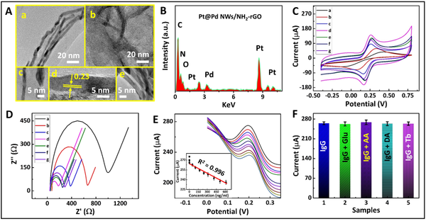
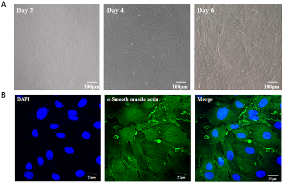
To save this page as a PDF, click this button and choose the PDF destination.

Poster Session 4

08:00 - 09:00 Thursday, 29th July, 2021

Presentation type Poster


P4.001 Sensitivity enhanced graphene-based field effect biosensor to detect tau protein for Alzheimer's disease diagnosis

S.S. Kwon, Y.J. Lee, S.H. Lee
Korea Institute of Science and Technology, Republic of Korea

Abstract

Graphene field effect transistors (FETs) are considered to be advantageous for use as sensors due to their excellent electrical and mechanical properties and simple structure. Sensing mechanisms that measuring the potential difference due to changes in species or concentration of ions in the electrolyte are known to be suitable for immunoaffinity (IA) biosensor applications. However, most graphene IA biosensor studies use pyrenebutanoic acid, succinimidyl ester (PSE) as a linker for antibody binding on graphene surface, but PSE has limited binding strength and charge transfer due to its molecular structure. In this work, we provide edge defects instead of linkers to enhance the sensitivity of the detection of tau and phosphorylated tau proteins, biomarkers in Alzheimer’s disease. Unsaturated carbon bonds at the graphene line defects provide a advantageous antibody binding sites even in the absence of linkers, and help improve sensitivity because they are directly connected.

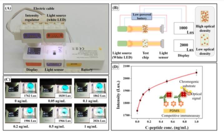
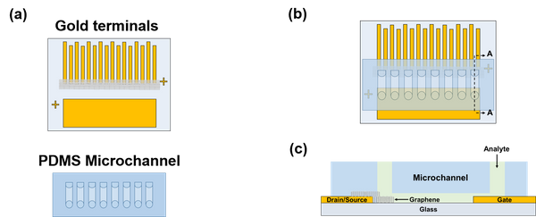
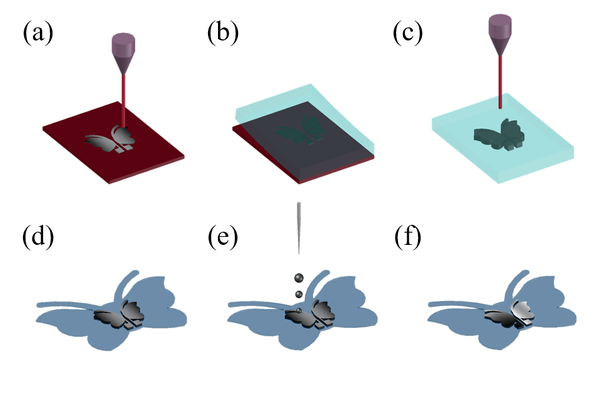
The control group (antibodies binding with linker, Fig 1(a)) and defective graphene group (prepatterned graphene without linker, Fig 1(b)) measure the electrical properties in the electrolyte-gate structure using a series of tau and phosphorylated tau protein solutions. The defective graphene group generated defects by patterning the transferred graphene instead of using a linker in the antibody immobilizing process. The characteristics of biosensor was evaluated by the shift of the lowest conductance point, called as Dirac point, for a series of tau and phosphorylated tau protein concentration changes.

Graphene purchased from ‘Graphene Supermarket’ and the biosensor was fabricated using a well-known process as shown in Fig 1(c) [1]. Since an electrical double layer is formed at the graphene-electrolyte interface, the current versus gate voltage (ISD-VG) curves of the graphene are more sensitive to the electrolyte gating than the back gating of the silicon substrate (Fig 1(d)). As shown in Fig. 2(a), the fabricated graphene glucose sensor shows a negative shift of the Dirac point with a glucose concentration increase. The defective graphene glucose biosensor without enzyme (Fig 2(b)) shows about 32% higher change than the control graphene biosensor structure with increasing glucose concentration (-40 mV/mM versus -53 mV/mM) [2].

This finding suggests that defective graphene biosensors can provide stronger immobilization of antibodies. These bonds can be greatly affected by the position and the density of antibody can be reduced, but it is advantageous in terms of the sensitivity of the graphene biosensor because it is directly connected to the graphene itself.

Uncaptioned visual

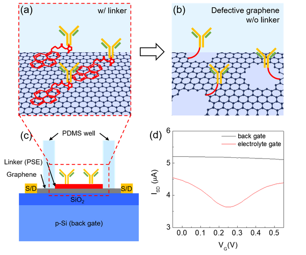
Fig 1. (a) Schematic images of graphene biosensors (a-c). The antibodies were binding on graphene surface using PSE as linker (a) or directly connected to the graphene edge defect (b). During the measurement, electrolyte was poured in to the PDMS well and reference electrode (Ag/AgCl) was inserted. (d) Current versus gate voltage curves of back gate (black) and electrolyte gate (red). In electrolyte gate, the Dirac point appears around 0.25 V, but in the back gate it is not visible in the range shown.

Uncaptioned visual

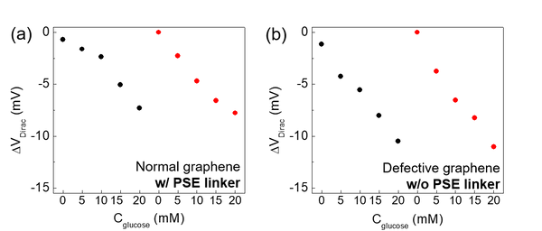
Fig 2. Dirac point shift with a series of glucose concentration changes (a, b). The graphene biosensors show sensitivity of -40 for normal graphene with PSE linker (a) and -53 mV/mM for defective graphene without PSE linker, respectively.

Reference

[1] S.S. Kwon et al., ACS Applied Materials & Interfaces, 8, 2016, 834-839

[2] S.S. Kwon et al., ACS Applied Materials & Interfaces, 9, 2017, 14216-14221


P4.002 In vivo monitoring of β-carotene in Euglena gracilis at single-cell resolution using a β-carotene-specific aptamer

C.R. Jin, J.Y. Kim, Y.E. Choi
Korea university, Republic of Korea

Abstract

Euglena gracilis is a unicellular phytoflagellate that synthesizes antioxidant vitamins, one of which is β-carotene, a precursor of vitamin A (retinol). Here, we detect the β-carotene in a live single cell of Euglena gracilis using an aptamer that specifically binds to β-carotene. The β-carotene aptamer was selected through graphene oxide assisted Systematic Evolution of Ligands by Exponential Enrichment (GO-SELEX). The fluorescein amidites (FAM)-labeled aptamers bound to the β-carotene exhibited ‘fluorescence on’, while FAM-labeled aptamers bound to the GO showed ‘fluorescence off’. Using the ‘fluorescence on/off’ switching properties associated with the β-carotene, we obtained an image of the fluorescent β-carotene in living cell by a confocal laser scanning microscopy (CLSM). Simultaneously, the content of β-carotene in the cell was quantified by fluorescence intensity. The result was commensurate with that obtained from existing methods of quantification with higher sensitivity and less time.


P4.003 Colorimetric biosensors based on smartphone and polydiacetylene for on-site food safety monitoring

Q.F. Li1,2,3, T.Q. Sun2, G.Ij. Salentijn3, B.A. Ning2, J.L. Bai2, D.P. Han2, Y. Peng2, Z.X. Gao2, Z.P. Wang1
1Jiangnan University, China. 2Tianjin Institute of Environmental and Operational Medicine, China. 3Wageningen University & Research, The Netherlands

Abstract

Introduction: Self-assembled polydiacetylene (PDA) vesicles, with the distinct advantages of low-cost materials, simple preparation as well as excellent chromatic, physical, and optical properties, can be perfectly combined with colorimetric strip or electrochemical chips for on-site inspection. The full development of this material can play an important role in the rapid screening and early warning of environmental pollution monitoring, food safety, and disease diagnosis.


Methods: Without involving expensive reagents and instruments, we achieved the fine control of the synthesis process of PDA vesicles and developed a visual colorimetric strip to semiquantitative and the quantitative detection of histamine, by analyzing the standard colorimetric card and gray value using image J and “Color Grab” in a smartphone. For a more sensitive and specific test, biotin-modified diethylstilbestrol (bio-DES) was prepared as a bifunctional ligand to couple with both PDA-streptavidin and DES antibody, achieving amplification of PDA response to biorecognition of DES for on-site smartphone detection.


Results: After optimizing the assembly conditions, the assay showed exhibited excellent stability and sensitivity, with a linear response to histamine within the range from 70 ppm to 2240 ppm. What’s more, we found a good linear relationship (R2= 0.9911; 0.4 to 1250 ng/mL) between the colorimetric response and the logarithm concentration of DES, with adequate specificity, accuracy, and precision. the visual paper-based biosensor for DES digital semi-quantitative analysis with a smartphone was performed, with a detection limit of 2 ng/mL. 


Discussion: The work we have done shows that PDA vesicles hold great potential in the intelligent system for on-site detection and real-time monitoring. Next, by combining with functional fluorescent materials and electrochemical detection platform, biomimetic fluorescent PDA vesicles will be extended to biosensor chips, which can be applied to the rapid screening and early warning of various risk factors in the environment, food, and disease diagnosis.

Uncaptioned visual


P4.004 Selective and multiplexed detection of Alzheimer’s disease biomarkers in human serum over SERS nanoprobes

I. Hwang, S. Lee, J. Kim
et al
Hanyang Univ., Republic of Korea

Abstract

Alzheimer’s disease (AD) is known to be caused by the deposition of amyloid beta (Aβ) plaques and neurofibrillary tangles in the human brain, leading to synaptic dysfunction and neurodegeneration. The earliest pathological stage of Aβ deposition in the brain can be identified by Aβ positron‐emission tomography (PET) or quantification of Aβ in cerebrospinal fluid (CSF). However, given that these two methods are costly and invasive, developing a cost‐effective and non-invasive alternative tool for early diagnosis of AD is urgently warranted. In that context, blood‐based AD biomarkers have been studied extensively for the non-invasive and early diagnosis of AD to date. It remains a big challenge to accurately quantify the amounts of amyloid peptides Aβ40 and Aβ42 as AD biomarkers in blood owing to their low levels. This has driven the development of sensitive and noninvasive sensing method for the early diagnosis of AD. Herein, an approach for the synthesis of Ag nanogap shells (AgNGSs) is reported as surface enhanced Raman scattering (SERS) colloidal nanoprobes for the sensitive, selective, and multiplexed detection of Aβ40 and Aβ42 in blood. Raman label chemicals used for SERS signal generation modulate the reaction rate for AgNGSs production through the formation of an Ag-thiolate lamella structure, enabling the control of nanogaps at one nanometer resolution. The AgNGSs embedded with the Raman label chemicals emit their unique SERS signals with a huge intensity enhancement of up to 107 and long-term stability. The AgNGS nanoprobes, conjugated with an antibody specific to Aβ40 or Aβ42, are able to detect these AD biomarkers in a multiplexed manner in human serum based on the AgNGS SERS signals. Detection is possible for amounts as low as 0.25 pg mL−1. The AgNGS nanoprobe-based sandwich assay has a detection dynamic range two orders of magnitude wider than that of a conventional enzyme-linked immunosorbent assay.


P4.006 Sandwich-type electrochemical aptasensor based on hollow mesoporous carbon spheres loaded with porous dendritic PtPd nanoparticles as signal amplifier for ultrasensitive detection of cardiac troponin I

Z. Wang, H. Zhao, K. Chen, M. Lan
East China University of Science and Technology, China

Abstract

Signal amplification is crucial to improve the sensitive for the detection of cardiac troponin I (cTnI) in electrochemical biosensor. Herein, we developed a novel signal amplification strategy to construct a sandwich-type electrochemical aptasensor for the detection of cTnI. PtPd dendritic bimetallic nanoparticles loaded on melamine modified hollow mesoporous carbon spheres (PtPd DNs/NH2-HMCS) was prepared as labels to conjugate with thiol-modification DNA aptamers probe (Tro6) for signal amplification. While introducing numerous amino groups, the melamine functionalized hollow mesoporous carbon spheres (NH2-HMCS) retains the edge-plane-like defective sites for the adhesion and electrocatalytic reduction of H2O2. With the unique characteristics of NH2-HMCS, it not only enhances the dispersity and loading capacity of PtPd dendritic bimetallic nanoparticles (PtPd DNs), but also improves the stability of bonding by the affinity interaction between PtPd DNs and amino groups. Meanwhile, the synergistic catalysis effect between PtPd DNs and NH2-HMCS significantly enhanced the electrocatalytic reduction of H2O2 and further amplified the signal. In addition, disposable screen-printing gold electrodes (SPGE) were used as thiolated single-stranded DNA aptamers (Tro4) carriers and sensing platforms to accelerate the electron transfer. Under optimal conditions, this aptasensor for cTnI detection displayed a wide dynamic range from 0.1 pg/mL to 100.0 ng/mL and a low detection limit of 15.4 fg/mL (S/N = 3). The sensor also successfully realized the analysis of cTnI-spiked human serum samples, meaning potential applications in acute myocardial infarction (AMI) diagnosis. 

Uncaptioned visual

Scheme 1. Diagram of the fabrication procedure for Tro6 label (A)and detection procedures of the aptasensor (B). 


[1]   Pourali A, Rashidi M R, Barar J, et al. Voltammetric biosensors for analytical detection of cardiac troponin biomarkers in acute myocardial infarction[J]. Trac-Trends in Analytical Chemistry, 2021, 134

[2]   Zhang H W, Noonan O, Huang X D, et al. Surfactant-Free Assembly of Mesoporous Carbon Hollow Spheres with Large Tunable Pore Sizes[J]. Acs Nano, 2016, 10(4): 4579-4586.

[3]   Fan F R, Liu D Y, Wu Y F, et al. Epitaxial growth of heterogeneous metal nanocrystals: From gold nano-octahedra to palladium and silver nanocubes[J]. Journal of the American Chemical Society, 2008, 130(22): 6949-+.


P4.007 Specific targeting of ErbB2 expressing breast cancer with ErbB2 peptide-conjugated liposomes

S. Ueno, T. Niidome, R. Lee
Kumamoto University, Japan

Abstract

ErbB2 is a type of receptor tyrosine kinase, which known to involve in tumorigenesis, tumor aggressiveness, and clinical outcome. ErbB2 is highly expressed in HER2 subtype breast cancer. Successful development of trastuzumab for targeting breast cancer has brought a great change in cancer treatment and diagnosis. However, despite its high affinity and specificity to the target, high immunogenicity and cost have led scientists to try new attempts. Compared to antibody, targeting peptide is cheap, stable for a long time, and has lower immunogenicity. Not surprisingly, this peptide has been extensively applied by conjugating with various drug delivery systems.

Herein, we fabricated an immunoliposomes, which are conjugated with tumor-targeting peptide and evaluated their functions. Physical and chemical characterizations (HPLC, MALDI-TOF, NMR, DLS and TEM) confirmed successful construction of ErbB2-target immunoliposomes. We synthesized both scrambled and ErbB2-target peptides, and verified their physicochemical characteristics were not different except targetability. ErbB2-target immunoliposomes exhibited better uptake in HER2-positive BT-474 cells than HER2-negative MDA-MB-231 cells as well as than non-targetable immunoliposomes with a scrambled peptide sequence.

This peptide-mediated targeting strategy is promising for improving the efficacy of chemotherapy in HER2-positive cancer. If anti-cancer drugs are delivered to ErbB2 overexpressing cells with the immunoliposomes, the cellular uptake of the drugs could be significantly improved through receptor-mediated endocytosis. We mainly focus on the characteristics of the immunoliposomes and targetability of peptide with suggesting the future perspective on breast cancer therapy.


P4.008 In-situ electrodeposition of antimony onto MXene (Ti3C2Tx)/MWCNTs nanocomposite for highly effective copper ions detection

X. HUI, M. Sharifuzzaman, X. XUAN, S. Sharma, A. Zahed, J.Y. Park
Kwangwoon University, Republic of Korea

Abstract

Recently, there has been an increasing interest in the determination of copper (Cu) ions in body fluids because the excess or deficiency of copper will have detrimental effects on human health and cause some diseases, such as Wilson’s disease, kidney failure, anemia, and impaired energy production [1]. However, accurate detection of Cu ions in body fluids is still a challenge due to the extremely low concentration. In order to improve the detection limit and sensitivity, some studies involved modifying electrodes with low-toxic metals (such as bismuth) to form alloys with detected metals. However, the potential overlap can also cause interference during detection. Therefore, reducing detection limit and increasing sensitivity are still a crucial trend of trace metal detection.

Here, we have newly proposed the layer-by-layer (LBL) combination structure of MXene-Ti3C2Tx and MWCNTs (MXene/MWCNTs) for Cu ions detection. In addition, the in-situ antimony (Sb) electroplating method was adopted for further increasing the sensitivity. MXene, a new class of 2D transition metal carbides and carbonitrides with excellent metallic conductivity, was prepared according to the MILD synthesis process [2]. When the MWCNT is incorporated with MXene as a spacer, it can not only help improve aggregation problem of MXene caused by hydrogen bonds and van der Waals interactions between layers effectively, but also enhance the interlayer conductivity.

The LBL-structure MXene/MWCNTs significantly increased the conductivity due to the combined synergistic effect and showed the best electrocatalytic performance characterized by CV and EIS (Fig. 1). Besides, the presence of Sb with optimized concentration improved the potential overlap problem and could aid in immobilization of more Cu as well (Fig. 2).

The combination of MXene/MWCNTs and in-situ electrodeposition of Sb exhibited quite low detection limit (0.1 ppb). Moreover, detecting Cu ions successfully in urine samples can provide a greater possibility for smart home-monitoring of trace metals.

Uncaptioned visual

Fig. 1 Schematic of MXene-MWCNTs nanocomposite synthesis as well as Cu2+ detection

Uncaptioned visual

Fig. 2 (a) CV of different modified electrodes (bare GCE, MWCNT/GCE, Mxene/GCE, Mxene/MWCNT/GCE) at a scan rate of 50 mV/s; (b) EIS of GCE with different modified conditions in 1.0 mM [Fe(CN)6]3-/4- solution.

Uncaptioned visual

Fig. 3 (a) Optimization of concentration of Sb3+ in ABS solution (0.1M pH 4.6) with the presence of 350 ppb Cu2+; (b) The concentration-current curve.

Uncaptioned visual

Fig. 4 (a) SWASV response of Cu2+ detection with different concentrations (10, 30, 50, 70, 90, 110 ppb) in ABS solution (0.1M pH 4.6) with the presence of 700 ppb Sb3+; (b) SWASV response of limit of detection of Cu2+ (0, 0.1, 0.2 ppb) in ABS solution (0.1M pH 4.6) with the presence of 700 ppb Sb3+; (c) SWASV response of Cu2+ detection with different concentrations (0, 100, 200, 300, 400, 500 ppb) in ABS solution (0.1M pH 4.6) with the presence of 700 ppb Sb3+; (d) SWASV response of Cu2+ detection with different concentrations (0, 10, 40, 70, 100, 130, 160 ppb) in the urine sample with the presence of 700 ppb Sb3+.

Table 1: Performance comparison of Cu2+ detection based on various materials.

Electrodes

Method

LOD (μg·L−1)

Linear Range (μg·L−1)

References

Mo6SxI9-x NWs/GCE

DPASV

0.2

0.80–240

[3]

Poly/GCE

SWASV

0.32

1.6-160

[4]

RGO/Bi/CPE

DPASV

26

20-100

[5]

Mxene/MWCNTs/GCE

SWASV

0.1

10-500

THIS WORK

References:

[1] W. Gao, H. Y. Y.Nyein, Z. Shahpar, H. M. Fahad, K.Chen, S. Emaminejad, Y. Gao, L. Tai, H. Ota, E. Wu, J. Bullock, Y. Zeng, D. Lien, and A. Javey, ACS Sens. 1 (2016), p. 866-874.

[2] Z. Li, L. Wang, D. Sun, Y. Zhang, B. Liu, Q. Hu, and A. Zhou, Materials Science and Engineering B 191 (2015), p. 33-40.

[3] H. Lin, M. Li, and D. Mihailovič, Electrochimica Acta 154 (2015), p.184-189.

[4] E. Pereira, B.L. Rivas, M. Heitzman, J.C. Moutet, C. Bucher, G. Royal, and E.S. Aman, Macromol. Symp. 304 (2011), p. 115.

[5] P.K. Sahoo, B. Panigrahy, S. Sahoo, A.K. Satpati, D. Li, and D. Bahadu, Biosen. Bioelectron. 43 (2013), p. 293

Acknowledgement:

This research was supported by the Bio & Medical Technology Development Program of the NRF grant funded by the Korean government (MSIT) (NRF-2017M3A9F1031270).


P4.009 A superhydrophobic chip integrated with an array of medium reservoirs for long-term hanging drop spheroid culture

B. Sun, G. Li
Chongqing University, China

Abstract

Hanging drop (HD) is one of the most popular methods used for spheroid culture. However, conventional HD systems are limited by the difficulty in exchanging the medium, and hence can only sustain spheroid culture for a few days. Here we present a medium-reservoir-integrated hanging drop (MRI-HD) chip for long-term spheroid cultures. This chip contains an array of medium reservoirs connected to hanging droplets via an array of thru-holes and consists of three layers: a top glass slide, a PDMS frame and a PDMS-base reservoir array layer integrated with a patterned superhydrophobic bottom surface (Figure 1). The MRI-HD chip can be facilely fabricated by using a desktop laser engraver and standard PDMS processing while its operation is simple (Figure 2). In contrast to hydrophobic surfaces in conventional HD systems, the superhydrophobic-patterned surface of this HD chip offers higher wetting contrast and thus can arrange quasi-spherical cell culture droplets (Figure 3), which is beneficial to formation of compact spheroids. More importantly, integration of medium reservoirs endows this HD device with the capability of long duration of continually replenishing media, which enables long-term culture of spheroids under undisturbed conditions. To demonstrate this advantage, a comparative experiment of MHCC97H liver cancer cell culturing was performed with a MRI-HD chip and a non-MRI HD chip, respectively. The results showed that the cultured spheroids in the non-MRI HD chip began to lose their integrity and broke into small satellite aggregates after 6 days of culturing (Figure 4), which was probably due to the depletion of the medium ingredients and the accumulation of metabolic substances. In contrast, the spheroids in the MRI-HD chip continued to grow even up to 30 days. In summary, our MRI-HD chip provides a simple and cost-effective platform for high-throughput preparation of microscale 3D cell constructs for drug discovery and regenerative medicine.

Uncaptioned visual

Figure 1 The medium-reservoir-integrated hanging drop device.

Uncaptioned visual

Figure 2 Schematic of fabricating and operating the medium-reservoir-integrated hanging drop device.

Uncaptioned visual

Figure 3 Hanging droplets formed on the proposed HD chip and the petri dish lid used in the conventional HD systems.

Uncaptioned visual

Figure 4 Comparison of spheroids cultured on the medium-reservoir-integrated HD chip and the HD chip without integrated medium reservoirs. Scale bar: 100 μm


P4.010 Rapid and ultrasensitive immunosensor of cancer biomarkers based on a fiber-type amplifier

M. Yamaguchi, R. Kase
Shinshu University, Japan

Abstract

Background: Measurement of cytokines in the blood of cancer patients offers the possibility of improved prognosis by facilitating early diagnosis and management. Thus, there is an urgent need for point-of-care testing devices that permit rapid determination of cancer biomarkers with ultrasensitivity in the pg/mL rangeIn this study, a novel signal amplification method for biomolecule detection has been developed based on an immunosensor featuring microfibers with micro/nanoscale surface topographies that provide an enhanced 3-D detection zone.

Materials and MethodsThe immunosensor consists of a glass microfiber bundle (fiber-type amplifier) and an optical sensor (Fig.1). An alkaline phosphatase (ALP)-labeled anti-interleukin (IL)-6 antibody conjugate was synthesized for the immunoassay. Both IL-6-bovine serum albumin (BSA) and a chemiluminescent substrate were immobilized on the microfibers. Micro/nanoscale surface topographies were fabricated on the microfibers using a femtosecond pulsed laser (wavelength 514 nm; pulse width 234 fs) to increase the reaction area and introduce hydrophilicity. When the fiber-type amplifier is attached to a blood droplet, 4 mL of sample is automatically drawn up by capillary action. Immediately thereafter, a competitive reaction occurs and the hydrolyzed chemiluminescence intensity is measured using the optical sensor.

Results and Discussion: After laser processing, the apparent contact angle of the surface-topography-modified microfibers decreased from 113.2° to 48.7°while the surface area of the microfibers increased by more than three fold. Linear regression analysis of the calibration curve yielded a correlation coefficient of 0.93. The relationship between the signal intensity, I, and the concentration of sample, s, was I = 2.88 × 10⁷ s + 1.30 × 10³ (count/gate) for concentrations in the pg/mL range (Fig.2).

Conclusion: The analytical performance of the immunosensor with the fiber-type amplifier permits determination of IL-6 levels within 10 min, which is compatible with point-of-care applications.

Uncaptioned visual


P4.011 Organic photo-electrochemical transistors with functionalized photosensitive gate electrodes for highly sensitive detection of salmonella

J. Song1, J. Guo1, W-W. Zhao2, P. You3, J. Hu1, X. Zeng1, F. Yan3, S. Chen4, P. Lin1
1Shenzhen University, China. 2Nanjing University, China. 3The Hong Kong Polytechnic University, China. 4City University of Hong Kong, China

Abstract

Detection of pathogenic bacteria plays a pivotal role in various important applications, including food safety, water monitoring and disease prevention. However, traditional detection techniques are suffering from certain limitations such as time-consuming protocols, intensive labour and high cost. High-performance biosensors are desired for the rapid identification of pathogens with high sensitivity and selectivity. Organic photo-electrochemical transistors (OPECTs) consisting of an organic channel and a photoelectrochemical gate electrode are an emerging candidate for developing high-performance biosensors, as they inherit the advantages of both organic electrochemical transistors (OECTs) and photoelectrochemical analysis. Herein, a novel immunosensor based on OPECT is achieved by functionalizing the photosensitive gate electrode with anti-Salmonella antibodies, which can specifically detect Salmonella down to the concentration of 100 cells/mL within one hour. The sensing mechanism is attributed to the photovoltage change caused by the steric hindrance effect of bacteria, which can be significantly amplified by an OECT due to the high transconductance, endowing the sensor with higher sensitivity than that of conventional electrochemical counterparts. Moreover, TEM characterizations clearly indicate that Salmonella bacteria can be modified by Au nanoparticles (NPs) via a simple mixing process, which can induce the exciton–plasmon interactions (EPI) effect and extend the detection limit down to 10 cells/mL. This work offers a highly sensitive, low-cost biosensor for the fast detection of bacteria and paves a promising way for exploiting high-performance immunosensors using the OPECT platform.


P4.012 An airway smooth muscle cells based bioelectronic tongue for screening of traditional Chinese medicine against asthma

C. Qin, Q. Yuan, C. He, Y. Pan, X. Wei, L. Zhuang, P. Wang
Zhejiang University, China

Abstract

Introduction: Airway smooth muscle cells (ASMCs) are the main effector cells that cause airway constriction and increase respiratory resistance in asthma. TAS2R14 and TAS2R10 are known to be the highest expression subtypes of TAS2Rs in ASMCs. The activation of them cause membrane hyperpolarization through a series of cell cascade reactions, leading to ASMCs relaxation and bronchiectasis (Fig.1). In this study, a ASMCs based bioelectronic tongue is developed to screen traditional Chinese medicines (TCM) against asthma. We explore the TCM relaxation effects on ASMCs through impedance changes, and construct three-dimensional ASMCs assays for verification (Fig.2).

Methods: Primary ASMCs from rats are isolated and cultured by enzyme-linked digestion and identified by immunofluorescence (Fig.3). Then, cells are planted into chips for electric cell-substrate impedance sensor (ECIS) detection. Meanwhile, the contraction and relaxation effects is reflected by the morphological changes of ASMCs collagen gel.

Results: The impedance shows different response to acetylcholine (Ach, bronchoconstrictor), isoproterenol (ISO, β2-adrenergic agonist) and quinine (bitter taste, which can relax bronchial), indicating that the screening model is successfully constructed (Fig.4 A,B). Compared with denatonium (DE), three TCM (tangeretin, nobiletin and picfeltarraenin IA), which can active TAS2R10 or TAS2R14 receptor, have better ASMCs relaxition effect, among which tangeretin works best. Besides, they work better when combined with β-adrenergic receptor drugs (Fig.4 C,D). Three - dimensional ASMCs gel spheres arrays are constructed and live/dead staining indicates that the ASMCs remain active in the gel (Fig.5 A). The Ach induced ASMCs gel contraction is inhibited by tangeretin, compared with the group adding PLC-β inhibitor U73122 to block the receptor activation signal pathway, which highlights tangeretin has strong relaxtion-inducing properties (Fig.5 B,C).

Discussion: The ASMCs based bioelectronic tongue responses to TCM which stimulate bitter taste receptors, it will lead to a new drug screening method for asthma or chronic bronchitis.

Uncaptioned visual

Fig.1 ASM Cellular relaxation signalling pathway.

Uncaptioned visual

Fig.2 Schematic of ASMCs based bioelectronic tongue and 3D ASMCs contraction assays.

Uncaptioned visual

Fig.3 (A) Primary ASMCs; (B) Immunofluorescence detection of primary cultured rat ASMCs.

Uncaptioned visual

Fig.4 (A,B) Impedance shows different response to cell contraction and relaxation ; (C,D) Selected TCM have strong effects of enabling ASMCs relaxation.

Uncaptioned visual

Fig.5 (A) Live/dead staining of ASMCs in the gel; (B,C) Three-dimensional ASMCs contraction assays demonstrate tangeretin can relax ASMCs.


P4.013 Multilayer MXene with chitosan assembled humidity sensor for human respiration monitoring

X. Li, G. Xu, Y.L. Lu, G. Liu, Z.H. Shi, Q.J. Liu
Zhejiang University, China

Abstract

Human respiration contains abundant physiological information. The successful detection of abnormal breathing including tachypnoea, respiratory distress and apnea can be used as indicators of diseases like cardiogenic pulmonary oedema, COPD and asthma. Since humidity changes dramatically between inspiration and expiration, we proposed a humidity sensor based on MXene/chitosan for human respiration monitoring. Through layer-by-layer assembly, MXene and chitosan were alternately adsorbed on laser-induced graphene electrodes as they are differently charged in corresponding solution. The multilayer structure was confirmed by SEM (Fig. 1a). Constant current was applied on the electrodes and resistance change caused by humidity was recorded. Fig. 1b shows the response curve under repeat wet-dry cycles, demonstrating that the sensor has short response/recovery time and good repeatability. Fig. 1c exhibits the stepwise resistance change under humidity ranging from 10% RH to 90% RH, indicating a wide response range and high sensitivity. The experiment proved that the sensor can track human respiration accurately and discriminate different respiration patterns (Fig. 1d). Derivation (red line) of the origin breath curve (blue line) was calculated to eliminate the baseline and quantify the inspiration/expiration time. The humidity sensor was fabricated on flexible substrate thus can be attached to arbitrary surface for convenience use, making it a promising candidate for POCT equipment. Furthermore, information like respiration rate, depth, ratio of inspiration time to expiration time can be extracted from the recorded curve, hopefully helping medical care personnel to predict and evaluate patients’ state of illness.

Uncaptioned visual

Fig.1. Preparation of MXene/Chitosan humidity sensor and SEM of the multilayer structure (a); Wet-dry cycle response showed fast response and recovery and good repeatability (b); Response from 10% RH to 90% RH (c); Response curve of different respiration patterns (d).


P4.015 Comparison of cultured cell attachment on a temperature-responsive polymer, poly-L-lysine, and collagen using modeling curves and a thermal-controlled quartz crystal microbalance

A. Alsaleem, S. Ito, K. Naemura, H. Muramatu
Tokyo University of Technology, Japan

Abstract

For investigating the effects of thermal responsive polymers to cultured cells, we have developed a quartz crystal microbalance (QCM) system with a micro-CMOS camera and a Peltier device so that the system enables microphotograph imaging and temperature controlling simultaneously with QCM measurement. This system was used in a CO2 incubator. The electrodes of the QCM were made of indium tin oxide (ITO) transparent electrodes that enable to obtain a transparent mode microphotograph.

After forming thermal responsive polymer poly(N-isopropylacrylamide) (PNIPAM), PLL and collagen films on the QCM, HepG2 cells were seeded on the QCM and cultured at 37℃.

The response of the QCM was monitored as the resonance frequency and the resonance resistance changes. At the same time, the micro photographs of the cells were recorded to observe the morphological change.

After injection of the cells, the resonance frequency decreased and resonance resistance increased, which meant that the cells attached on the quartz crystal surface. Then, the resonance frequency and resonance resistance changes saturated after 24h. By observing with the micro-CMOS camera, the number of cells and the adhesion of cells increased. The response curve of the resonant frequency and resonant resistance varied depending on the surface treatment of the quartz crystal.

We have built a modeling equation for the response curves and estimated the time constants for the response curves. Analysis of the fitting curves showed that the time constants of the first-lag response were 11 h for PLL,16 h for collagen,and 38 h for PNIPAM films. These findings were supported by photographic images showing wider cell spread on PLL and collagen than on PNIPAM.

Resonance frequency analysis can provide analysis of the mass and viscosity of cells that are difficult to obtain through the microscopic measurements only. Totally, this study provides information about adhesion behavior in kinetical perspective.


P4.016 Sensitive label-free detection of CD44 protein, a promising cancer biomarker, using grating-free optical fiber sensor tip

A. Bekmurzayeva, Z. Ashikbayeva, Z. Myrkhiyeva, A. Nugmanova, M. Shaimerdenova, T. Ayupova, D. Tosi
Nazarbayev University, Kazakhstan

Abstract

CD44 protein level in serum of cancer patients is a marker of tumor progression and metastasis in many cancers including breast, colon, gastric and blood cancers. To date, biosensors developed to detect this biomarker are based on photo/electrochemical sensing mechanisms. Using optical fiber as a transducer in biosensing, instead, offers the possibility of miniaturization, application in vivo due to its small size and inertness to electromagnetic interference. Herein, we propose detection of this important tumor biomarker using fiber optic tip sensor fabricated on a single mode fiber by a commercial splicing machine. The fabrication of this type of sensor is a fast and easy process once the appropriate program is established; and does not require the inscription of gratings which reduces its cost significantly. In this work, fiber optic tip sensors were fabricated, interrogated using optical backscatter reflectometry and then calibrated in solutions with different refractive indices (RI) reaching the sensitivity of 95.76 dB/RIU. The sensors sensitive to RI change were then employed for the construction of a biosensor for CD44 detection by immobilizing anti-CD44 antibody. Exposure of the functionalized fiber surface to different CD44 protein concentrations showed spectral amplitude increase with increasing concentrations of the analyte. On the other hand, detecting two control proteins with the biosensor did not show significant change in the obtained signal. The proposed immunosensor displayed a good performance in a wide concentration range of the protein (20 pM - 100 nM) with an achieved limit of detection of 20 pM thus offering a novel promising way in the detection of protein biomarkers with possible in situ applications in the future.


P4.018 Ultrasensitive and simultaneous detection of multiplex prostate cancer

A.R. Gul, K.Y. Park, J.P. Shin, S. Gosh, T.J. Park
Chung-Ang University, Republic of Korea

Abstract

Prostate carcinoma (PCa) has been being an utmost serious threat to elderly men around the world as it contributes to thousands of deaths every year. PCa is the fifth most common cancer among men in the world, and 11.4% is the annual percent increase in prostate cancer which is the second-largest rise following thyroid cancer. Although well-established clinical tests could provide early diagnosis, access to these tests is limited in developing countries, where a relatively higher incidence of prostate cancer is present. This has prompted an urgent need for developing a rapid, cost-effective, and robust diagnostic tool for point-of-care (POC) detection of prostate cancer. Lateral flow immunoassay (LFA) has found widespread applications in POC diagnostics. However, the low sensitivity of LFA limits its ability to detect important PCa biomarkers [e.g., prostate-specific antigen (PSA)], resulting in a false-positive and false-negative indication that leads to several unnecessary biopsies. To address this issue, we developed an improved LFA by optimizing the gold nanoparticle (AuNP)–antibody conjugate conditions (e.g., the conjugate pH and the amount of added antibody), the diameter of AuNP, and modifying the structure of the test strip (multiple target detection strip). Through these improvements, the proposed test strip could be able to detect multiple biomarkers with enhanced sensitivity over conventional lateral flow assay. The developed LFA can rapidly and specifically detect PCa with the naked eye, offering the incredible potential for POC assessment and personalized medication.


P4.019 Liver diagnostic sensor for fibrosis and cirrhosis progression using light-emitting diode for Raman spectroscopy

J.H. An1, K.J. Lee2
1KC University, Republic of Korea. 2Daejeon University, Republic of Korea

Abstract

A new raman measurements to use the light-emitting diode (LED) for the analysis of a liver lesions induced cirrhosis. The levels of liver ALT and AST were increased by TAA treatment, and the level of expression of SAM-alpha and TNF-beta were enhanced in cirrhosis liver. The signal of the Raman spectroscopy in the a male Wistar rat fresh liver tissues were showed at 1594 cm−1 from a 532 nm, 785nm laser detector , and the signal of LED spectroscopy were observed at 1587 cm−1 from a 540 nm detector. The line width of the LED emission spectrum was about 2000 cm−1 and the intrinsic width of the detected raman feature was about 1000 cm−1. The signal of the laser Raman spectroscopy was not detected peak in the male Wistar rat paraffin embedded liver tissues, but the peak of LED spectroscopy showed a slight shift to the long wavelength. From this Raman difference spectroscopy method, the LED spectroscopy analysis in liver lesions was detected at 505 and 552 nm, and showed N shaped patterns. In the 9 week in liver lesions, where the liver cirrhosis had progressed slightly, the peaks observed at 494,520, and 552 nm. In the 30 week, where the liver tissues were showed clearly cirrhosis, the spectrum had a W shape, and decreased at 494 nm, and increased at 535 nm. These results demonstrate that the LED spectroscopy might be a useful monitoring for the diagnosis of tissue than that of laser raman spectroscopy.


P4.020 Hormone detection with movable plastic membrane enzyme-linked immunosorbent assay (ELISA) sensor by alkaline phosphatase

A. Go, Y. Park, E. Jin, M. Sun, M. Lee
Chung-Ang University, Republic of Korea

Abstract

Protein hormones in plasma have been assayed by sensitive immunoassay techniques. The modified ELISA (Enzyme-Linked Immunosorbent Assay) was developed for the enhancement of sensitive responses. A new plastic 3D structure membrane that is capable of performing immunoassay with an electrochemical sensor has been designed and manufactured. We could successfully characterize the membrane and quantify hormones including thyroid-stimulating hormone (TSH), parathyroid hormone (PTH) and cortisol. For the measurement of those hormones, we have also built the well-type cartridge in which a reaction buffer, washing buffer and substrate was placed. The 3D membrane with a cylindrical shape (0.38Ⅹ1.2 cm) can be transported to each well of the cartridge. The current coming from platinum electrode is measured after the reaction with alkaline phosphatase (ALP) and p-aminophenylphosphate (p-APP) which include nicotinamide adenine dinucleotide (NADH) for an enhanced current signal. The measured signal to noise level using NADH showed 2-fold higher than without using NADH. Our developed ELISA system was applied successfully to the monitoring of the 1pg/ml ~ 100ng/ml TSH, 10pg/ml ~100ng/ml PTH and 1pg/ml ~ 100ng/ml cortisol and provided a linear calibration curve of each target. The biosensor exhibited a correlation coefficient of r = 0.944 (TSH), 0.940 (PTH) and 0.976(cortisol). In addition, for the accuracy of the proposed new ELISA system, quality control materials were examined using the differential pulse voltammetry (DPV) method.


P4.021 A thin-film transistor active matrix array for 2D real space electrical imaging of heart cell cultures

A.C. Eiler, J. Sugita, S. Ihida, H. Toshiyoshi, K. Fujiu, A. Tixier-Mita
The University of Tokyo, Japan

Abstract

Introduction

In vitro multielectrode array (MEA) technology is becoming a fundamental tool in cardiac electrophysiology for drug discovery [L.D. Garma et al., PLoS One, 2019]. However, this technology does not provide a whole 2D study of cell cultures because of its low density of microelectrodes and limited surface area. This paper reports for the first time the 2D electrical real-space imaging of heart cell culture activity with a new active matrix array device using the Thin-Film Transistor (TFT) technology. This transparent device can provide a high spatial resolution of cell culture activity by surpassing currently available MEAs in terms of density of transparent microelectrodes, and its cm-sized measurement surface area.

Methods

The device consists of an array of 150 x 150 Indium Tin Oxide (ITO) microelectrodes (100 μm x 100 μm), individually controlled by an array of addressing switches; the TFTs. The electrical signals were measured through a measurement system (MCS USB-ME32-FAI-System) and optical observation was performed using an inverted microscope (Figure 1). Heart cells were isolated from neonatal mice and cultured for 3 days on the TFT device without surface treatment.

Uncaptioned visual

Figure 1: (a) Setup of the experiment; (b) Description of the TFT array and the activation of the microelectrodes; (c) Comparison of the TFT device with standard MEAs in terms of density of microelectrodes and measurement surface areas.

Results

Cell electrical activity of ~240 beats per minute was successfully measured and confirmed by optical observation performed in parallel. The positive and negative deflections of the electrical signals usually detected on electrocardiograms were clearly observed. The optical images were superposed to the electrical data (Figure 2) and a 2D mapping of the peak-to-peak voltage amplitude was built (Figure 3) for confirmation of the regions with strong and low electrical activity of the heart cell culture observed with an inverted microscope.

Uncaptioned visual

Figure 2: Culture of heart cells: cardiomyocytes and fibroblasts. Scanning of the cell electrical signals was performed over 7 rows of 8 microelectrodes. For each row, the electrical activity was measured for 5 seconds on each microelectrode successively. The regions of active cardiomyocytes could be distinguished from the non-active fibroblasts.

Uncaptioned visual

Figure 3: Culture of cardiomyocytes only. 2D mapping of the cell electrical activity was built based on the average of the peak-to-peak voltage amplitude measured on the microelectrodes. Only a limited number of microelectrodes were connected. An improved version of the platform could allow measurements up to 150 x 150 microelectrodes (15 mmrecording area).

Discussion

These results confirm that TFT devices could be used to precisely provide a 2D real space electrical imaging of heart cell cultures. We believe that such device can become a key tool for research in electrophysiology on-chip.


P4.022 Skin gas monitoring device with ethanol biochemical gas sensor and gas concentrator

T. Arakawa, T. Aota, K. Toma, K. Mitsubayashi
Tokyo Medical and Dental University, Japan

Abstract

We develop a biochemical gas sensor using the enzymatic reaction of alcohol dehydrogenase (ADH) to target ethanol in skin gas. By introducing a gas concentrator using liquid nitrogen, we construct a highly sensitive system for skin gas measurements. To measure skin gas using the bio-sniffer, we establish a system for measuring concentrated gases by connecting the sensor with a gas concentrator and introducing concentrated skin gas to the sensing surface. This suppresses diffusion of the concentrated gases to achieve maximum fluorescence intensity by optimizing the measurement system. The calibration curve from obtained peak values shows ethanol gas can be measured over 1–3100 ppb, which includes skin gas concentrations during alcohol consumption. Finally, when applied to measurements of ethanol in skin gas following alcohol consumption, the output is found to be dependent on concentration, similarly to using standard gases. Thus, the system can be used for non-invasive percutaneous evaluation of VOCs in blood.

A highly sensitive and selective biochemical gas sensor and skin gas monitoring system with skin gas concentrating device and skin-cell for the skin ethanol concentration determination was demonstrated. This bio-sniffer measured the concentration of ethanol according to the fluorescence intensity change of nicotinamide adenine dinucleotide (NADH), which was produced by an enzymatic reaction of alcohol dehydrogenase. The NADH detection system was employed an UV–LED as the excitation light, and a highly sensitive photomultiplier tube as a fluorescence intensity detector. The calibration range of the ethanol bio-sniffer with a concentrating device was confirmed from 1.0 to 3100 ppb. 6 minutes is required for making concentrated gas from original gas. The concentration ratio can be adjusted 250–1000 times.

Next, "skin-cell" was prepared in order to continuously collect and measure skin gas (Fig.1). A carrier gas was introduced into the cell, and then skin gas was continuously delivered to the sensitive part of the bio-sniffer. In the ethanol measurement experiment, ethanol was ingested by drinking alcohol (0.4 g/kg body weight), and the sensor output was monitored. As a result of experiments on the wrist part, an increase of ethanol skin gas concentration was confirmed after drinking. Continuous measurement by intermittent (every 6-minute) of ethanol in skin gas using concentrating device was possible. Accordingly, this highly sensitive and selective bio-sniffer could be used to measure the ethanol in skin gas, and it is expected to apply for breath and skin research and investigation of biomarkers for clinical diagnosis.

Uncaptioned visualUncaptioned visual

Figure. 1 (a) Experimental system of skin gas measurement by skin-cell. (b) Cross-sectional image of experimental setup of simultaneous measurement of sweating and skin gas.

Figure. 2 (a) The output response of the ADH bio-sniffer to various concentrations of ethanol gas using gas concentrator. (b) calibration curve of the skin gas system.


P4.023 Gut-on-a-chip modeling and simulation for drug development

L. Cambruzzi Dalló1, A. Schuck2, P. Lora1, C.A. Cima1
1Universidade do Vale do Rio dos Sinos, Brazil. 2Sungkyunkwan University, Republic of Korea

Abstract

Currently, in the pharmaceutical industry, the use of animals in experiments for the development of new drugs is indispensable. However, ethical considerations, high cost and physiological differences between species are factors that strongly affect these experiments. Even with the use of cell cultures for in vitro assays, we are still far from a reliable representation of human physiology. The two-dimensional presentation and the absence of fluid flow are factors that contribute to the non-translational findings. There are organ-on-a-chip systems that can work with three-dimensional cell cultures and simulate the flow of fluid in the human body. These technologies come from the semiconductor industry, specifically microfluidics, which has become an important tool for numerous applications, especially in the field of bioengineering. This work aims to present the modeling and simulation of a human intestine through a microfluidic device, thus enabling the analysis of biological interactions in this system. The methodology used was the modeling and simulation of the device geometry in the multiphysics COMSOL software thus obtaining the necessary deformation for emulation of the human intestine physiology. From the simulations performed, it was possible to measure a pressure around -0.25 atm results in a maximum deformation of 11.3 % and the displacement was 20 μm. As a conclusion in the simulation context, the device presents a viable solution. The use of mechanical device simulation was a useful design tool, allowing the refinement of the structure and ensuring that its physical parameters are within a range compatible with those observed in real human organs. This is expected to decrease the overall manufacturing time of an effective device as well as its likelihood of operation after the first round of manufacturing.

Fig. I. (a) Gut-on-a-chip modeling; (b) modeling of membrane pores.

Uncaptioned visual Uncaptioned visual

Fig. II. (a) Deformation gradient simulation; (b) total displacement simulation.

Uncaptioned visual


P4.024 Detection of lactate concentration using a graphene-based field-effect transistor with common-gate configuration and coplanar electrode array

A. Schuck, H.E. Kim, Y-S. Kim
Sungkyunkwan University, Republic of Korea

Abstract

High concentration of serum lactate (> 2 mM) in the blood may be associated with several clinical conditions, e.g., hemorrhage, septic shock, and tissue hypoxia [1–3]. We developed a graphene-based field-effect transistor (GFET) to detect the lactate concentration based on an electrochemical reaction [3,4].

The terminals were fabricated on glass using photolithography coupled with gold deposition by thermal evaporation. Graphene was transferred to the substrate using a PDMS layer. Then, PDMS microchannel was integrated onto the substrate. The GFET with a common-gate configuration and coplanar electrode array is shown in Fig. 1. Standard lactate was mixed with a buffer solution and the lactate enzyme. After injecting each sample in the channels, transfer characteristics were measured by a parameter analyzer.

First, phosphate-buffered saline (PBS) solution was used to create a reference Dirac point (VDirac = 1 ± 0.03 V; n = 16). For the lactate experiment in a buffer solution, the VDirac shifted while the lactate concentration was increased (Fig. 2(a)). Additionally, pristine human plasma and plasma mixed with the lactate enzyme were measured in the devices (Fig. 2(b)). A higher VDirac shift was noticed in the treated sample when compared with the pristine plasma.

The lactate dehydrogenase (LDH) enzyme converted L-lactate to pyruvate and reduced the nicotinamide adenine dinucleotide (NADH) (Fig. 3) [3]. Consequently, a doping effect occurred on the graphene surface. The change in the charge concentration is related to the shift of the VDirac. Our GFET device was able to detect serum lactate concentration (0 to 10 mM) considering the pH level changes. After a further investigation of the detection of the lactate concentration in human plasma, the next step is an interference experiment using biochemicals (e.g., ascorbic acid, CaCl2, KCl) that can generate false-positive results.

References

[1]K. Rathee, V. Dhull, R. Dhull, S. Singh, Biosensors based on electrochemical lactate detection: A comprehensive review, Biochem. Biophys. Reports. 5 (2016) 35–54. https://doi.org/10.1016/j.bbrep.2015.11.010.

[2]S.M. Lee, W.S. An, New clinical criteria for septic shock: serum lactate level as new emerging vital sign, J. Thorac. Dis. 8 (2016) 1388–1390. https://doi.org/10.21037/jtd.2016.05.55.

[3]F. Alam, S. RoyChoudhury, A.H. Jalal, Y. Umasankar, S. Forouzanfar, N. Akter, S. Bhansali, N. Pala, Lactate biosensing: The emerging point-of-care and personal health monitoring, Biosens. Bioelectron. 117 (2018) 818–829. https://doi.org/10.1016/j.bios.2018.06.054.

[4]H.E. Kim, A. Schuck, J.H. Lee, Y.-S. Kim, Solution-gated graphene field effect transistor for TP53 DNA sensor with coplanar electrode array, Sensors Actuators B Chem. 291 (2019) 96–101. https://doi.org/10.1016/j.snb.2019.03.080.

Uncaptioned visual

Fig. 1 – Schematic of the GFET with a common-gate configuration: (a) glass substrate with the active layer (graphene) and Ti/Au terminals, and PDMS microchannel; (b) integration of the microchannel and the substrate; (c) cross-section view of the GFET device.

Uncaptioned visual

Fig. 2 – Experiments using the fabricated common-gate GFET device: (a) shift of the Dirac point (ΔVDirac) for different concentrations of lactate (0 to 10 mM). Inset: transfer curves of each lactate concentration and buffer solution; (b) transfer characteristics of the GFET devices using pristine human plasma and plasma treated with lactate enzyme.

Uncaptioned visual

Fig. 3 – Electrochemical reaction for the lactate assay occurring in the GFET device.


P4.025 A novel peripheral neuron stimulation system to cure myelination diseases based on highly uniform electrical field

A. Intisar, W-H. Kim, H.Y. Shin, S.J. Lee, M.Y. Kim, Y.J. Mo, Y.S. Kim, Y-I. Lee, M.S. Kim
DGIST, Republic of Korea

Abstract

Introduction: The myelin sheath of neurons plays a vital insulating role in making the saltatory nerve conduction possible, along with maintenance of the nerve conduction velocity. Demyelination of peripheral neurons, therefore, incites significantly poor performance of the neurons, resulting in reduced conduction velocity that can lead to neuropathies. There has been, unfortunately, no established treatments available to humans for such peripheral demyelinating neuropathies. To explore an innovative intervention, we developed a novel peripheral neuron stimulation system with highly uniform electrical field. In this novel study, we have revealed that electrical stimulation (ES) can enhance myelination in the peripheral nervous system (PNS).

Methods: A custom-designed electrical stimulation device was employed, and was able to generate a uniform electrical field across the entire stimulation region via COMSOL Multiphysics simulation. Based on previous screening, we applied a bipolar 20 Hz stimulation for 1 hour to embryo dorsal root ganglion (DRG) at the beginning phase of myelination.

Results: ES significantly enhanced the percentage of neurons undergoing myelination and the average length of myelin sheath at the mature phase of myelin. In further testing we found that ES upregulates numerous genes directly responsible for the processes of myelination, Schwann cell mitochondrial metabolism and lipid biosynthesis. Given that there is a surge in lipid required by the Schwann cells during the myelin development, the effect of ES on lipid biosynthetic pathway by enhancing mitochondrial metabolism is an important and novel approach.

Discussion: We aimed to build on this novel approach and eventually validate the mechanistic pathway that leads to the aforementioned enhancement. Nucleated from this critical outcome, we are planning to explore the effectiveness of ES in ameliorating the PNS demyelinating phenotype in disease models in further investigations. By properly establishing a background, we expect ES to become an effective treatment option for demyelinating neuropathic conditions.


P4.026 Graphene Quantum Dots (GQDs) based homogeneous assay for the detection of Ochratoxin A (OTA)

A. Bonanni, W.L. Ang
Nanyang Technological University, Singapore

Abstract

Graphene Quantum Dots (GQDs), are used in this work as biosensing platform for the recognition of the major food contaminant Ochratoxin A (OTA), with a fluorescent-labelled DNA aptamer (FAM OTA Aptamer) functioning as the biorecognition element. The principle of the detection lies in the formation of non-covalent interactions between the FAM OTA Aptamer and GQD surface, and the consequent fluorescence quenching. The further change in the fluorescent signal, induced by the formation of FAM OTA Aptamer/OTA conjugate during the detection step, could then be correlated to the presence and concentration of the target analyte. We observed in this work that upon tuning the concentration of GQDs, a switch in the biorecognition mechanism occurred. Specifically, while a lower GQD concentration (0.060 mg/mL) resulted in a restoration of the fluorescence intensity upon incubation with OTA, a higher GQD concentration (0.150 mg/mL) provided a further quenching of the final fluorescence intensity. Upon further calibration study, it was discovered that the latter mechanism provided a better option in terms of linearity of response, detection limit and selectivity.

Uncaptioned visual

Figure 1: Schematic of the bioassay


P4.027 Interdigitated electrode design and analysis using COMSOL multiphysics simulations

Y-S. Zhan, K. Settu
National Taipei University, Taiwan

Abstract

Interdigitated Electrodes (IDEs) have been widely used in gas sensor and biosensor applications, owing to its simple structural design. The sensitivity can be improved based on the shape of the geometric parameters. In this study, two designs of interdigitated electrode with varied electrode shapes; rectangular and circular shape were modeled and analyzed. The analysis was done using COMSOL Multiphysics, finite element analysis software to study the intensities of electric fields generated on the electrodes. The electrode size is 8mm x 5mm, the thickness is 0.05mm, and the electrode material is Ag-silver, the substrate material is Polyethylene terephthalate (PET) with a thickness of 0.15mm. The simulation results show that the electric field intensity generated on the circular electrode is higher than that of the rectangular interdigitated electrode with the same working area. The average electric field for the circular IDEs is 1.61 kV/m and 1.4 kV/m for rectangular shaped IDEs. From the cross-sectional view, it is seen that the electric field of the circular electrode is concentrated and uniform than the electric field of the rectangular-shaped interdigitated electrode. Moreover, a circular interdigitated electrode is better suited for fluid detection since it can receive dispensed circular sensing layer dots. Whereas in the rectangular-shaped interdigitated electrode, these fluid dots fail to cover the entire working area, thereby wasting sensing material. Thus, the use of a circular electrode with high electric field strength can improve the detection sensitivity, reduce the design area and achieve more detection efficiency.


P4.029 Timing controlled three-dimensional surface-modified paper-based analytical device for portable immunoassay applications

Y.S. Chien, C.A. Chen, Y.D. Li, C.F. Chen
National Taiwan University, Taiwan

Abstract

According to the report from the World Health Organization, infectious disease caused millions of people died globally. Due to the majority of newly infected cases is in the resource limited environments, a rapid, simple, low cost, and highly accurate on-site detection platform for infectious diagnosis is needed for better disease control. In this study, we developed a three-dimensional microfluidic paper-based analytical device integrated with timer function (3D-sotPAD) (Figure 1). We use different viscosity solutions to generate different flow rates and fluidic time, resulting in a time delay phenomenon without electronic timer. In this way, solution contacts with sucrose and pullulan pre-dried onto the designed paper in two separate flow channels that cause a time difference. The regions of pre-dried dye in the device are then rehydrated by the passing reagents flow at different times, and thus flow up to the top layer and precisely shows visualized color. Meanwhile, we use three dimensional vertical flow immunoassay design in the hydrophilic cellulose flow channels as the detection system, which can greatly reduce dead volume of reagents compared with lateral flow systems. Furthermore, the combination with 3D-printed packaging device can help multi-layer paper to contact more closely and reduce time differences and detection error caused by the reagent flow among different layers. To verify the applicability of the timer-based paper platform, we apply to the detection of Human immunodeficiency virus (HIV) capsid protein p24. The results show that the limit of detection is 0.01 ng/mL, with a total turnaround time of 7 min (Figure 2). We expect this high sensitive, timer-combined, portable paper assay device meets the World Health Organization criteria that provides advantages of affordable, sensitive, specific, user-friendly, rapid and robust, equipment-free and deliverable in resource-limited regions.

Uncaptioned visual

Figure 1. 3D-Paper-Based Analytical Device

Uncaptioned visual

Figure 2. Paper-Based Analytical Device for HIV diagnosis


P4.030 Isolation of paramecia using a modernized electromigration chip

A.P. Shunmugam, J.G. Fernandez
Singapore University of Technology and Design, Singapore

Abstract

Introduction:

Paramecium is a well-known single celled microorganism that lives in freshwater, brackish and marine water bodies. These are usually 50-300 microns in length and are fully covered with hair-like structures called cilia all around the cell. They are widely been used in schools to study basic cell biology and laboratories as model organisms in various research fields like genetics, evolution biology and cell biology. Recently, paramecium has been studied closely to understand their movement to be bio-mimicked into innovative robots. Even though paramecium has been studied well for years, their isolation process is very old and tedious method. The aim of this project is to improve the isolation process of paramecium using a modernized microfluidic channel and thereby demonstrating the manipulation and control of the movement of paramecium.

Methods:

Galvanotaxis is the innate response of paramecium to an external electric field applied which has been utilized in the isolation method. When electric field is applied in the microfluidic channel, paramecium gets segregated from the rest of the sample and can be collected separately.

Results:

Uncaptioned visual

This is the image of the fabricated microfluidic chip to segregate paramecia from the rest of the microorganisms.

Uncaptioned visual

The above image shows the efficiency of electromigration chamber. (a) Graph showing the inlet paramecia percentage, trapped paramecium percentage of 72.7%. (b) images of the inlet samples showing many other organisms along with paramecium (taken at 5X). (c) images of the outlet samples showing trapped paramecium (taken at 5X).

Conclusion:

The proposed chip was successfully designed and studied using Field element analysis. Also, several quantitative analyses were done to find the optimized experimental conditions to segregate paramecia with trapping efficiency of 73%.


P4.031 MUC1 impedimetric aptasensing based on interdigitated array electrode chip using a novel diffusion element

C-Y. Lai, T-S. He, W-C. Huang, L-C. Chen
National Taiwan University, Taiwan

Abstract

Uncaptioned visual
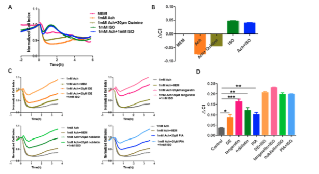
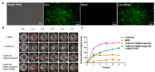
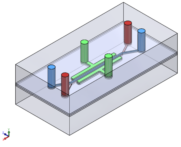
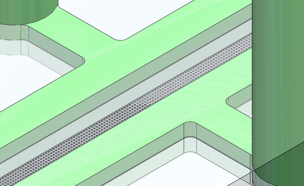
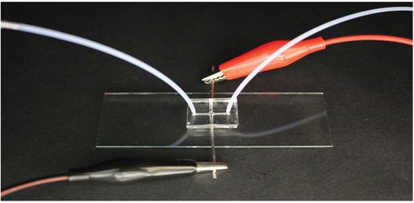
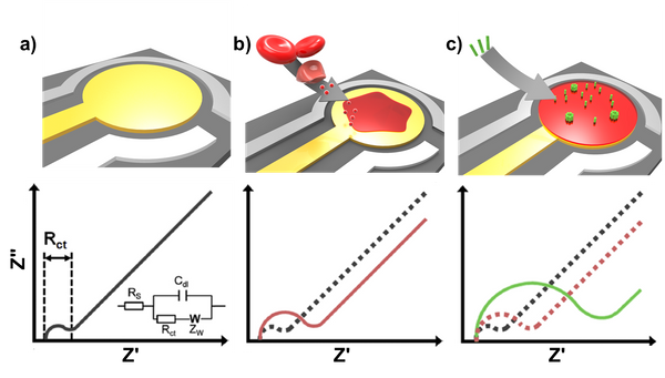
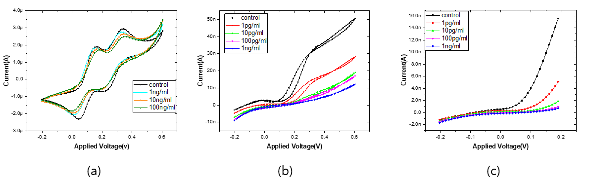
Interdigitated array (IDA) electrode-based impedimetric aptasensors are emerging devices, with several combined advantages (e.g. sensitive, robust, miniaturized … etc.) facilitating them to thrive in a variety of applications. However, as IDA electrodes exhibit finite diffusion, the electrochemical impedance spectroscopy (EIS) data of such aptasensors lacks an appropriate circuit element for characterizing the low frequency region. In this study, impedimetric aptasensors for detection of tumor marker MUC1 are fabricated based on IDA chips using an element specialized for parameterizing the IDA diffusion impedance. For chip construction, a 3D-printed fixture is used to clip a PDMS microwell onto the IDA electrode. For equivalent circuit fitting, the IDA diffusion element is used for characterizing the diffusion impedance, whereas using a Warburg element would have failed [1]. For MUC1 detection, a novel aptamer (K2 aptamer) selected by our team is used, which its specificity is confirmed using SPR (K2: 7.30±0.62RU and 30mer random ssDNA: 2.90±0.08RU), and Fe(CN)63-/4- does not hinder the binding event of K2 and MUC1. Using the method above, regenerable and selective MUC1 aptasensors using IDA chips are constructed. These low cost, regenerable and miniaturized chips along with their analysis methods have great potential for further medical use and commercialization. (Regenerable thrombin aptasensors are also fabricated and a baseline equation is further derived for quantifying charge transfer resistance (Rct) related to non-specific binding events. A significant proportional change of Rct between detection of 20nM thrombin (ΔRct/Bss = 30.0±7.92%) and 15μM HSA (ΔRct/Bss = 6.20±2.19%) is present, and EIS detection from 1.37~333nM is performed to confirm the binding affinity with a maximum ΔRct/Bss (Bmax) of 55.7% and a dissociation constant (KD) of 18.4nM.)

Figure 1. Graphical abstract for IDA chip-based MUC1 aptasensor and circuit analysis method.

[1] Lai, C.-Y., Weng, J.-H., Shih, W.-L., Chen, L.-C., Chou, C.-F., & Wei, P.-K. (2019). Diffusion impedance modeling for interdigitated array electrodes by conformal mapping and cylindrical finite length approximation. Electrochimica Acta, 320, 134629.

DOI: 10.1016/j.electacta.2019.134629


P4.032 Detecting Anomalies in Blood Glucose Regulation using Machine Learning Techniques on Data from Wearable Photoplethysmography Sensors

Bohan Shi1,2, Marcus Soo2, Shih-Cheng Yen1, Arthur Tay1, Seng Bin Ang3
1National University of Singapore, Singapore. 2Activate Interactive Pte Ltd, Singapore. 3KK Women's and Children's Hospital, Singapore

Abstract

Advances in wearable technologies have gradually changed our daily life as many people have been using wrist-worn wearable every day for fitness and health monitoring. In recent years, the high-end consumer wearables have integrated green light reflection photoplethysmography (PPG) sensor into their products, which mainly monitors the change of heart rate. These wearables have the potential to vastly enhance the reach of other public health concerns and chronic illness management, such as diabetes mellitus (DB).

The objective of our study was to evaluate the feasibility and accuracy of using the PPG signals from a consumer wearable to detect abnormal blood glucose regulation (> 7mmol/l). Eventually, this detection model will be incorporated into a mobile App to continuously and non-invasively evaluate a user’s risk of developing DB.

In the study, we recruited 413 volunteers (Mean Age 35.4 ± 10.8; Age range 16 to 74), and they were instructed to sit still for eight minutes while wearing a PPG-enable fitness tracker (Glo, Actxa), which have been used for several national health campaigns in the past. Blood samples were drawn from all volunteers prior to the experiments to determine the volunteers’ blood glucose levels using a standard glucometer (ACCU-CHEK Performa, Roche). We derived a set of promising features and trained a machine learning model to distinguish the normal and abnormal blood glucose level. To the best of our knowledge, this is a pilot study that systematically investigates the use of consumer wearable to detect the blood glucose anomalies on a large scale.

Currently, our Support Vector Machine model achieved a result of F1 score of 86.3 and accuracy of 85.1%. This preliminary result indicates the possibility of a low-cost alternative method to detect abnormal blood glucose levels in a non-invasive and continuous manner.


P4.033 Mass fabrication of various types of multicellular breast tumor spheroid using droplet-based microfluidic system

S.W. Cho1, H.S. Kim2, J.S. Lim1, B.S. Kwak3
1Yeungnam University, Republic of Korea. 2Kwangwoon University, Republic of Korea. 3Dongguk University, Republic of Korea

Abstract

The in vivo tumor is in the three-dimensional shape inside patients body. The evaluation of anti-cancer drugs efficacy is conventionally performed using two-dimensional cell monolayer platforms. However, the two-dimensional platform tends to overestimate the efficacies due to the absence of complex tumor characteristics, such as high cell packing density, extracellular matrix, and hypoxic core. Recently, methods to fabricate in vivo like multicellular tumor spheroids(MCTSs) have been developed to overcome the addressed limitations, but they are still in formidable challenges due to the low productivity.

In this study, we have developed a droplet-based microfluidic system capable of fabricating large amounts of MCTS within a short period of time. The droplet-based microfluidic system can encapsulate up to 280 cancer cells in a single droplet with a generation speed of 16-18Hz. From 1ml of cell solution, the system can generates 10,000 ~ 30,000 droplets. To form a MCTS, we have incubated the cell encapsulated droplets under cell incubation conditions and the cancer cells in droplet to form an one single MCTS with 100 to 150μm in diameter. Here, this shown the figure(1) was utilized to form MCTSs using four different types of breast cancer cell lines (BT-474, MCF-7, SK-BR-3, and MDA-MB-231). Two breast cancer cell lines (BT-474, MCF-7) are known to form a MCTS whereas the others (SK-BR-3, MDA-MB-231) are not due to their metastatic characteristics. The non-metastatic cancer cells formed a very circular MCTS whereas the metastatic cancer cells formed a cell aggregation model.

Uncaptioned visual

Figure1. Fabrication of multicellular tumor spheroids (MCTs) using 4 different breast cancer cell lines using the droplet based microfluidic system


P4.034 Cell isolation in microfluidic pore array

Jing-Yao You1, Chun-Yi Huang1, Sheng-Wen Liu1, Chien-Chong Hong1, Chie-Pein Chen2, Ally Huang3
1National Tsing Hua University, Taiwan. 2Mackay Memorial Hospital, Taiwan. 3IVD R.E.D. Microfluidic Chip Service & Support Co. Ltd., Taiwan

Abstract

This study presents novel passive microfluidic cell isolation chips with micromachined micropore array for single cell isolation of BeWo. Currently, several tools, such as flow cytometry, serial dilution, manual cell picking, and cell printer, are applied for cell isolation and analysis However, reagents and professionals are required for the procedure. In order to simplify the sample preparation, culture, and analysis.

Uncaptioned visual

Fig. 1 Schematic of cell isolation microfluidic chip for BeWo cells.

Here, we develop laser micromachining technique for plastic substrates and apply the technique to make 50 μm-micropore array. Five layers of plastic layers are assembled to realize the microfluidic chip (Fig. 1) to isolate BeWo cells. The micropore structures have narrow pores with wide pores at the bottom. 5-μm filter papers are bonded at the fourth layer of the chip to keep cells for further culture. COC plastic, which is biocompatible to cells, is used as the chip material. The cross section and top view of the fabricated device is shown in Fig 2a and b. The cell samples are injected into the chip manually. The cells will flow into the micropore and reach the fourth layer for further culture.

Uncaptioned visual

Fig. 2 Fabrication of the microfluidic chip. (a) cross section, (b) top view of the fabricated cell isolation chip.

Figure 3 shows the different culture results on traditional culture disk and COC plastic chip overnight. Compared with the results of the culture disk, the sterilized COC chip can get good results for overnight culture. The analysis of the cells will be studied in the near future.

(a)

Uncaptioned visual

(b)

Uncaptioned visual

Fig. 3 BeWo cell culture overnight on (a) traditional culture disk and (b) COC plastic chip.

In summary, the developed microfluidic chip with micromachined micropore array provides an easy-to-use and high performance method for rapid isolation of BeWo cells.


P4.035 Development of a new micro pupil–based colorimetric method for the detection of faecal indicator in tap water

Didem Rodoplu1, Cherng–Shyang Chang1, Cheng-Yuan Kao1, Chia-Hsien Hsu1,2,3
1National Health Research Institutes, Taiwan. 2National Tsing Hua University, Taiwan. 3National Chung Hsing University, Taiwan

Abstract

Development of simple, user–friendly and portable bacteria detection methods are of great importance for ensuring microbial safety in drinking water. This study presents the development of a simple microfluidic based colorimetric method for the feacal indicator Escherichia coli (E. coli) detection in tap water. A new imaging method was developed by taking the advantage of monitoring color changes from the transparent polydimethylsiloxane (PDMS) microfluidic inlet and outlet holes. Because the amount of light entering the human eye is restricted by the narrow pupil for imaging purposes, we called narrow imaging microfluidic inlet holes as µ-pupil of our method. First, magnetic particle–captured target bacteria were collected inside the µ-pupil with an external magnetic field. Then, resazurin reagent was used for the time dependent detection of viable bacteria after incubation at 1 h intervals. The horizontal and vertical optical setups were utilized for capturing frontal and lateral views to visualize color transitions from blue to pink inside the micro pupils. These images were analyzed by using a MATLAB software. The correlation between the optical density and bacteria concentration showed linearity in the range of 103–105 CFU/ml E. coli after 6 h of incubation, and 100–104 CFU/ml E. coli after 12 h of incubation. Culture plating and qPCR tests were performed in parallel as a control for conformation of bacteria detection. The specificity of the detection method was validated by the blank signal of Lactobacillus gasseri. It is an original idea to collect bacteria captured particles at the bottom of the µ-pupil in order to observe time-dependent colorimetric changes by simple optical setups. The results of this study revealed the potential of micro pupil–based imaging, and the applicability of the detection method for future simple and on–site bacteria detection in drinking water when there are limited resources. 


P4.036 Digital isothermal amplification system based on nicking and extension chain reaction and its application for quantification of respiratory infectious virus

Y. Ju1, Y. Song2, S.J. Lee2, K.G. Lee2, H.G. Park1
1Korea Advanced Institute of Science and Technology (KAIST), Republic of Korea. 2National NanoFab Center (NNFC), Republic of Korea

Abstract

We herein describe a novel digital isothermal amplification system for the quantification of respiratory infectious virus by utilizing our recently developed nicking and extension chain reaction system-based amplification (NESBA), an advanced version of nucleic acid sequence-based amplification (NASBA) technique. The NESBA technique was integrated into the microfluidic droplet device implementing all the steps to realize digital nucleic acid detection including generation and compartmentalization of nanoliter-sized droplets, isothermal incubation, and our newly developed miniaturized fluorescence monitoring systems (mFMS). By simultaneously conducting isothermal NESBA reactions in the localized ~ 104 droplet microreactors in our system, the genomic RNA (gRNA) extracted from respiratory syncytial virus A (RSV A) as a model target was successfully identified down to 10 copies within a sample. Due to the intrinsic advantages of digital isothermal amplifying methods including the elimination for the requirement of thermalcycling device and the much shorter amplifying time, the digital NESBA platform technique equipped with mFMS would serve as a promising platform system to achieve point-of-care molecular diagnostics for various infectious diseases.


P4.038 Fluorescence resonance energy transfer based aptasensor for the detection of phthalic acid esters

S.W. Lee1, S-W. Lee1, E-H. Lee2,3
1Seoul National University of Science and Technology, Republic of Korea. 2Ewha Womans University, Republic of Korea. 3Pusan National University, Republic of Korea

Abstract

Phthalic acid esters (PAEs) have been widely used as plasticizers (e.g. in polyvinyl chloride) to increase their flexibility, since the 1920s. PAEs are a class of organic contaminants, and they can be easily detected in the environment due to their extensive use. More importantly, some of them are classified endocrine-disrupting chemicals. In this study, we presented fluorescence resonance energy transfer (FRET) based aptasensor for highly sensitive detection of PAEs. The aptasensor consists of the group specific PAE-binding DBP-1 aptamer and signaling probe DNA, which were labelled with 1-pyrenebutyric acid N-hydroxysuccinimide ester (PBN) and cyanine-3 (Cy3) as a FRET pair, respectively.

The FRET performance was demonstrated by measuring donor-acceptor fluorescence changes. Using the fluorescence spectra, the FRET efficiency (E) and peak intensity ratio (I575 of F-Cy3/I450 of F-PBN) was calculated. As a result, the FRET efficiency was increased from 0.08 to 0.8 at molar ratios (Cy3-singaling probe/PBN-aptamer) of 0 to 0.077, and then saturated above molar ratio of 0.08. This demonstrates that a novel fluorophore pair of PBN and Cy3 is capable of FRET, and its FRET efficiency is acceptable for applications into sensor platform.


P4.039 Mild thermal annealed graphene oxide platform for enhancing circulating tumor DNA detection.

C.Y. Chen1, C.Y. Huang1, S.J. Cheng1, K.Y. Hsieh1, S.L. Chen1, C.H. Hsieh2, G.Y. Chen1
1National Chiao Tung University, Taiwan. 2Chang Gung Memorial Hospital, Taiwan

Abstract

Introduction: Circulating tumor DNA is a promising biomarker in liquid biopsy for cancer mutation profiling. However, the low concentration has limited clinical use. To deal with the challenge, we developed a novel platform interfaced with annealed graphene oxide (GO) for modify fluorophore-labeled DNA probe to specifically detect ctDNA for thyroid cancer with BRAF V600E mutation in a highly sensitive manner.

Methods: In this platform, GO absorbed single-stranded DNA probe and quenches the fluorescence in dye labeled DNA probe. Upon adding a target DNA, the probe desorbed from the GO surface forming double-stranded DNA and the fluorescence signal was recovered. In order to enhance the sensitivity of this platform. We induce a phase transformation in GO through mild thermal annealing, resulting in the oxygen clustering on GO. Then the DNA probes 5’-end were modified on the GO oxygen groups through stable amide bond and then the entire probe will be absorbed on the GO surface. Finally, we compared the sensing performance by using confocal microscopy.

Results: The phase transformation of annealed GO form oxygen clusters and without losing oxygen content. After DNA probe modification step, annealed GO has a lower background fluorescence signal compare to GO. With the addition of target DNA, the fluorescence was intensified along with increase of the target DNA concentration and annealed GO had a higher signal-to-background ratio. The annealed GO also appeared to be more resistant to nonspecific DNA displacement.

Discussion: The annealed GO platform not only improve the immobilization efficiency of the DNA probes but also create more aromatic regions for pi-stacking with DNA bases and made annealed GO adsorb probe more tightly. The fluorescence quenching efficiency was further improved after annealed process. Such tighter adsorption and stronger fluorescence quenching made annealed GO a highly potential platform for ctDNA detection.

Uncaptioned visual


P4.040 Nanobiosensors: Exosome separation and capturing by graphene oxide based microfluidic chip for diagnostic applications

S.L. Chen, C.Y. Huang, S.J. Cheng, K.Y. Hsieh, C.Y. Chen, G.Y. Chen
National Chiao Tung University, Taiwan

Abstract

Introduction: Exosome are cell-derived vehicle that size between 50-200 nm, which carry diversity biomarkers on surface and important proteins moreover some have been indicated as cancer markers. Herein, we create an exosomes separating device which consists a microfluidic channel for isolating nano size particles, followed by an immune-affinity-based purification that use graphene oxide as a nano-bio interface. This method avoids damages to exosomes since no high speed centrifugation is needed during the separating process, thus allowing brings higher purity and recovery rates than current methods.

Methods: We have improved the design of the Y-shape structure according to the method mentioned in the published article to capture the exosomes in the groove by purely physical methods, so as to avoid the damage to bio-information in exosome during centrifugation. Targeting makers on exosome’s surface. Due to GO’s high biocompatibility and ease of surface modification, we applied it as a nano-bio interface for click chemistry reaction to bind antibody in single domain to enhance the efficiency of specific capturing.

Results: At this stage, this system can stuck micro-particles which like circulating tumor cells (10 µm), make EVs can pass to second part, which have immobilized antibodies on GO’s surface by click chemistry in single domain to enhance efficiency of specific capturing. During exosomal separation, our system avoids damages on them by preventing high speed centrifugation also in single domain antibodies have enhance the efficiency of exosome specific capturing.

Discussion: In summary, we have developed a system to isolate exosome by physical method. During Exosomal separation, our system avoids damages on them by preventing high speed centrifugation. This highly reduce the loss of important bio-information in exosome for clinical diagnosis.

Uncaptioned visual


P4.041 Real time FET monitoring of UV-induced single Keratinocyte cell damage for skin health study

S.Y. Wu, P.H. Chen, Y.S. Huang, Y.L. Wang
National Tsing Hua University, Taiwan

Abstract

Cells can respond to the outside stimulation either by tolerating it, or restoring through some mechanisms, or undergoing cell death. One of the most prominent fatal factors to cells is UV light, since it may cause the DNA impairs, cell apoptosis and skin disorder. Therefore, if we could detect the critical UV dose for cells, then it is possible to prevent people from skin cancer.

Uncaptioned visual

Based on the above statement, we have fabricated a highly sensitive and selective array (Fig.1) to a whole cell resolution and it becomes very possible to exam the cell viability by our sensor because if the cell viability decreases, the binding conditions upon the sensor would not be good as healthy cells.

In this research, the unique electrical double layer (EDL) is utilized to sense the change of the capacitance which is caused by the cells and then we can exert double layer Capacitance (Cdl) to alter the current gain. Throughout this study, we have used current gain as the sensor signal instead of absolute drain current. Current gain is defined as the difference of transistor drain current before and after applying gate voltage Vg which are 0V and 1V respectively.

Uncaptioned visual

The current gain of FET sensor will be affected by transmembrane potential change, especially there will be a clearly opposite trend before and after UV exposure. Since the cells will adhesive on the sensor surface, causing the Cdl to change. Once the Cdl change, the potential of FET will also change. In terms of cells suffering from the damage, the cell membranes cannot adhesive on the sensor anymore, the corresponding current of Cdl will get smaller compared to intact one. (Fig.2) All in all, we can distinguish the cells are healthy or not under different stimulations by this novel and rapid platform to exam.


P4.042 Screening of DNA aptamers to airborne bacteria through the centrifugation based-partitioning method

H.R. Kim1,2, B.C. Kim1,2
1Korea Institute of Science and Technology (KIST), Republic of Korea. 2University of Science and Technology (UST), Republic of Korea

Abstract

The airborne bacteria has widely effected to human health such as infectious diseases, toxic effects, allergies, and respiratory symptoms. To prevent the outbreak of exposure in airborne bacteria, we need a more rapid method to obtain specific receptors. The aptamers are short oligonucleotides in high affinity and specificity to the targets. In general, the isolation method of the bacteria-specific aptamer is Whole Cell-SELEX (Whole Cell-Systematic evolution of ligands by exponential enrichment). However, this method has disadvantages of time-consuming and requiring highly skilled labor. Here, we used the centrifugation based-partitioning method for rapid isolating aptamers that are specific to dominant airborne bacteria species Micrococcus luteus (KCTC 9857), Bacillus cereus (KCTC 3711), and Staphylococcus xylosus (KCTC 3342)). The centrifugation based-partitioning method is a rapid screening process compared to SELEX. The serial removal of unbound DNAs to the bacterial cells from an initial mixture of bacteria and DNA libraries through serial centrifugation, one-time separation, and further one-time amplification of DNA bound to the target bacterial cells applied in this non-SELEX-based method allows successful aptamer isolation. As a result, we could isolate the DNA aptamers that bind to each airborne bacterium specifically (Kd: 14 ~ 300 nM). The centrifugation based-partitioning method can reduce the time to isolate the aptamers compared to the conventional SELEX and apply to the other targets.


P4.044 A bioelectrochemically modulated redox sensor: cellular redox stress driven gene expression for production of commodity compounds

H. Khandelwal, S. Mutyala, Y.E. Song, J.R. Kim
Pusan National University, Republic of Korea

Abstract

The bacterial redox sensor has been highlighted since the monitoring and control of cellular redox state influence the cell metabolism and target metabolite production. In Pseudomonas putida, fpr ferrodoxin NADP+ reductase gene senses the ROS/oxidative stress and gives transcriptional response to defend against toxic compounds. Pseudomonas putida KT2440, fpr regulation is known to be induced by the LysrR type transcriptional factor FinR. Such transcriptional regulation is helpful for bacteria to sense the oxidative stress created by the environmental factors which could be induced by redox mediators or environmental pollutants. This mechanism could be applied to upregulate gene of interest placed downstream of the fpr gene. In this study, a dual plasmid was made to harbour finr-fpr gene and fluorescent proteins into the Pseudomonas putida KT2440. The redox stress/ ROS generated by various redox mediator like pyocyanin, Paraquat, HNQ and HQ and was carried out under anaerobic and aerobic conditions. The redox sensor was activated by use redox cycling drugs and generated response was measured. The fluorescence intensity was quantified, and protein expression levels were analyzed. In further studies this property will be applied for induction of the gene of interest using the redox mediated stress in a Bio-electrochemical system (BES), which can be used for controlled modulation of the downstream gene expression by applied potential for higher production of the biofuels or valuable chemical synthesis. These result indicated that monitoring and controlling of bacterial redox stress can be used as a parameter to regulate gene expression and metabolite production.


P4.045 Droplet-based microfluidic system for fabricating 3D tumor spheroids

Jaehun Lee1, Jaewon Park2, Hyun Soo Kim3
1Korea Institute of Machinery and Materials, Republic of Korea. 2Southern University of Science and Technology, China. 3Kwangwoon University, Republic of Korea

Abstract

Conventionally, the study of tumor cells in vitro was performed using cells cultured in a 2D monolayer. However, tumors in vivo exist in three-dimensional structures, and thus, there are limitations in studies using 2D monolayer tumor cells due to the difference in physical properties. To date, three-dimensional tumor has been fabricated using several methods, such as hanging drops, scaffold structures, and multi-well plates, but these methods are somewhat difficult to operate and also limited in low yields as well as irregular sizes. To overcome these limitations, we have developed a droplet microfluidic system that can generate large quantities of three-dimensional tumors, having similar sizes to in vivo tumors, all in high-throughput as well as short periods of fabrication time.

The developed system is manufactured through a 3D printer. The device utilizes a cross junction design, where fast-flowing carrier oil solution break cell-suspended solution into uniform sized droplets. Cancer cell lines from various organs, such as breast, liver, lung, and pancreatic, were tested to form 3D tumors.

The developed microfluidic system can produce droplets with a diameter of 1350 μm at a speed of 25 droplets/min. In addition, incubating droplets at a concentration of 1x10⁷ cells/ml for 24 hours resulted in a 3D tumor formation with a diameter of 500 μm or more. After fabricating 3D tumors inside droplets, these tumors were recovered and further analyzed through immunofluorescence staining (ZO-1, E-cadherin, collagen type Ⅳ). The results of ZO-1, E-cadherin, and collagen type Ⅳ staining clearly showed that in vitro 3D tumors generated in droplets have similar characteristics compared to in vivo tumors.


P4.046 Development of graphene-based enzymatic electrochemical biosensor for urea detection

Y-M. Huang, K. Settu
National Taipei University, Taiwan

Abstract

Urea detection has been researched greatly owing to its importance in food science, environmental monitoring, and the functional condition of human organism. In this work, we presented a low-cost screen-printed graphene electrode (SPGE) biosensor modified with enzymes (urease and glutamate dehydrogenase) for specific detection of urea. The electrode was fabricated by screen-printing graphene paste onto a pre-stabilized PET substrate. Then, it was cured in an oven at 120C for 30 min. Next, the SPGE was soaked in sodium hydroxide (NaOH) solution at optimum conditions to remove insulating polymers and to increase surface roughness. The SPGE surface morphology was observed with scanning electron microscope, the result shows that the NaOH-treated surface exhibits a more porous structure compared to the bare ones. The SPGE characterization was carried out with capacitance measurement and cyclic voltammetry (CV). The capacitance remains constant from 1 kHz to 1 MHz with ~2% deviation, which indicates the high stability of our electrodes. CV result shows a linear relation between the peak current and the square root of scan rate which suggests the electrode is diffusion controlled, and it possesses an electron transfer kinetics faster than the mass transport in the K3[Fe(CN)6] redox probe solution. Finally, the electrode surface was drop-coated with prepared enzyme solution and dried at 55C for 15 min. The preliminary urea detection was carried out using chronoamperometry (CA). The measured CA curves of urea is significantly different from PBS. This evidences that the developed urea biosensor is capable for urea detection. Further the sensing conditions will be optimized to improve the sensitivity with the goal of detecting urea in human samples with low detection limit and wide range.


P4.047 Highly sensitive and selective detection of α-toxin using PEGylated erythrocyte membrane-functionalized impedance biosensor

I. Kim, G. Lee, D.S. Yoon
Korea University, Republic of Korea

Abstract

The α-toxin is a pathogenetic exotoxin secreted by Staphylococcus aureus (S. aureus). The water-soluble monomers (33.2 kD) bind to human blood cells (i.e., erythrocytes, platelets, monocytes, and lymphocytes) via unidentified cell surface receptors and assemble to form heptameric complexes, transmembrane pores, on the target cells. The pores induce cell death via lipid bilayer permeabilization to ions, water, and small molecules, and cell lysis. Recently, it is reported that methicillin-resistance S. aureus (MRSA) is most often causes of skin and lung infection which can cause sepsis, an extreme bodily response to an infection.

To selectively detect α-toxin, we devised erythrocyte camouflaged biosensor via functionalization of erythrocyte membrane (EM) onto electrochemical impedance biosensors (EM@EIB). The EM was purely extracted, PEGylated and functionalized onto the EIB with vesicle fusion method. The EM functionalization was optimized and verified with fluorescence imaging and electrochemical impedance spectroscopy (EIS). The α-toxin selecrively binds to EM. On the other hand, blood proteins hardly bind to EM.

As a result, EM@EIB was highly sensitive and selective to α-toxin from 0.0001 to 1 mg/ml concentration. Also, the selectivity of EM@EIB was verified with human serum albumin (HSA), gamma globulin (GG), and fibrinogen (Fib). Also, EM@EIB was sensitive to α-toxin spiked in human serum containing a number of proteins, molecules, and ions.

We developed a novel erythrocyte camouflaged nano-biosensors that functionalized with PEGylated EM. EM, the camouflaging layer, selectively binds to α-toxin against HSA, GG, Fib. It is confirmed that the selectivity was ~10 folds enhanced by EM coating onto the EIB.

Uncaptioned visual

Figure 1. Schematic illustrations of preparation of EM@EIB and its α-toxin measurement.

Uncaptioned visual

Figure 2. Selectivity of EM coated EIB compared with uncoated EIB.


P4.048 Optimizing smartphone-based colorimetrics for various intensity and colour change-based assays used for food contaminant analyses

J.L.D. Nelis1,2, L. Bura3, Y. Zhao1, K.M. Burkin4, K. Rafferty1, C.T. Elliott1, K. Campbell1
1Queen's University Belfast, UK. 2CSIRO, Australia. 3University of Parma, Italy. 4Lomonosov Moscow State University, Russia

Abstract

Smartphone-based colorimetric assay quantification is increasingly reported and has potential to cause a paradigm shift in food testing. However, a complete characterisation of the performance of existing colour spaces and single channels for optimum colour/intensity change quantification is still absent while inter-phone variation issues are often overlooked. Moreover, it has not been ascertained if utilizing existing colour spaces is necessary to reach optimal assay quantification. In this study performance of ΔRGB and all single channels from RGB, HSV and CieLab colour space were characterized utilizing existing software to extract single channel values (fig. 1a). Performance of all non-repeating random combinations of 2 and 3 channels of these colour spaces was equally characterised using a developed smartphone application and machine learning (fig. 1b). pH-strips, gold and carbon black nanoparticle-containing paper strips, ELISA assays and commercial lateral flow assays (LFAs) were used for these characterisations. For single channels, CieLAB and HSV channels never outperformed the best RGB channels. Moreover, RGB channels allowed effective quantification of domoic acid in shellfish, gluten in buffer, bovine milk in goat milk and goat cheese with LFAs (R2>0.90; prediction errors <40%) and soil pH prediction (R2>98 prediction errors <3%) with pH-strips. Inter-phone variation was high for LFAs but low using pH-strips (prediction errors <10%; six phones compared). Interestingly, novel channel combinations showed great promise in terms of prediction error and inter-phone variation reduction, outperforming classic RGB, HSV and CieLab colour spaces (fig. 1b). For LFAs the channel combination BSA was optimum (prediction error 36 ± 6%; R2=0.97). For pH prediction RLC was optimum (prediction error 1.31 ± 0.02%; R2=0.997). This study showed that colour change-based assays and random channel combinations are promising concepts that should be considered for smartphone-based analysis.

Uncaptioned visual

Figure 1: Workflow and primary results using single channels and random channel combinations.


P4.049 An immune-sensor based on surface acoustic wave for urine level detection of a stimulant drug

M. Jeon1,2, Y. Kwak1, B. Ju2, K. Kim1
1Korea Electronics Technology Institute, Republic of Korea. 2Korea University, Republic of Korea

Abstract

Recently, the number of crimes committed while using stimulant drugs has been increasing. Therefore, sensors for detecting stimulant drugs are becoming more important. In this paper, we have developed an immune-biosensor based on surface acoustic wave that can sensitively detect target materials in liquids from the amount of shift from the center frequency. The substrate used 36° lithium tantalate, which is suitable for the generation of a Love wave with low loss in a liquid environment. Al electrodes were deposited and patterned as interdigital transducers. Then, SiO2 film was deposited and patterned as a guiding layer. It reduced the wave velocity and increased the mass sensitivity. We conducted an experiment to determine the optimum thickness among 1, 3, and 6 µm for the effect. The biotin SAM was immobilized using an Au layer deposited on the guiding layer. Streptavidin at a concentration of 50 ppm was introduced and the frequency shift that occurred when the streptavidin was bound was 12.5 kHz at 1 and 3 µm thickness. However, at 6 µm thickness, the frequency shift was 22.5 kHz. These results show that the thicker the guiding layer, the greater the mass sensitivity. Based on this optimized thickness result, we fabricated a methamphetamine detection sensor by immobilizing anti-methamphetamine. When methamphetamine was dropped to a concentration of 150 ppm, the average observed frequency shift was 68 kHz. This was a threefold higher frequency shift than at a concentration of 50 ppm, which was consistent with the results tested with a threefold greater mass. The concentration of methamphetamine in a drug abuser’s urine can be as little as 100 ppm, which means that our developed immuno-sensor is sufficiently sensitive to detect methamphetamine in human urine. In addition, this sensor can be applied to detect various stimulant drugs with small molar mass.


P4.050 E faecalis produces a detectable electrochemical signal at an electrode but it is not its main metabolic strategy

J. Hinks, E.J.Y. Han, S. Wuertz
Singapore Centre for Environmental Life Sciences Engineering, Singapore

Abstract

Lactic acid bacteria are an important group of organisms, their detection and quantification is relevant in food, medical, and environmental settings. They are typically considered to be non-respiring, fermentative organisms characterized by an incomplete respiratory pathway resulting from their inability to produce heme and therefore cytochromes with reactive centers.

E. faecalis is an important lactobacilli, it is pathogenic and is an indicator of the microbial quality of recreational water. Recent reports suggest E. faecalis can partake in direct extracellular electron transport making it suitable for bioelectroanalytical detection. Better ways to detect and quantify E. faecalis are iindustrially relevant. Given that it E. faecalis can reduce an electrode under aerobic or microaerophilic conditions it would simplify bioelectroanalytical detection as achieving anaerobic conditions in field settings would not be necessary. Recent trends towards microscale detection systems pose even more of a challenge in this regard as oxygen diffusion becomes increasingly problematic with diminutive volumes.

Recent bioelectroanalytical techniques using redox-active enzyme-specific glycosides have enabled specific detection of target organisms. However, fermentative metabolism has been implicated in poor detection times using heme deficient mutants of E. coli suggesting that respiratory metabolism is important to achieve good bioelectroanalytical detection. Here we explore the electrochemical detection of E. faecalis using microscale systems.

We show that E. faecalis is amenable to bioelectroanalytical detection approaches and that it produces a rapid and measurable bioelectric current with the addition of a bespoke electrochemical glucoside as a specific detection compound and without the need for degassing (Fig. 1). We further explore the metabolic strategy and electron transport efficiency and show that inducing respiration does not improve the bioelectrochemical signal from E. faecalis and, furthermore, that the electrical output is not a quantitatively significant part of the electron flux in E. faecalis.

Uncaptioned visual

Figure 1. Bioelectroanalytical detection of E. faecalis.


P4.053 A versatile colorimetric thermo-sensor based on peptide nucleic acid and PEGylated graphene oxide

J. Lee
Korea Institute of Toxicology, Republic of Korea

Abstract

Temperature is an important parameter in various fields, including biology, chemistry, medical sciences, environmentology and industry. Unfortunately, the detection of temperature using colorimetric methods has not been sufficiently explored. Here, we developed a guanine (G)-rich DNAzyme (Dz)-based colorimetric thermosensor using peptide nucleic acid (PNA) and polyethylene glycol-functionalized graphene oxide (PEG-GO). The Dz served as a DNA template for thermosensitive nanostructures and as catalytic DNA for colorimetric assays. Using the combination of PNA and PEG-GO, we were able to control Dz activity in a thermosensitive manner, resulting in colorimetric visualization of temperature. The temperature-sensing range of this system could be simply tuned by designing a PNA strand based on the melting temperature of the Dz/PNA duplex. The programmable Dz/PNA structure with PEG-GO enabled sensitive, rapid, temperature-dependent responses. Moreover, this design permitted recall of the target temperature, enabling visualization of the temperature at a later time point. This robust system may be used as a rapid tool for practical temperature-sensing applications, such as in health diagnostics and food safety, and could be a valuable resource for basic and applied nanobiotechnology research.


P4.054 Metal enhanced fluorescence-based caspase-3 detection by using DNA/peptide-functionalized Au nanoparticles

J.H. Choi, J.W. Choi
Sogang University, Republic of Korea

Abstract

Fluorescence-based biosensors for the early diagnosis of several diseases such as cancer have widely utilized due to it enables straightforward and intuitive detection by naked eyes. However, there has been limitation to detect small amount of biomarkers, which lead to early diagnosis and increase to chance of the full recovery, comparing other sensitive analytical methods such as electrochemical and surface-enhanced Raman spectroscopy (SERS)-based detection. In this study, we developed metal enhanced fluorescence (MEF)-based highly sensitive detection system by using DNA and peptide-functionalized Au nanoparticles (AuNPs) for the caspase-3 detection, which closely related diverse diseases. AuNPs were functionalized by thiolated DNA, maintaining the optimal distance for MEF, DEV-containing short peptide strand, and fluorophore. Caspase-3 could sense the specific peptide sequences (DEV), and the distance between Au surface and fluorophore becomes approximately 8 nm, generating MEF. This system could improve the sensitivity of the caspase-3, covering from 5 pg/ml to 10 ng/ml. We propose that the highly sensitive and straightforward fluorescence detection by MEF using Au nanoparticle can be widely applied for sensitive detection of proteolytic biomarkers and early diagnosis of several diseases.

Acknowledgments: This research was supported by the National Research Foundation of Korea (NRF) grant funded by the Korea government (MSIT) (No.2019R1A2C3002300) and Basic Science Research Program through the National Research Foundation of Korea (NRF) funded by the Ministry of Education (No.2016R1A6A1A03012845) and the Basic Science Research Program through the National Research Foundation of Korea (NRF) funded by the Ministry of Education (NRF-2018R1A6A3A11051465).

Reference

[1] Choi, J. H., Kim, H. S., Choi, J. W., Hong, J. W., Kim, Y. K., & Oh, B. K. (2013). A novel Au-nanoparticle biosensor for the rapid and simple detection of PSA using a sequence-specific peptide cleavage reaction. Biosensors and Bioelectronics, 49, 415-419.


P4.055 Label-free and washing-free adenosine triphosphate detection method based on personal glucose meter

J.K. Ahn1, H.G. Park2
1Korea Institute of Industrial Technology, Republic of Korea. 2Korea Advanced Institute of Science and Technology, Republic of Korea

Abstract

Herein, we have presented a label-free and washing-free system for biomolecular detection using a personal glucose meter (PGM). ATP has been chosen as a model target, and cascade enzymatic reactions supported by hexokinase and pyruvate kinase have been adopted to relate the amount of ATP to glucose detectable by handheld PGMs. The existence of target ATP, in theory, helps hexokinase to catalyze the transfer of glucose to glucose 6-phosphate by delivering glucose to a phosphate group, thereby reducing the volume of glucose in relation to the amount of ATP. Additionally, pyruvate kinase enzyme recovers adenosine 5′-diphosphate (ADP), which is produced after hexokinase-catalyzed enzymatic reaction, to ATP. The regenerated ATP is again used to catalyze several rounds of enzymatic cascade reactions, contributing to an amplification of the signal. As a result, the increase in the amount of glucose which is inversely proportional to the amount of ATP is clearly determined by a handheld PGM. Using this strategy, we successfully determined ATP with high selectivity down to 49 nM even in real samples such as tap water, human serum, and bovine urine. Importantly, the established device does not need costly modification and washing procedures but is worked comfortably with a readily accessible PGM which will pave the way for the development of a simple and cost-effective sensing method.

Uncaptioned visual


P4.056 3D pyramid sensor for dopamine detection in Parkinson's disease model

K.J. Lee1, K.P. N'deh2, G-J. Kim3,4, J-S. Shin1, J.H. An3
1Daejeon University, Republic of Korea. 2Seoul National University of Science and Technology, Republic of Korea. 3KC University, Republic of Korea. 4Sogang University, Republic of Korea

Abstract

Dopamine (DA) is an important neuromodulator in the brain which is linked to several neurological diseases. Low DA levels or depletion in DA levels have been linked with Parkinson’s disease (PD). In this study, we reported a highly sensitive three-dimensional (3D) pyramid DNA sensor system based on a combination of DNA and bead-based immunoassays for detecting DA with surface-enhanced Raman spectroscopy (SERS). A three-floor hierarchical 3D pyramid microstructure was fabricated using two-photon stereolithography, and gold nanoparticles (Au-NPs) particles were coated onto the 3D pyramidal structure. The 3D microstructure of our pyramidal biosensor provided a high surface area, increasing the amount of antibodies to be bound on coated Au-NPs. Our method relies on the coated Au-NPs with adsorbed antibodies and Au-NPs that are encoded with DNA and antibodies which are able to squeeze the target protein captured by the coated-AuNPs-bound antibodies. The 3D pyramid DNA sensor was used to detect DA in the serum of PD patients. The biosensor could detect dopamine in standard DA down ≈ 0.1 pM, a lower limit detection value compared with that found in recent literature for DA biosensor (≈ 1 pM). In addition, DA levels in the serum of ten patients with parkinsonism had significantly lower DA concentrations (≈ 1 nM – 9 nM) than that of three healthy subjects (≈ 10 nM – 40 nM). This finding strongly implies that the 3D pyramid DNA sensor is reliable and highly sensitive for the detection of DA levels in PD patients. Thus, our biosensor can be used for the diagnosis Parkinson’s disease.


P4.057 Electrical control of stem cell differentiation using nanostructured biohybrid material

J. Lim, J. Yoon, J-W. Choi
Sogang University, Republic of Korea

Abstract

Due to the huge potential for regenerative medicine and stem cell therapy, control of the neural differentiation has attracted lots of interests in neurobiological field. For this reason, various researches for accurate neural differentiation have been reported such as use of nanoparticles or nanopatterned electrode. However, these methods still suffer from undesired side effect caused by nanoparticles and low differentiation specificity and efficiency. Therefore, it is necessary to develop a new method with high biocompatibility and specificity to induce the neural differentiation. In this study, the nanostructured biohybrid material based on recombinant azurin (AZU), DNA and Au nanoparticle (AuNP) for the efficient neural differentiation was developed for the first time. The nanostructured biohybrid material showed high biocompatibility due to the use of biomaterials such AZU and DNA. In addition, introduction of the AuNP allowed the electrical controllable complex with the differentiation inducible agent to be connected to the nanostructured biohybrid material without the use of the chemical linker. The result indicates that the neural differentiation was successfully conducted due to the release of the differentiation inducible agent from electrical controllable complex by applied electrical stimulation. Furthermore, by adjusting the time and location of applying electrical stimulation, it was possible to increase the efficiency of cell differentiation over a long period of time. In conclusion, proposed nanostructured biohybrid material can be applied as a novel platform that can effectively differentiate stem cells into the desired type of cells for regenerative medicine and stem cell therapy.

Acknowledgements: This research was supported by the National Research Foundation of Korea (NRF) grant funded by the Korea government (MSIT) (No. 2019R1A2C3002300) and by the Ministry of Education (No. 2016R1A6A1A03012845).


P4.058 Human platelet analysis using serotonin oxidation by electrochemical collision method on an ultramicroelectrode

J. Lee1,2, S. Hong1, B-K. Kim2
1Sookmyung Women’s University, Republic of Korea. 2Ewha Womans University, Republic of Korea

Abstract

In order to continuously monitor human health, a method for rapidly and accurately measuring platelet levels within a significant range of the presence and concentration of serotonin in platelets is needed. To overcome the time-consuming and pretreatment problems of conventional serotonin release assays, this study proposes to directly detect serotonin in a single platelet using the single-entity electrochemistry (SEE) method. Statistical analysis was performed on data based on the oxidation reaction of serotonin obtained by performing cyclic voltammetry and current-time measurements. The serotonin concentration takes into account the amount of charge obtained by integrating the current signal generated by the collision with an electrode at the individual particle level. In particular, the collision frequency was calculated to perform a stochastic treatment to determine the regression curve of platelet concentration. Our SEE methods will be a significantly novel method that will overcome the limitations of existing sampling, analysis time, and analysis environment to enable the development of high-performance sensors capable of on-site diagnosis and real-time diagnosis.


P4.062 3D PLA scaffold promotes proliferation and differentiation of human neural stem cell: A platform to drug screening in neural tissue engineering

Y.H. Lee1,2, G.J. Kim1,3, K.J. Lee4, J.W. Choi3, J.H. An1
1KC University, Republic of Korea. 2Seoul National University of Science & Technology, Republic of Korea. 3Sogang University, Republic of Korea. 4Daejeon University, Republic of Korea

Abstract

In this study, we fabricated a 3D scaffold using industrial polylactic acid (PLA) material to promote proliferation and differentiation in human neural stem cell (F3.Olig2 cells) and for drug screening. 3D PLA scaffold with a square shape pattern was fabricated via computer aided design and printed via FDM technique. F3.Olig2 cells are cultured in 3D PLA scaffolds to investigate their ability to differentiate into oligodendrocyte. The morphology of F3.Olig2 cultured in 3D PLA scaffold was formed 3D cells. Our results showed that the 3D PLA scaffolds enhance proliferation of F3.Olig2 compared to 2D plate. Also, when F3.Olig2 cells were treated with sonihedgehog(Shh) protein for 7days, the 3D PLA scaffolds  showed high expression of motor neuron cell type-specific phenotypes HB9 compared to 2D plate. To investigate whether F3.olig2 cultured in 3D PLA scaffold exhibit a resistance to mitoxantrone drug, F3.olig2cells were exposed to a range of drug (0.5 – 3 µM). 2D cultured cells treated with 3 µM mitoxantrone had 87.86% cell viability compared with untreated cells in 2D plate, while cell cultured in 3D PLA scaffolds 73.64% cell survival compared to untreated 3D scaffolds scaffold. Depending on the 3D environment and cell–cell and cell–matrix interactions, cells may become resistant or sensitive to certain drug treatments. Our results showed that F3.Olig2 cells cultured on 3D scaffolds increased drug  sensitive compare to 2D culutred F3.Olig2 cells. Therefore, we suggest that the newly engineered industrial PLA scaffolds is an innovative 3D scaffold  for cell proliferation, 3D formation, differentiationand drug-screening applications.


P4.063 Electrochemical biosensor based on single-cytosine using gold electrode modified with functionalized molybdenum disulfide

W.S. Seo, J.H. Kim
Korea University, Republic of Korea

Abstract

Introduction

Silver ions are widely used in industries such as food, cosmetics and pharmaceuticals due to their antibacterial effect. However, recent studies have found that Ag ions are harmful to the human body. They cause a cytopathogenic effect by inactivating sulfhydryl enzymes in combination with various metabolites. Therefore, detection of Ag ions is very important, and various methods for detecting this ion have been presented. Compared with other detection methods, electrochemical method has the advantages of simple and fast detection time and detection capability at low concentrations, but modifying electrode with DNA is expensive. In this study, we introduce a method for detecting Ag ions with single-cytosine (SC) instead of DeoxyriboNucleic Acid. Furthermore, functionalized molybdenum disulfide (MoS2) is grafted to the electrode for improving sensitivity.

Methods

The three-electrode system produced by the semiconductor process consists of Au working electrode, Pt counter electrode and Ag/AgCl reference electrode. MoS2 grown by CVD was dry-transferred on the working electrode. The MoS2 film with sulfur vacancies by plasma treatment was functionalized with 3-mercoptopropionic solution and modified to SC. When measuring Ag ions, additional SC was dropped for forming the mismatching cytosine-Ag+-cytosine.

Results

The quantitative determination of Ag ions was confirmed by square wave voltammetry (SWV). As shown in Fig.1, as the Ag ions increase, the peak current increased because the bound Ag ion performs a direct path role in the electron transfer.

Uncaptioned visual

< Figure 1 >

Discussion

Compared with previous work, our strategy using SC provides cost-effectiveness, widely measurable range, simplicity and rapidity, and especially low detection limit (LOD). Our system has a lower LOD than previous studies because the bound Ag ions act as a pathway for electron transfer. The proposed stratagem is more suitable for on-site diagnostics than previous studies because detection of Ag ions at extremely low concentrations is important.


P4.064 Electrochemical multiplex detection of Pb2+ and Cd2+ using amyloid

C.H. Kim, J.H. Park, W. Kim, S.J. Jo, W.C. Kim, M.W. Kim, H.J. Park, W.S. Lee, J.S. Park
Korea University, Republic of Korea

Abstract

Recently, harmful substances can flow into the body through various routes due to fine dust and yellow sand, which has become a major international threat. In particular, heavy metal ions can be very harmful to the human body, and if these substances accumulate in the body, they can cause serious illnesses such as Alzheimer's disease. In this study, we are conducting research on a sensor for detecting heavy metal ions based on the binding process that occurs between heavy metal ions and amyloid. The reaction between heavy metal ions and amyloid detects Pb and Cd through the influence of electrochemical techniques. Using amyloid of β-lactoglobulin, Lysozyme, and Bovine serum albumin, we are proceeding with the electrochemical analysis of heavy metal ions. We succeeded in detecting Pb and Cd up to 500 nM through β-lactoglobulin.


P4.065 A novel high throughput screening platform for screening the effects of electrical stimulation with in vitro Charcot-Carie-Tooth (CMT) disease cell model

W-H. Kim, S. Kim, A. Intisar, M.Y. Kim, H.Y. Shin, Y-I. Lee, S.J. Lee, M.S. Kim
DGIST, Republic of Korea

Abstract

Introduction: Electrical stimulation (ES) has been used as a therapeutic method in various diseases. However, the mechanisms of ES in remyelination have not been fully understood. Overexpression of peripheral myelin protein 22 (PMP22) gene is associated with demyelination of peripheral nerves, which is the cause of Charcot-Marie-Tooth type 1A (CMT1A) disease. Many portions of CMT1A patients have disrupted myelin sheaths by impaired Schwann cells (SC), which cannot wrap around the axons. Impaired SC has abnormal myelination-related genes, such as MBP, MPZ, MAG, etc. These myelination-related genes are mainly regulated by Sox10 and Krox20 transcription factors. Therefore, it is important to grasp how these genes are expressed in CMT1A disease condition. In this study, we developed a novel high throughput screening platform to investigate the effects of ES with in vitro CMT1A disease model. This HTS platform could be contributed to determine optimal ES conditions for myelination from various parameters of ES including frequency, voltage and duration.

Methods: In vitro CMT1A disease model was established with lentiviral infection to induce overexpression of PMP22 in rat Schwannoma cell line. Evaluations of ES on myelination-related genes were performed with quantitative real-time PCR, luciferase assay and fluorescence analysis from the HTS platform.

Results: We found abnormal gene expressions of Sox10 and Krox20 in PMP22-overexpressed SCs. In addition, we confirmed that abnormal gene expressions were normalized via the proper ES condition.

Discussion: Given that the conditions of ES are very diverse, HTS-based ES platform is critical to search an appropriate ES condition. Based on the developed HTS platform and in vitro CMT1A disease model, we could see potentials to improve the genes associated with CMT1A. These results suggest that ES can be a powerful intervention method to relieve CMT1A disease.


P4.066 Selection and characterization of aptamers for the airborne fungal spore detection

J.Y. Kim, J.J. Oh, J.W. Seo, C.R. Jin, D.H. Kim, Y.E. Choi
Korea University, Republic of Korea

Abstract

Airborne fungal spores cause infectious diseases and, allergic reactions including asthma and rhinitis. To protect people from fungal-related diseases, real-time detection of airborne fungal spores is necessary. However, current methods such as culture-based and DNA based methods are time-consuming to measure fungal aerosol in situ. In this study, to detect the airborne fungal spores, we selected fungal spore-specific aptamers through the systematic evolution of ligands by exponential enrichment (SELEX) process. The fungal aerosol samplings were carried out in various places and the species composition and concentrations of fungal aerosol were confirmed. The SELEX was conducted with dominant fungal species and successfully screened the aptamer candidates. 


P4.067 Thermally stable phosphorothioated DNA-functionalized quantum dots for FRET-based DNA assay

J.C. Park1, C.A. Hong2, Y.S. Nam1,3
1Korea Advanced Institute of Science and Technology, Republic of Korea. 2Yeungnam University, Republic of Korea. 3KAIST Institute for Nanocentury, Republic of Korea

Abstract

Quantum dots (QDs) are regarded as a promising Förster resonance energy transfer (FRET) donor for DNA assay due to their superior optical properties including high quantum yield, large absorption cross section, and excellent photostability. However, the sensitivity and specificity of QD-based FRET assay are reduced by poor thermal stability and non-specific DNA adsorption during DNA hybridization, which impedes the practical application of QDs to bio-sensing platform.

In this study, we demonstrate the importance of ligand stabilization on the surface of QDs with phosphorothioated single-stranded DNA (pt-ssDNA) in an aqueous milieu for sub-nanomolar FRET-based assay. To this end, we synthesized highly luminescent, pt-ssDNA functionalized CdTe/CdS QDs (ptDNA-QDs) and compared the thermal stability and FRET performance with conventional monothiolated ssDNA-functionalized QDs (mtDNA-QDs). mtDNA-QDs exhibited particle aggregation and photoluminescence (PL) quenching during DNA hybridization at 70 oC, resulting in decreased sensitivity. In contrast, the ptDNA-QDs maintained their colloidal stability and PL properties at the elevated temperature. As a result, the limit of detection of the ptDNA-QDs was > 30 times lower (0.47 nM) than that of the mtDNA-QDs while maintaining the high specificity to a target sequence.Uncaptioned visual


P4.068 Electrokinetics-enhanced, label-free, selective, and electrical quantification of bacteria using a microfluidic sensor platform

C-H. Han, J. Jang
Ulsan National Institute of Science and Technology, Republic of Korea

Abstract

Continuous monitoring of pathogenic microorganisms is critical in many applications. In this study, we present an integrated microfluidic sensor platform for label-free, selective, and continuous electrical monitoring of bacteria. This sensor platform involved serial electrokinetic processes of electrohydrodynamic (EHD) focusing and dielectrophoretic (DEP) concentration, and an antibody-functionalized single-walled carbon nanotubes (SWCNTs)-based electrical sensor.

The EHD focusing induced cross-sectional (yz-) AC electroosmotic vortices and negative DEP pushing forces from the bottom surface; hence, particles were confined at a particular stagnant point during the continuous transport through the main channel direction (x-). The confined and transported particles were then concentrated onto the detection areas of the electrical sensor by positive DEP forces. For initial measurements, 380-nm diameter fluorescence beads in 0.001x phosphate buffered saline (PBS) were infused, and dramatically more (15 times) beads were concentrated on the detection area than those without EHD focusing.

With the demonstrated principle, label-free, continuous, and selective measurement of S. aureus bacteria in 0.01x PBS (similar electrical properties to tap water) was tested. A good linear relationship between a relative increase of the electrical conductance of the SWCNTs caused by the bacterial concentration and logarithmic S. aureus concentration was shown, with a detection time of 40 min and a limit of detection of 150 CFU/mL, where the bacterial solution was infused at a constant flow rate of 200 nl/min corresponding to averaged flow speed of 340 μm/s. A selectivity test against E. coli K-12 and B. subtilis showed that this microfluidic sensor platform was highly specific to S. aureus. This system has the potential for real-time measurement of airborne and waterborne pathogenic bacteria.

Uncaptioned visual

Uncaptioned visual

Uncaptioned visual


P4.069 Development and validation of quality control samples for a highly sensitive multiplex immunoassay

G. Djoko1,2, B. Govind1, N. Gordon1, S. Murugan3, A. Abhyankar4, S. Vashist3
1Pictor Ltd, New Zealand. 2University of Liège, Belgium. 3Pictor, Germany. 4Pictor, India

Abstract

Quality control (QC) samples are important for the development, manufacturing and use of immunodiagnostic kits. Their development is easy for singleplex immunoassays like ELISA, which determines only one analyte. However, the preparation of QCs is challenging for a multiplex immunoassay (MIA) as a single sample may not be positive for all the analytes. The commercial singleplex QCs have low open vial stability and are expensive. Therefore, the development of cost-effective QCs for MIA is a critical requirement.

We have indigenously developed QC samples for our ToRCH MIA, which can determine eight ToRCH (Toxoplasma gondii, Rubella, Cytomegalovirus, Herpes Simplex Virus1/2) analytes (i.e., IgG and IgM). The QC for IgMs was prepared by spiking humanized monoclonal IgM antibodies against Toxoplasma gondii, Rubella, Herpes Simplex Virus 1/2, and a sample positive for Cytomegalovirus (CMV) in a negative sample for all analytes. On the other hand, the QC for IgGs was developed by pooling the patient samples that show positive results against the ToRCH analytes. The developed QCs demonstrated a signal over cut-off of 7.2, 6.2, 6.7, 5.4, 1.9, 4.7, 2.9 and 2.4 for T. gondii IgG, Rubella IgG, CMV IgG, HSV1/2 IgG, T. gondii IgM, Rubella IgM, CMV IgM, HSV1/2 IgM, respectively. The validation of QCs on three manufacturing batches of PictArrayTM ToRCH kit demonstrated good repeatability for the QC samples, with a within batches repeatability of <15% for T. gondii IgG, Rubella IgG and CMV IgG and ~18% for HSV1/2 IgG. For IgM assays, the within batches repeatability were <13% across all the analytes. The repeatability between batches were 15%, 19%, 19%,19%,13%,9%,16% and 14% for T. gondii IgG, Rubella IgG, CMV IgG, HSV1/2 IgG, T. gondii IgM, Rubella IgM, CMV IgM, and HSV1/2 IgM, respectively.


P4.070 Rational design and development of bienzyme nanoparticles-based total cholesterol electrochemical sensors

M.W. Kim, C.Y. Park, Y.H. Kim, R. Stephanie, T.J. Park
Chung-Ang University, Republic of Korea

Abstract

In this study, we demonstrate the first report of bienzyme nanoparticles (BENP)–based cholesterol electrochemical sensor. As a nano-sized agglomerated enzyme particle, the developed BENP consists of two enzymes, cholesterol oxidase (ChOx) and horseradish peroxidase (HRP). An alternative expression system of the former is also firstly introduced through this work by constructing its production system in Pichia pastoris exploiting ChOx expressing gene taken from Streptomyces sp. The optimal ChOx expressing conditions were investigated by modulating the host strain, inducer types and concentration, and the induction time. Afterward, the obtained ChOx was combined with HRP at different ratios to synthesize the BENP with an enhanced mutual synergistic enzymatic activity. To utilize the material for electrochemical sensors, the BENP is furtherly modified with cysteamine to graft thiol (-SH) group that facilitates BENP immobilization on gold electrode through Au-S bond. The kinetic study of the BENP was conducted using cholesterol and H2O2, which are substrates of the BENP constituents, ChOx and HRP, respectively. Moreover, its characterizations, catalytic activity, specificity, and selectivity toward cholesterol also has been investigated. The developed BENP-based cholesterol electrochemical sensor exhibited a long dynamic range (0-675 mg/dl) with a low detection limit (0.18 mg/dl), high selectivity, and long-term stability (> 98% of its initial activity over 25 days).


P4.071 Bio-sensing of molecular interaction of viral entry using optical force microscopy

G. Lee, Y. Lee, S. Oh, K. Kim
Korea Brain Research Institute, Republic of Korea

Abstract

Bio-sensing of the molecular interactions between the viral glycoproteins and their host receptors is important for a better understanding of virus entry into host cells. In particular, the binding between the viral Spike glycoprotein and ACE2 receptor is a critical first step in SARS-CoV-2 infection and constitutes a major drug target in the current search for treatments of COVID-19. We have been using an optical force microscopy method to investigate the mechanisms by which the S-glycoprotein binds to the ACE2 receptor. Here, we demonstrate how to detect the molecular interactions between the S-glycoprotein coated on the microbead and the ACE2 receptor expressed on the surface of living cells, by utilizing spatial manipulation and binding force measurement of the target objects in the optical force microscope. We look forward to the use of this method in research to develop binding inhibitors that target the early stage of viral entry.


P4.072 Doping two dimensional materials in peptide-imprinted conductive polymers for the ultrasensitive determination of C-reactive protein

M.H. Lee1, K.H. Liu2, Z.X. Zhang3, C.Y. Lin3, C.H. Yang3, H.Y. Lin3
1I-Shou University, Taiwan. 2Zuoying Branch of Kaohsiung Armed Forces General Hospital, Taiwan. 3National University of Kaohsiung, Taiwan

Abstract

C-reactive protein (CRP) is an annular (ring-shaped), pentameric protein found in blood plasma, whose circulating concentrations rise in response to inflammation. Molecularly imprinted polymers (MIPs) have been developed to replace antibodies for the recognition of target molecules (such as antigens), and have been integrated into electrochemical sensing approaches by polymerization onto an electrode. Recently, useful peptide epitopes of proteins were found via molecular simulation or empirical methods, and used as templates in molecular imprinting. Peptide-imprinted polymers were characterized and found to achieve both high capacity and selectivity. Currently, however, there are no general rules for optimal peptide selection. In this work, several 2D (conductive) materials were employed to improve the performance of MIP sensors. Screen-printed electrodes were coated by the electropolymerization of aniline and metanilic acid, commingled with three peptides of CRP and various 2D materials. Tungsten disulfide (WS2) with an average particle size of 2 m was found to increase the sensitivity of detection of peptide-imprinted conductive polymer-coated electrodes to CRP. The sensing range and limit of detection of this peptide-imprinted polymer-coated sensor are from 0.01 to 10 pg/mL and around 1.0 fg/mL. (This work was supported by MOST 106-2221-E-390-013-MY3, MOST 107-2923-M-390 -001-MY3 and MOST 108-2923-B-390 -001-MY3 from the Ministry of Science and Technology of ROC, Taiwan.)

References:

  1. M.-H. Lee, J. L. Thomas, Y.-C. Chang, Y.-S. Tsai, B.-D. Liu and H.-Y. Lin*,” Electrochemical Sensing of Nuclear Matrix Protein 22 in Urine with Molecularly Imprinted Poly(ethylene-co-vinyl alcohol) Coated Zinc Oxide Nanorod Arrays for Clinical Studies of Bladder Cancer Diagnosis,” Biosensors and Bioelectronics, 79, 789-795, 2016.
  2. M.-H. Lee, J. L. Thomas, C.-L. Liao, S. Jurcevic, T. Crnogorac-Jurcevic and H.-Y. Lin*,” Polymers imprinted with three REG1B peptides for electrochemical determination of Regenerating Protein 1B, an urinary biomarker for pancreatic ductal adenocarcinoma,” Microchimica Acta, 184 , 6, 1773-1780, 2017.
  3. M.-H. Lee, J. L. Thomas, C.-L. Liao, S. Jurcevic, T. Crnogorac-Jurcevic and H.-Y. Lin*,” Epitope Recognition of Peptide-imprinted Polymers for Regenerating Protein 1 (REG1),” Separation and Purification Technology, 192, 213-219, 2018.

P4.073 Measurements of secretion molecules in single cell level using aptamer based functional microcup

C.M. Joe1, H.C. Kim2,3, T. Kamata4, D. Kato4, M.B. Gu1
1Korea University, Republic of Korea. 2Graduate School of Engineering, Tokyo University of Agriculture and Technology, Japan, Japan. 3Cellular and Molecular Biotechnology Research Institute, National Institute of Advanced Industrial Science and Technology (AIST), Japan. 44Health and Medical Research Institute, National Institute of Advanced Industrial Science and Technology (AIST), Japan

Abstract

Even within the same cell group, each cell has heterogeneous property in its gene expression patterns, signal networks, and gene regulatory mechanisms and so on. So, single cell analysis has drawn a heavy attention because of its possibility to understand each cellular function, environmental adaptation, and their molecular mechanisms. However, current single cell assays present limitations in measuring varieties of secretion metabolites in single cell level depending on cell states, types, and surrounded environments. Thus, our purpose is to develop analytical method which can capture single cell in cell network and measure each cell secretion varieties depending on different cell cycle stages and environments. In this research, microcup, semispherical bowls consisting of two different nanometer-thick material, has been fabricated with nanocarbon inner and nickel outer layers. Fluorescence-modified aptamer was immobilized in the nanocarbon inner layer. When this functional microcup does not capture a cell or cell does not secrete any target molecules against the aptamer probes, the fluorescence is quenched by the adhesion of fluorescent aptamers onto the nanocarbon surface. When this microcup captures a cell, and aptamer binds to target secretion molecules, the aptamer conformation changes and the fluorescence recovers. By using this new aptamer-based functional microcup, we successfully detected a secretion molecule, ODAM (Human odontogenic ameloblast-Uncaptioned visualassociated protein), which is considered to be potential biomarker for periodontal diseases.

Figure 1: scheme of Aptamer based functional microcup


P4.075 Electrical antimicrobial susceptibility test through measuring growth of bacteria

K. Lee1, S. Lee1, J. H. Song1, J. Oh2, I. H. Park3, M. Han3, D. Yong3, K. J. Lim1,2, J. Shin1,3, K. Yoo1
1Yonsei University, Republic of Korea. 2ProteomeTech Inc., Republic of Korea. 3Yonsei University College of Medicine, Republic of Korea

Abstract

Emerging antibiotic resistance bacteria, rapid antibiotic susceptibility testing(AST) is required to avoid misdiagnosis and misuse of antibiotics. However, conventional methods for bacterial detection and AST require more than 16h, leading to delays that have contributed to an increase in antibiotic-resistant bacteria. Here, we report an electrical AST (e-AST) system that provides the AST results within 6 h. The proposed e-AST system is composed of 60 aptamer-functionalized capacitance sensors, measuring equipment and pattern matching algorithm. For the evaluation of e-AST, 30 clinical isolates strains from septic patents were tested. The comparison with broth micro-dilution results showed a categorical agreement of 97% with a minor error off 2.2%, major error of 0.38%, and very major error of 0.38%.


P4.076 Characterization of the interplay between toxic metal ions and amyloids

Wonseok Lee1,2, Joohyung Park1, Woong Kim1, Seongjae Jo1, Minwoo Kim1, Hyunjun Park1, Chihyun Kim1, Woochang Kim1, Jinsung Park1
1Korea University, Republic of Korea. 2Korea National University of Transportation, Republic of Korea

Abstract

Introduction: Amyloids which deposited on the brain have been known as important biomarker for neurodegenerative diseases such as Alzheimer's and Parkinson’s disease. Recently, it has reported that toxic metal ions are able to promote and inhibit the formation of amyloid aggregates. More recently, from the reaction between metal ions and amyloids, amyloid aggregates were used as a conductive nanomaterial. Additionally, amyloids were used in various nano/bio applications such as enzyme sensors and water purification filters. Despite of this importance of interplay between metal ions and amyloids, it is still unclear. In this study, we characterized the interplay between amyloids and toxic metal ions using the electrochemical device. Method: To investigate the interplay between amyloid fibrils and heavy metal ions we performed the electrochemical analysis (i.e., cyclic voltammetry analysis). Various metal ions (e.g., palladium, mercury, copper, and cadmium) were treated to amyloids deposited on the electrode within different concentrations. Meanwhile, the binding energy between amyloids and metal ions was analysed by simulation (i.e., molecular dynamics). Results: First of all, the results measuring by the electrochemical device well matched with the results of the simulation analysis. We observed that palladium ions had stronger reactions with amyloids than the other metal ions. Discussion: From our results, we demonstrated that the interaction between amyloid fibrils and heavy metal ions. Our results can be helpful in the field of nano/bio techniques using amyloids and metal ions.


P4.077 Exploring the effective capacitive circuit of Si nanowire bioFETs

C.W. Chiang1, M.P. Lu2, Y.Y. Weng1, Y.S. Yang1
1National Chiao Tung University, Taiwan. 2Taiwan Semiconductor Research Institute, Taiwan

Abstract

Nanowire bio field-effect transistors (NW bioFETs) have the advantages of label-free, real-time, ultra-high sensitive and specific detection. In the past decades, NW bioFET has been intensively developed for versatile biological application purposes. In the NW bioFET system, several electrical characteristics associated with the capacitive circuit of bioFET, such as the magnitudes of the transporting current, the NW conductance, and the threshold voltage are commonly used as the sensing signals to monitor the electrical changes after the biomolecules adsorption. However, there is still a lack an understanding of the capacitive circuit of the NW bioFETs from the device physics viewpoints. In this study, we have adopted a CMOS-compatible process technique to fabricate the NW bioFETs for narrowing the technical gap to the commercial application. The ca. 107 nm-width single-crystal Si NWs were fabricated by using the E-beam lithography technique to define the nano-lithography patterns on the SOI wafer. The operational polarity of carriers in NW was set as p-type through the ion implantation process. We modified HBsAb as our bio-probes for capturing the target proteins, HBsAg, where HBsAg has the pI within acidic range. We found that the changes in the threshold voltage of Si NW bioFET may relate to the charge polarity of the chemical/biological molecules on the NW surface. In addition, the changes in the subthreshold swing of the NW bioFETs revealed the relationship with the adsorption of the chemical/biological molecules. Thereby, the effective capacitive circuit of the Si NW bioFETs can be extracted according to the changes in both the subthreshold swing and the threshold voltage. This report provides useful information about the capacitive system of Si NW bioFETs for biosensing and bioelectronics applications.


P4.078 Synthesis of shape engineered multicolor carbon quantum dots for multicolor bioimaging and sensing application

M. Moniruzzaman, K. Jongsung
Gachon University, Republic of Korea

Abstract

   Little progress has been achieved relating to the preparation of shape-specific carbon quantum dots (CQDs) with a well-ordered edge structure and multi-color fluorescence from a single precursor by monitoring and controlling the reaction time. Selecting phloroglucinol (having suitable three-fold symmetry, C3h; symmetry elements: E, C3, C32, σh, S3, S3-1) as a precursor of CQDs is useful for monitoring the shape and structure of CQDs during dehydration mediated controlled growth, which assists to better focus on their formation and PL emission mechanism. We report the rapid synthesis of novel shape-specific (trilateral and quadrilateral) CQDs with multi-color fluorescence emission [blue (B-CQDs), green (G-CQDs), and yellow (Y-CQDs)] by controlling the reaction time. The mechanism of controlled bottom-up growth involves six-membered ring cyclization of the single precursor (phloroglucinol) through the elimination of neighbouring active -OH and -H groups in a sulfuric acid medium. Interestingly, wide-range multi-color fluorescence emission of non-nitrogenous CQDs is achieved based on solvatochromism. We consider that the evolution of the tunable photoluminescence (PL) emission can be attributed to both the size of the conjugated domain and oxygen-/sulfur-containing edge electronic states. Furthermore, the multi-color fluorescence CQDs are successfully used as propitious fluorescent probes for multi-color cell (HeLa) and zebra fish imaging owing to an effective intracellular distribution and good biocompatibility.

Uncaptioned visual

Fig 1 Schematic elucidation for controlled growth mechanism of shape specific (trilateral and quadrilateral) multi-fluorescence [Blue (B- CQDs), Green (G- CQDs) and Yellow (Y- CQDs)] carbon quantum dots.


References

[1] F. Yuan, T. Yuan, L. Sui, Z. Wang, Z. Xi, Y. Li, X. Li, L. Fan, Z. A. Tan, A. Chen and M. Jin, Nature commun., 2018 9, 1-11.


P4.079 Multi-array electrochemical immunosensor on the detections of Ovarian cancer cell

H.C. Wang1, W.L. Chen1, C.C. Yuan2, F.M. Wang1
1National Taiwan University of Science and Technology, Taiwan. 2Cheng Hsin General Hospital, Taiwan

Abstract

Carcinoma antigen 125 (CA125) is a common biomarker for Ovarian cancer detection; however, CA125 cannot clearly distinguish the different from pregnancy, endometriosis, reproductive age, and other gynecological diseases. It therefore could lead to false-positive results with wrong judge. Currently, there are several biomarkers in promoting the accuracy of Ovarian cancer detection such as Cytokeratin 19 (CK19), Carcinoma antigen 153, and Bruton’s tyrosine kinase (Btk).

In this research, multi-array electrochemical immunosensor that combines a detection of CA125 and CK19 has been established. In order to measure the electrochemical response on CA125 and CK19, Toluidine blue and Prussian blue are adopted as the electrochemical mediates. Figure 1 shows the electrochemical response of multi-array electrochemical immunosensor in terms of the concentration changes of CA125 and CK19, which those antigens are released from ES-2 cell line. According to the result, the electrochemical responses on CA125 and CK19 are taking place at -0.2 V and 0.1 V, respectively. The regression curve shows high detection rate that this multi-array electrochemical immunosensor is eligible for the detection of multi-biomarkers on single test. The limit of detection of this sensor is around 10-7 μg mL-1, which can be further applied to the early detection of serum and tracking of clinical Ovarian cancer patients. This research demonstrates that this new electrochemical immunosensor enables high measurement accuracy on cell line.

Uncaptioned visual

Figure 1 The electrochemical response of multi-array electrochemical immunosensor.


P4.080 Performance evaluation of double-layer microfluidic enzymatic biofuel cell at different electrode positions

H. Khan, A. Ullah, H.J. Choi, G. M. Kim
Kyungpook National University, Republic of Korea

Abstract

Enzymatic biofuel cells (EBFCs) that utilize enzymes to harvest energy from abundantly available biological fuels can be a feasible alternative to non-renewable energy sources. Due to their operation in mild conditions, miniaturized EBFC can be a competitive candidate to apply as a small-scale power source for implantable and portable electronic devices. In membraneless microfluidic EBFC (typically Y-shaped) the fuel and oxidant flow in two streams without mixing due to co-laminar flow thereby eliminate proton exchange membrane. The microfluidic EBFC with electrodes at the bottom of the microchannel has been exclusively employed. However, the EBFC with other electrode architectures i.e. anode and cathode at different positions in the microchannel is rarely reported. 

In this work, a new perception of analyzing different configurations of electrodes in the microfluidic EBFC was presented. To accomplish this study, a double-layer EBFC with electrodes at both the top and bottom of the microchannel was fabricated which required emerging fabrication methods such as soft-lithography, vapor etching, and stenciling. Four configurations were investigated which were termed randomly according to different positions of anode and cathode in the microchannel. The performance of EBFC was evaluated by polarization and power curves using Linear sweep voltammetry (LSV) where the device was linearly polarized from OCP to 0.03 V at a slow scan rate of 1 mV.sec-1. Surprisingly, the EBFC with 2nd configuration (cathode at the top and anode at the bottom of microchannel) indicated a 73 % increment in performance compared to typically used 1st configuration based EBFC (both cathode and anode at the bottom). This can be attributed to the phenomenon that the lightweight oxygen molecules go upward while the high molecular weight glucose tends to go down in the microchannel. This study can be extended to analyze the performance of microfluidic EBFC with a simpler one flow channel design.


Uncaptioned visual


Fig. 1. The diagram illustration of four electrode’s configurations (A) and schematic and optical image of double-layer microfluidic EBFC 

Uncaptioned visual


Fig. 2. Polarization (dotted lines) and power density (solid lines) curves of four configurations of electrodes in microfluidic EBFC composed of FAD-GDH attached bioanode and laccase attached biocathode operating in phosphate buffer at pH 7 with 100 mM glucose at the anode and oxygen saturated phosphate buffer at pH 4.5 at the cathode.


P4.081 Retroreflective cTnI immunosensor based on the biospecific particle movement and time-lapse imaging in microchannel

H.J. Chun, K.R. Kim, D. Lee, Y.C. Yu, H.C. Yoon
Ajou university, Republic of Korea

Abstract

We introduce a biosensing platform for immunosensing based on the specific particle movement and a time-lapse image process using retroreflective Janus particle (RJP). Generally, to remove the unreacted optical probe, a washing process must be performed essentially. Although this process can minimize signal interference resulting from nonspecific binding, it has a disadvantage that the additional step should be performed for measurement. To overcome this limitation, we tried to develop a wash-free immunosensing system, and focused on the physical properties of the RJP that the RJP was sinking to the bottom by gravity in the solution. After completion of the antigen-antibody reaction, inverting the channel causes particles that do not react with the antibody on the sensing surface to sink to the bottom. This phenomenon suggests that the washing process can be removed via inverting the channel upside-down. At this time, there is a possibility that signals will also be observed in particles that sink to the bottom of the channel. Since these particles rotate in the solution, they are observed as shiny signals and these signals could act as a noise signal. Thus, to eliminate these signals, a time-lapse image process was employed. To confirm the applicability of the developed sensing system for the immunoassay, cardiac troponin I (cTnI) immunosensing was performed. The number of RJPs bound to the sensing surface is proportional to the amount of the cTnI so that the concentration of the cTnI can be quantitatively analysed by counting the number of observed RJPs. Consequently, the quantitative analysis of the cTnI was successfully accomplished using the human serum samples via a simple optic system and measurement step. Considering that the immunosensing is possible with a simplified measurement process, we believe that the developed wash-free sensing system will be a promising tool under the resource-limited environment conditions.

Uncaptioned visual


P4.082 Hierarchical manganese-copper nanoflower: a strong candidate to replace natural laccase in sensitive detection of phenolic compounds and dye decolorization

T.N. Le, T.D. Tran, M.I. Kim
Gachon University, Republic of Korea

Abstract

Due to the clinical significance of several phenolic compounds, their simple, reliable, and sensitive determination has gathered a continuous interest. In the present work, we have developed hierarchically-structured MnO2-Cu3(PO4)2 hybrid nanoflowers (H-Mn-Cu NFs) through the initial formation of MnO2 nanoflowers by rapid reduction of KMnO4 using citric acid, followed by time-dependent anisotropic growth of the petals of the flowers, which were composed of Cu3(PO4)2 crystals, consequently yielding H-Mn-Cu NFs. The resulting H-Mn-Cu NFs had higher laccase-like activity than that of free laccase, yielding 5-fold lower Km with 20-fold higher Vmax compared with those of free laccase, possibly due to their much larger surface area with relieved mass transfer limitation by their porous nature. By utilizing H-Mn-Cu NFs as laccase mimics, target phenolic compounds such as epinephrine and 2,4-dichlorophenol were successfully detected with detection limits of 1.32 µM and 0.79 µM, respectively, both of which were lower values than those from free laccase. Furthermore, the H-Mn-Cu NFs were much more stable in a range of pH, temperature, and ionic strength, as well as long-term storage condition. In addition, H-Mn-Cu NFs were successfully applied to decolorize several dyes including crystal violet, neutral red, and rhodamine B within 1 day of incubation, at the same time, over 3 days of incubation should be required with free laccase. This study established a potent biosensing and bioremediation platform for the phenolic compounds by replacing natural laccase into nanoflower-based laccase mimic, which could be further applied in diverse biotechnological fields.

Uncaptioned visual

Figure 1. Schematic illustration of hierarchically-structured MnO2-Cu3(PO4)2 hybrid nanoflowers (H-Mn-Cu NFs) for colorimetric determination of phenolic compounds.


P4.083 An IoT-based optical platform for on-site multiplexed detection of Haematocrit and β-hCG

N. H. Bhuiyan, J. H. Hong, M.K. Yoo, M.Jalal Uddin, Joon S. Shim
Kwangwoon University, Republic of Korea

Abstract

In this work, an IoT device based optical platform as a colorimetric analyser capable of real-time data uploading to the server via internet connectivity for disposable rapid diagnostic chip has been designed, implemented and characterized. To dodge the over-exposure effect on image due to direct reflection of light, a unique light-diffusing model has been proposed and to reduce ambient lighting impact a strategic distance has been maintained. Finally, with the image processing algorithm, the proposed device has been successfully applied for the detection of blood hematocrit and β-hCG with a limit-of-detection (LOD) of 0.1% and 10 mIU/mL respectively [1].

Fig. 1(a) illustrates the conceptual design of the platform. Fig. 2(a) shows the real figures of the imaging platform and fig. 2(b) shows the picture of the PDMS diffusers and LED array used. Fig. 3(a-c) shows the differences in lighting conditions at different conditions showing the LED array. Fig. 4(a-c) demonstrates the phenomena that exhibit the uniformity under different inclinations of the LED. Fig. 5(a) shows the standard deviations from the relative intensity of demonstrative biomarker using two different PDMS diffuser along with diffuser free measurement. Fig. 5(b,c) shows the baseline intensities and relative intensities at different conditions.

Fig. 6(a-c) shows images of the acrylic made microchannel containing blood hematocrit of 13 different levels of concentrations (0.001% to 45%) with corresponding relative intensities and LOD shown respectively and fig. 7(a-c) represents the images for multiplexed measurement of β-hCG biomarker for different concentrations (0.001 to 1000 mIU/mL) with corresponding relative intensities and LOD shown respectively using the colorimetric platform.

In conclusion, an IoT based point-of-care (POC) testing device for smartly executing calorimetric analysis was successfully developed and demonstrated along with the optimization of its illuminating condition. Then the device was successfully tested for multiplexed detection of blood hematocrit and β-hCG biomarker.

[Reference]

  1. Uddin M. Jalal, Gyeong Jun Jin, Kyu Shik Eom, Min Ho Kim, Joon S. Shim, Bioelectrochemistry 122 (2018) 221-222.

Uncaptioned visual


P4.084 Detection of cortisol in saliva using aptamer functionalized gold nanoparticles based on localized surface plasmon resonance

S. Jo, W. Kim, J. Park, W. Kim, M. Kim, C. Kim, H. Park, W. Lee, J. Park
Korea University, Republic of Korea

Abstract

Depressive disorder is one of the most common mood disorders and is caused by a variety of factors, including neurobiological changes, combinations of interacting genes, the environment, or the life cycle of an individual. In general, mental health or psychological tests are performed to identify depressive disorders. The above method can lead to inaccurate results due to differences in patient's personal preferences and responses. It is therefore difficult to distinguish between depression and psychological reasons.

Cortisol is a well-known biomarker of depressive disorder. By measuring cortisol in your body, you can diagnose your condition more accurately. Typically, cortisol was measured using antibodies by various indirect methods such as enzyme-linked immunosorbent assay (ELISA), induction of redox reaction by BSA-cortisol, glucose oxidase labeled cortisol and fluorescence by cortisol-horseradish peroxidase.

In this study, we have detected cortisol using aptamer activated gold nanoparticles with a substrate based on localized surface plasmon resonance. We synthesize gold nanoparticles of various sizes to find the optimal conditions for aptamer functionalized substrates. We analyzed the size and array spacing of gold nanoparticles and found that 80nm gold nanoparticles were most efficient for aptamer attachment. The detection limit (LOD) is 0.1 nM as a direct measurement method using gold nanoparticle substrates acting as aptamer. It also has excellent selectivity for steroid hormones whose molecular weight and structure are similar to cortisol. In addition, the possibility of measuring cortisol in plasma could be confirmed by using real human saliva. Subsequently we measured cortisol in saliva in patients with mood disorders and compared it with ELISA results and significance values were obtained.


P4.085 A point-of-care microfluidic-electrical device for counting CD4+ T cells using anti-CD4 antibody coated magnetic-gold nanoparticles

N.H. Nam1,2, D.Q. Loc1, P.T. Huong1, L.M. Quynh1, V.D. Ngoc1, T.T. May1, T.T.P. Huyen1, P.T. Huong1, N.T.V. Anh1, B.T. Tung1
et al
1Vietnam National University, Viet Nam. 2Vietnam Japan University, Viet Nam

Abstract

Counting CD4+ T cells is necessary assay in making decision to provide antiretroviral therapy to HIV/HCV-positive patients. A novel, simple, and affordable device for counting CD4+ T cells is still urgently needed in developing countries. The objective of this study is to develop a novel point-of-care microfluidic-electrical device (the schematic diagram is shown in Fig.1) for specifically counting isolated CD4T cells using magnetic-gold nanoparticles coated with anti-CD4 antibody. The carboxyl functionalized magnetic-gold (Fe3O4/Au-COOH) nanoparticles with size of 100-200 nm were conjugated with goat anti-mouse IgG antibody via EDC cross-linking with -NH2 group of the antibody. The antibody conjugated nanoparticles, then further coated with a sandwich layer of mouse anti-human CD4 IgG antibody to ensure directionally binding of Fc fragment on the nanoparticle’s surface and outward exposure of Fab fragment for further interaction with CD4 receptor on CD4T cells. The coated composite was incubated with 50 µL blood sample, then the CD4+ T cells were captured by an electromagnet while other non-specific cells were washed away. The CD4+ T cells were then concentrated and pumped through a microfluidic channel with integrated three-electrodes differential capacitive sensing element (Fig. 2) that can produce electrical peaks of approximetly 100 mV for a single cell over the background of medium of ~ 20 mV (Fig. 3). Total CD4+ T cells were therefore counted based on accumulate events of electrical signal and analysis using in-house built software. The device was applied for counting blood samples in comparison to the gold-standard flow fluorescent based cytometry assay using FACS Canto system. Our data demonstrates that the point-of-care device is reproducible and gives stable results over a wide range of CD4+ T cell concentrations (1000–200 CD4 T cells/µL blood) with good correlation with the standard assay.

Figure 1

Uncaptioned visual

Figure 2

Uncaptioned visual

Figure 3

Uncaptioned visual


P4.086 Lectin conjugated multifunctional Janus nanoparticles for the sensitive detection of exosomes

Y. Choi1, H. Koo2, K. Kim3, J. Choi1
1Chung-Ang University, Republic of Korea. 2Seoul National University of Science and Technology, Republic of Korea. 3Dongguk University, Republic of Korea

Abstract

Introduction: As cancer progresses, the degree of glycosylation increases, so it would be advantageous to detect sugar chains rather than surface proteins whose profile changes with cancer progression. In this study, the detection of exosomes with these cancer cell surface sugar chains was demonstrated using lectins.

Methods: A total of 26 lectin candidate groups were identified by microarray with high sugar chain and high binding potential. Among the lectin candidate groups, exosomes secreted from three pancreatic cancer cell lines and two of the most strongly binding lectins were selected. Janus nanoparticles were prepared and covalently bound to lectin on one surface and characterized. The microfluidic channel assay confirmed that these exosomes were captured using Janus nanoparticles on the surface. The presence of exosomes secreted from pancreatic cancer cells in clinical samples were confirmed by the reported lectin-Janus nanoparticle assay.

Results: Among the lectin candidates, conA and SNA were selected. Compared with the degree of binding affinity of the CA19-9 antibody, lectin binding strength to surface proteins of cancer cell lines was not inferior. The lectin-Janus nanoparticles were about 300 nm in size and stable in solution. As a result of confirming the degree of binding of nanoparticles to the concentration of exosomes in the liquid phase through the secondary antibody, it showed that the detection ability similar to the results using the existing CA19-9 antibody. It was confirmed that exosomes in the fluid flowing on the surface can be detected on the microfluidic channel. Three pancreatic cancer patient blood samples were applied to the microfluidic channel to confirm whether the exosomes were detected, and compared with the conventional antibody detection method.

Discussion: This study selects lectins that bind to surface sugar chains of exosomes and reports the capture of exosomes using sugar chain-lectin binding.


P4.087 Rapid molecular diagnostic sensor based on ball-lensed optical fibers

S.R. Lee1, B.J. Park1, J.S. Kim1, K.L. Lee1, S.J. Hong1, H.J. Bang1, S.H. Seo1, Y. Shin2, S.H. Kim2, B.H. Gu2
et al
1Osong Medical Innovation Foundation, Republic of Korea. 2Asan Medical Center, Republic of Korea

Abstract

Given the fatal health conditions caused by emerging infectious pathogens, such as severe acute respiratory syndrome coronavirus 2, their rapid diagnosis is required for preventing secondary infections and guiding correct treatments. Although various molecular diagnostic methods based on the nucleic acid amplification have been suggested as gold standards for identifying different species, these methods are not suitable for the rapid diagnosis of pathogens owing to their long result acquisition times and complexity. In this study, we developed a rapid bio-optical sensor that uses a ball-lensed optical fiber (BLOF) probe and automatic analysis platform to precisely diagnose infectious pathogens. The BLOF probe is easy to align and has a high optical sensing sensitivity (1.5 fold) and large detection range (1.2-fold) for an automatic optical sensing system. Automatic signal processing of up to 250 copies/reaction of DNA of Q-fever-causing Coxiella burnetii was achieved within 8 min. The clinical utility of this system was demonstrated with 18 clinical specimens (9 Q-fever and 9 other febrile disease samples) by measuring the resonant wavelength shift of samples positive or negative for Coxiella burnetii DNA. The results from the system revealed the stable and automatic optical signal measurement of DNA with 100% accuracy. We envision that this BLOF probe-based sensor would be a practical tool for the rapid, simple, and sensitive diagnosis of emerging infectious pathogens.


P4.089 Fluorescent chemical sensor array based on indolizine core skeleton for discriminating sub-decimal pH changes (0.2 unit)

H. Kim, SK. Choi, J.S. Min, E. Kim
Ajou University, Republic of Korea

Abstract

Owing to several advantageous features, fluorometric sensors have been widely used especially for chemical sensing. Understanding of structure photophysical relationship of given fluorophore and development of supramolecular chemistry synergistically allowed to discover multiple different useful fluorescent sensors. Intermolecular interactions between analytes and fluorescent sensors, cause a perturbation of the photophysical properties of a given fluorophore, used as a signal for analysis. On the other hand, most of the living organism have sensing system to aware molecular environmental changes, including the light, odorants, sounds, mechanical forces, pathogens and etc. Interestingly, natural evolution of our sensing system does not had made individual specific receptor for corresponding individual stimulus. Instead, pattern recognition of our brain system interprets the combined response from the several hundred receptors, which eventually discriminates the analytes highly specifically. In this way, our sensing system efficiently recognize the diverse environmental changes with the limited receptors, rather than develop unique individual receptors for the multiple each different environmental stimulus. In this context, nature inspiring-optical array system based on fluorescent compounds could pave the new way to develop an accurate molecular sensing system for discriminating multiple different analytes. Since specificity of the system is arising from combined response of the fluorescent compounds, we can bypass the difficulty of development for highly specific fluorescent probe. More importantly, taking advantage of recent advance in machine learning algorithm could dramatically increase fidelity of the system. Here we present a fluorescent chemical sensor array for pH sensing system. Based on the pKa values of the functional groups, we designed 30 different fluorescent compounds, responding to pH changes. A spotting of fluorescent compounds on wax printed cellulose filter paper allowed us to produce a single fluorescent chemical array, which could discriminate the sub-decimal pH changes (0.2 unit) in the sample via analysing fluorescent array images, captured by conventional smartphone.


P4.090 Detection of biochemical molecules using vertical nanogap arrays assembled with nanoentities

N.H. Minh, J.S. Yoon, D.H. Kang, Y-E. Yoo, K. Kim
Korea Institute of Machinery and Materials, Republic of Korea

Abstract

Nanogap biosensors show an ultra-sensitive detection of target biomolecule with high on/off signal. In this work, we report the novel design and fabrication of vertical nanogap arrays for the detection of biochemical molecules based on the biotin-streptavidin binding. A large number of nanogaps are facilely built by manipulating and assembling nanoentities over interdigitated electrodes. Conductive nanoparticles conjugated with streptavidin molecules are bound to the biotinylated nanoentities and bridge over the nanogaps, which results in an abrupt change in the conductivity. The concentration of the target molecules can be statistically measured with a large and randomly distributed nanogap array. We can precisely control the size of the nanogaps and easily reconfigure the device with different bioreceptors. This makes our nanogap device a versatile platform for highly sensitive biochemical detection and could be applied to lab-on-a-chip architectures and point-of-care diagnostic devices.


P4.093 The protein corona formation and implications for nanoparticle-based sensing performance

Parisa Moazzam, James Webb, Richard D Tilley, J Justin Gooding
The University of New South Wales, Australia

Abstract

Clinical diagnosis requires measurements that are rapid, sensitive and reproducible. One strategy to achieve great sensitivity with a fast response time is to confine the sensing volume to the nanoscale, as used in nanoparticle-based sensors. However, interaction of the nanoparticles with surrounding biomolecules after exposure to complex biofluids complicates these measurements. Protein corona is a complex interface between a nanoparticle and biomolecules in a sample solution. The behaviour of the protein corona is of particular importance for dispersible electrodes, a concept we have pioneered using surface modified gold-coated magnetic nanoparticles. These modified nanoparticles capture target antigen within a biofluid and then from a classic sandwiched immunoassay with sedcondary receptor antibodies. The sandwiched nanoparticles upon application of a magnet these nanoparticles are reassembled into a macroelectrode for electrochemical detection of the target antigen. After exposure to the major biofluids including plasma, serum and whole blood, a protein corona begins to form around the nanoparticles electrodes which can negatively impact sensing performance. Using dynamic light scattering, zeta potential and transmission electron microscopy we have measured size, charge and shape of nanoparticles before and after spiking in plasma, serum and whole blood. Through this work we were able to observe the effective aspects of protein corona formation and implication for the sensing application of these gold-coated magnetic nanoparticles.

This research was approved by the UNSW Human Research Ethics Panel Executive under a Negligible Risk human ethics status at UNSW (application HC200319). 


P4.094 Multifunctional microfluidic device for blood typing and blood disease primary screening

J.H. Lin, T.T. Tsai, Q. Zeng, J.Y. Guo, C.C. Chang, C.F. Chen
National Taiwan University, Taiwan

Abstract

In critical moments, such as when a patient is bleeding profusely or unresponsive, medical staff members are unable to quickly determine the blood type of patients, especially uncommon blood types (e.g., ABO subgroups). The missed ABO subgroups subject patients to the risk of severe hemolytic transfusion caused by ABO incompatibility. Consequently, it is an arduous task to differentiate the weak ABO subgroups. Several methods have been developed for this purpose, including gel testing, serum glycosyl transferase studies, and molecular techniques. However, gel testing requires relatively many samples of blood (50 μL). Furthermore, special procedures and sophisticated equipment are required in serum glycosyl transferases studies and molecular techniques, limiting their applicability in resource-limited regions of the world.

In this work, we demonstrate a multifunctional, portable, and disposable microfluidic device for blood typing and primary screening of blood diseases by observing the length of red blood cells agglutination. The preloaded antibodies (anti-A, anti-B, and anti-D antibodies) interact with injected whole blood cells to result in an agglutination reaction that blocks a microslit in the microfluidic channel to accumulate red blood cells and form a red line. Moreover, the different blood density and property of agglutination of normal and subtype blood groups, as well as blood diseases, including anemia and polycythemia vera, cause different lengths of blood agglutination. The blood sample is incubated with the antibodies in three separate microchannels that cause agglutination and shows the red lines in the observation area. In addition, several blood samples, including polycythemia vera, anemia, and ABO subgroups, were successfully screened by observing different lengths of red lines on the device (Fig. 1). This method of observing red lines caused by blood agglutination to distinguish blood types and diseases is both affordable and feasible, suggesting its promise for use in resource-limited regions.


Uncaptioned visual

Fig. 1. Typical red line caused by the hemagglutination reactions between anti-D and blood samples that featured (a) normal ABO groups, (b) erythrocytosis (PV), (c) anemia (thalassemia), (d) ABO subgroups.


P4.095 Identification of ligand/receptor interactions based on aptamer-assisted protein-induced fluorescence enhancement

J. Song, H. Kim, J. Yoon, H.G. Park
KAIST, Republic of Korea

Abstract

We herein describe a novel method to identify ligand/receptor interactions, called aptamer-assisted protein-induced fluorescence enhancement (AptPIFE). In this method, a detection probe consisting of ligand-specific aptamer-incorporated strand and Cy3-labeled DNA strand holds the target ligand in proximity to the Cy3. The corresponding receptor protein then binds to the ligand very near to Cy3, consequently stimulating Cy3 to emit significantly enhanced fluorescence based on PIFE phenomenon. By employing this simple design principle, we successfully identified the interaction of a model target thyroid hormone (TH) with its corresponding thyroid hormone receptor (TR), down to 19.5 nM with excellent specificity within 10 min. The practical applicability of this method was also successfully verified by reliably determining TH in human serum and screening thyroid hormone antagonist.


P4.097 An extracellular matrix-immobilized FRET reporter for visualizing protease activity secreted from living cells

H. Lee, S.J. Kim, H. Shin, Y.P. Kim
Hanyang University, Republic of Korea

Abstract

Despite the diverse roles of cell-secreted proteases in the extracellular matrix (ECM), classical methods to analyse protease activity have not been explored at the cell culture site. Here we report a stable, matrix-sticky sensor for extracellular protease to visualize and monitor proteases secreted from living cells. The reporter is composed with a collagen-binding protein and a fluorescence resonance energy transfer (FRET) coupler of enhanced green fluorescent protein (EGFP) and small dye molecule. The extracellular FRET reporter via split intein-mediated protein trans-splicing is able to adhere to collagen matrices, leading to fluorescence changes by matrix metalloproteinase-2 (MMP2) activity during living cell culture without impeding cell viability. The reporter was able to detect the MMP2 activity in both two- and three-dimensional cell culture systems. When a proMMP2 mutant (Y581A) was altered protease secretion and activity was transfected into cancer cells, the reporter revealed a dramatic reduction in MMP2 activity, compared with cells transfected with wild-type proMMP2. Our reporter is immediately amenable to monitor protease activity in diverse ECM-resident cells as well as to study protease-related extracellular signalling and tissue remodeling.


P4.098 A screen printed cloth-based analytical device for electrochemiluminescence detection of salbutamol in pork samples

N. Praoboon, J. Senabut, T. Tangkuaram, S. Sangsrichan, S. Kuimalee, S. Phaisansuthichol, P. Pookmanee, S. Satienperakul
Maejo University, Thailand

Abstract

The fabrication of cloth-based analytical devices (CAD) combine with electrochemiluminescence (ECL) detection were established using screen printing technology to create carbon electrodes. A simple hand-coloring method was employed to make the hydrophobic barrier of electrochemical chambers on the hydrophilic cotton cloth. Further modifications on working electrode surface were carried out by drop casting method by using platinum nanoparticles (PtNPs) and chitosan solution. The enhancing of ECL signal of tris(2,2′-bipyridyl)ruthenium (II) complex in Britton-Robinson buffer pH 9.5 exhibited the potential quantitative method for salbutamol detection on a cloth-based analytical device. The ECL signals were easily recorded via a red sensitive photomultiplier tube which made the CAD-ECL detection readily to be use as low-cost disposable sensors and portable format for food safety monitoring. Under the optimal condition, the CAD-ECL sensor has illustrated two linear calibration curves for the determination of salbutamol at concentration ranges of 0.05 to 500 µg.L-1 and 5x10³ to 1x10⁶ µg.L-1 (r2 > 0.995). The limit of detection was observed at 6.8 ng.L-1. The proposed method has been successfully applied to measure salbutamol in illegally adopted pork samples and the method validation have been compared versus LC-MS method. 


P4.100 Cationic polyelectrolyte encapsulated, -NH2 rich highly stable black phosphorus-polyallylamine composite for selective and ultrasensitive immunosensing platform

S.C. Barman, M.A. Zahed, M. Sharifuzzaman, H. Kim, J.Y. Park
kwangwoon University, Republic of Korea

Abstract

Nowadays, two-dimensional (2D) layered black phosphorus (BP) has drawn great attention after graphene in the field of biosensor due to its unique features such as 2D layered structure, excellent surface activity, biocompatibility, and high carrier mobility. The practical applications of BP are still challenging due to the degradation in the ambient condition.[1] On the other hand, cancer- preventing will be the most challenging issue in the upcoming days.[2]

Current electrochemical immunosensors have several problems such as lower stability of the sensing matrix and lack of anchoring sites for the detection elements, which consequently provide confusing results. Therefore, in order to improve their sensitivity and selectivity, new materials are necessary to be tailored by maintaining sufficient number of anchoring sites and electron transferability.[3] Thus, the materials having biomolecule clipping functional groups are desired for the fabrication of highly sensitive and reliable immunosensors.[4]

In this study, we have successfully encapsulated BP with polyallylamine (PAMI) by electrostatic interaction to overcome BP degradation problem, to increase electron transfer rate, and –NHfunctional groups for the immobilized biomolecules.

The BP:PAMI composite synthesis process and field emission scanning electron microscopy (FE-SEM) image of BP was shown in Figures 1 and 2a, respectively. The composite showed better electrochemical performance (Figures 2c and 2d). As a proof of concept, anti-IgG was immobilized. Under optimized conditions, the developed electrode showed a linear range of 0.006-438 ng/mL and limit of detection (LOD) of 6 pg/mL (Figures 2d and 2e) as well as better selectivity (Figure 2f) for IgG detection.

Due to the PAMI encapsulation of BP, we can confirm that the proposed sensing platform offer highly populated binding sites, excellent stability, and higher selectivity compared to the other reported works (Table 1). We also expect that it is highly useful for detecting other cancer biomarkers after treating with relevant antibodies.

Uncaptioned visual

Figure 1: PB:PAMI composite synthesis route.

Uncaptioned visual

Figure 2: a) FE-SEM image of black phosphorus, b) Cyclic voltammetry curve of GCE and GCE/BP:PAMI, c) stability measurement using CV for 50 segment d) detection of IgG biomarker using DPV technique, e) Corresponding calibration curve, f) Selectivity measurement in presence of blank, IgG (15 ng/mL), ascorbic acid (AA, 0.1 µM/mL), prostate specific antigen (PSA, 25 ng/mL)and thrombin (throm, 25 ng/mL). All electrochemical test were carried out in the presence of 5 mM of [Fe(CN)6]-3/-4 redox probe.

Table 1: Comparison table for IgG detection with other reported works.

Substrate

Electrode materials

LOD (ng/mL)

Linear Range
(ng/mL)

Ref.

*SPCE

AuNPs-OPPy

0.02

0.5-125

[4]

SPCE

Poly(2-aminobenzylamne)

0.15

1-50

[5]

GCE*

CNT/Fc-IL-CHO/Ab2

0.01

0.05-30

[6]

GCE

BP-PAMI/Anti-IgG

0.006

0.006-438

This work

*GCE=Glassy Carbon Electrode, DPV=differential pulse voltammetry, SPCE=Screen Printed Carbon Electrode, CNT=Carbon Nano tube, EIS=Electrochemical Impedance Spectroscopy.

References

[1]C. R. Ryder, J. D. Wood, S. A. Wells, Y. Yang, D. Jariwala, T. J. Marks, G. C. Schatz, M. C. Hersam, Nat. Chem. 2016, 8, 597.
2]S. C. Barman, M. F. Hossain, H. Yoon, J. Y. Park, Biosens. Bioelectron. 2018, 100, 16.
[3]S. Sharma, H. Byrne, R. J. O’Kennedy, Essays Biochem. 2016, 60, 9.
[4]M. A. Amouzadeh Tabrizi, M. Shamsipur, A. Mostafaie, Mater. Sci. Eng. C 2016, 59, 965.
[5]T. Putnin, W. Jumpathong, R. Laocharoensuk, J. Jakmunee, K. Ounnunkad, Artif. Cells, Nanomedicine Biotechnol. 2018, 46, 1042.
[6]Y. Shen, G. Shen, Y. Zhang, Int. J. Electrochem. Sci. 2018, 13, 8905.

Acknowledgements

This research was supported by the Technology Innovation Program (20000773) by the Ministry of Trade, Industry & Energy (MI, Korea) and the Bio & Medical Technology Development Program (NRF- 2017M3A9F1031270) by the Korean government (MSIT).


P4.101 Thermally drawn fibers modified with carbon nanotubes for electrochemical sensing

M. Wang1, H. Abe1, I. Richard2, T. Yoshinobu1, F. Sorin2, Y. Guo1
1Tohoku University, Japan. 2Ecole Polytechnique Fédérale de Lausanne (EPFL), Switzerland

Abstract

Nowadays, bioelectronic devices are evolving from rigid to flexible materials, among which thermally-drawn-fiber-based devices have attracted a great attention thanks to their inherent flexibility and seamless integration of multi-functionalities. Such fiber-based bioelectronic devices have greatly advanced current biological studies, particularly neuroscience, by enabling multimodal investigation. However, fiber-based electrochemical sensing is still an unexplored area, as it imposes new demands for material properties – both electrochemical property for sensitivity and thermomechanical property for compatibility with the fiber drawing process.  Here we designed and fabricated fibers made of carbon nanotube (CNT) composites, developed fiber-based microelectrodes and evaluated their electrochemical sensing performance. The carbon-black-impregnated polyethylene (CB-CPE) was chosen as the base material, into which CNT was loaded in a concentration range of 3.8 to 10 wt%. Subsequently, fiber-based electrochemical microelectrodes were fabricated by thermally drawing fibers from a macroscopic preform, in which CNT composite was arranged at a prescribed position surrounded by polyetherimide (PEI).  Figure 1a shows pictures of the preformed structure and thermally drawn fibers. The microscopic cross-section of the fiber shows that it preserves the prescribed architecture and that the electrode size is about 90×105 µm2.  Impedance spectroscopy shows remarkable decrease of the resistance with the increased CNT loading ratio, suggesting that CNTs notably increased effective electrical current pathways inside the composites. In addition, the performance of fiber-based microelectrodes was characterized for detection of dopamine (DA), an important neurotransmitter in the brain. Dopamine was successfully detected with a low detection limit of 60 nM and a linear sensitivity of 0.00572 pA∙µm-2∙µM-1. Later on, leveraging the non-conventional fiber drawing process, both a reference electrode and a counter electrode can be converged together with CNT composite into a single fiber, which realizes a standalone miniature fiber-based electrochemical detector with a great potential for point-of-care sensing applications.

Uncaptioned visual


P4.102 Vertical-dual-layer-adapted homeostatic integrated nanogap sensors for quantitation of E.coli

S. Kim, C. Lee, R. Yu, S. Oh, S. Kim, W. Yun
Sungkyunkwan university, Republic of Korea

Abstract

The real samples that biosensors target are highly diverse and various substances may be contained within a single sample, therefore, sensors for application to real samples face limits on selective and accurate quantitation. To overcome this problem, we have introduced the vertical-dual-layer (VDL) which consists of a lower layer, directly contacting the sensor and maintaining a constant composition, and an above sample layer of various substances to maintain homeostasis of the sensor. The formation of the VDL was observed by using a dye or an electrochemical signal measurement. The sample layer can be utilized to have multiple components and compositions, while the homeostasis of the sensor is still intact for all tested samples. The target for measurement by biosensor is included in the sample layer, dielectrophoresis can be used to selectively induce the target to the surface of the sensor. We were able to observe the operation of biosensor with homeostasis maintained by using the integrated nanogap sensors to measure gold nanoparticles and applied to detect Escherichia coli. It is expected that this technique may be applied and utilized to real samples instead of purified sample immediately to detect targets.


P4.103 Nanobiosensor for the non-invasive measurement of stress level using ligand binding domain of human glucocorticoid receptor and carbon nanotube

S. Cho, D.J. Lee, H. Park, S. Hong, H.J. Ko, T.H. Park
Seoul National University, Republic of Korea

Abstract

Stress is related to various diseases such as diabetes, high blood pressure, stroke, and cardiovascular disease. In modern society, people are under a lot of stress through the complex environment. Conventionally, measuring methods of stress level include electrocardiogram, electroencephalogram, and self-diagnosis and so have the limitation that expensive equipment and expert are necessary. When a person is stressed, the blood level of cortisol increases, which causes the weakness of immune system and diseases by high blood glucose and pressure level. Therefore, measuring cortisol can be a useful tool for indicating stress level. Cortisol exists in body fluids such as blood, sweat, and saliva. Non-invasive and simple measurement of cortisol level is an unmet need.

In this study, a nanobiosensor was developed to selectively detect the cortisol in saliva using the ligand binding domain of human glucocorticoid receptor, NR3C1 which is a nuclear receptor protein. NR3C1 and its binding domain were produced in E. coli and purified. Nanobiosensors were fabricated by immobilization of the purified NR3C1 and ligand binding domain on the carbon nanotube field-effect transistor and could detect cortisol in concentration-dependent manner but the nanobiosensor using ligand binding domain was more sensitive to cortisol than using NR3C1.

All the take together, we developed the nanobiosensor measuring human stress level with high selectivity and sensitivity and this nanobiosensor can be useful for the prevention of stress-related diseases by the measurement of stress level in real time.


P4.104 Neat electrochemical tiling of 2D MXene-Ti3C2Tx nanosheets on interdigitated dual-microelectrodes functionalized by task-specific ionic liquid tag for multiplexed bladder cancer detection

M. Sharifuzzaman, N.J. San, S.C. Barman, M.A. Zahed, J.Y. Park
Kwangwoon University, Republic of Korea

Abstract

In this context, a controlled and cost-effective growth mechanism of 2D MXene-Ti3C2Tx nanosheets (MXNSs) onto miniaturized gold interdigitated dual-microelectrodes (IDDμEs) via electrochemical deposition (named as ElectroMXenition) have been reported for the first time. Furthermore, a task-specific ionic liquid (TSIL), 4-amino-1-(4-formyl-benzyl) pyridinium bromide (AFBPB) has been newly synthesized and exploited for the substantial binding of enzymes as well as MXNSs. The resulting MXNSs-AFBPB film modified IDDμEs exhibits 7 folds higher redox current and could be utilized to fabricate a multiplexed immunoassay for the simultaneous detection of Apo-A1 and NMP 22 as model bladder cancer analytes with detection limits down to (LOD) down to 0.1 and 0.3 pg mL − 1, respectively.

The deposition of homogeneous MXNSs on the specific working interface for biosensing is still facing challenges. Electrochemical deposition is an adaptable solution to cope with these shortcomings.1 Besides, the aldehydic group (-CHO) of AFBPB could help direct attachment of the antibody and the amine (-NH2) group would attach strongly with the phenolic (-OH) group of MXNSs.2

The gold IDDμEs (width = 5 μm, thickness = 2 μm and gap = 10 μm) were fabricated using the micro-fabrication technique on Si/SiOwafer. ElectroMXenition was conducted using CHI workstation (applied potential ± 3 V, scan rate at 0.1 V/s, and scanned for 30 cycles). Finally, we assembled the AFBPB on MXNSs.

Figure 1. shows the schematic diagram. Figure 2. shows the fabrication process, electroMXenition, FESEM of MXNSs. Optimized concentration (1.5 ng mL-1) and pH (7.4) for electroMXenition are shown in Figure 3. Cyclic voltammetry (CV) and electrochemical impedance spectroscopy (EIS) analysis are shown in Figure 4. Figure 5. shows the differential pulse voltammetry (DPV), calibration (inset) and selectivity analysis. Tables 1 and 2 show the comparison of Apo-A1 and NMP 22 immunosensors. We anticipate that the electroMxenition method has great potential.

Uncaptioned visual

Figure 1. Schematic display of the different steps involved in the preparation and functioning of the dual Apo-A1 (W1) and NMP 22 (W2) immunosensor.


Uncaptioned visual

Figure 4. (A) Cyclic voltammetry and (B) EIS of the modified electrode in K3[Fe(CN)6]/K4[Fe(CN)6]

Uncaptioned visual

Figure 5. DPV and calibration (inset) curves of (A) Apo-A1 (W1) and (B) NMP 22 (W2) at different concentrations from 0.1 pg mL-1 to 50 ng mL-1 and 0.3 pg mL-1 to 50 ng mL-1 in a (5.0 mM k3/K4 +PBS (pH 7.4)) redox probe. Selectivity test of (C) Apo-A1 (W1) and (D) NMP 22 (W2) against different interferents, respectively.

Uncaptioned visual

Figure 3. Optimized conditions for electroMXenition solution (A) MXNSs concentration (1.5 ng mL-1) and (B) pH of PBS (7.4).

Uncaptioned visual

Figure 2. (A) Fabrication process of IDDμEs, final sensor photograph and FESEM of IDμEs. (B) ElectroMXenition technique of MXNSs on IDDμE, FESEM of MXNSs on IDμE and cross-sectional image of MXNSs (zoom view).

Table 1. Comparison of Apo-A1 immunosensors

Sensors

Methods

Linear range

LOD

Ref.

eELISA-ITO

CC

1 pM-100 nM

1 pM

3

ELISA (Commercial)

-

6.25 ng/mL-100 ng/mL

6.25 ng/mL

abcam

IDDμEs/MXNSs-AFBPB

DPV

0.1 pg/mL-50 ng/mL

0.1 pg/mL

This work

* eELISA = electrochemical enzyme-linked immunosorbent assay, CC = chronocoulometry

Table 2. Comparison of NMP 22 immunosensors

Sensors

Methods

Linear range

LOD

Ref.

rGO-TEPA/AuNPs-PtNPs-MOFs

DPV

0.005 ng/mL-20 ng/mL

1.7 pg/mL

4

NH2-GS/Au@Pd/Ag

SWV

0.010 ng/mL-18 ng/mL

0.3 ng/mL

5

IDDμEs/MXNSs-AFBPB

DPV

0.3 pg/mL-50 ng/mL

0.3 pg/mL

This work

* rGO-TEPA = reduced graphene oxide-tetraethylenepentamine, AuNPs-PtNPs-MOFs = gold nanoparticles‑platinum nanoparticles-metal organic frameworks, NH2-GS = amination graphene,

References

1Q. Chen, D. Liu, L. Lin and J. Wu, Sensors Actuators B Chem., 2019, 286, 591–599.

2D. Manoj, K. Theyagarajan, and K. Thenmozhi, Biosens. Bioelectron., 2018, 103, 104–112.

3S.-E. Kim, Y. J. Kim, S. Song, K.-N. Lee and W. K. Seong, Sensors Actuators B Chem., 2019, 278, 103–109.

4S. Zhao, Y. Zhang, S. Ding, J. Fan, Z. Luo, W. Liu and G. Zang, J. Electroanal. Chem., 2019, 834, 33–42.

5N. Li, Y. Wang, Y. Li, W. Cao, H. Ma, D. Wu, B. Du and Q. Wei, Sensors Actuators B Chem., 2014, 202, 67–73.

Acknowledgments

This research was supported by the Technology Innovation Program (20000773) by the Ministry of Trade, Industry & Energy (MI, Korea) and the Bio & Medical Technology Development Program (NRF- 2017M3A9F1031270) by the Korean government (MSIT).


P4.105 Uric acid sensing based on sandwich carbon electrode sets decorated with gold nano particles

S. Jung, J. Lee, H. Shin
Ulsan National Institute of Science and Technology, Republic of Korea

Abstract

We developed an enzymatic uric acid sensor with enhanced electrochemical sensing performance based on the structural advantage of sandwich type suspended carbon mesh electrode (SME) and substrate bound interdigitated electrodes (IDE) and effective charge transfer of gold nanoparticles. As shown in Figure 1A, the H2O2 molecules produced by the reaction between uricase and uric acid can be consumed completely by the SME as the SME covers the underlying carbon IDE. Otherwise, the generated H2O2 can be diffused into the bulk solution without reacting with the sensing electrode. As the surface area of the IDE is larger than the planar electrode, electrodeposition of gold nanoparticles is improved. Electrochemical reactivity of the electrode surface with or without gold nanoparticles is evaluated using an IDE (Figure 1B).

The electrode structure was fabricated using simple and cost-effective technique known as carbon-microelectromechanical-systems (C-MEMS). The polymer structure was patterned using successive photolithography and converted into carbon electrodes by vacuum pyrolysis. The fabricated IDE has a thickness of 600nm, width of 450nm, and gap of 1.9μm. The SME has a thickness of 900nm and gap of 1μm from the top surface of the IDE (Figure 2).

The uricase enzyme was selectively immobilized to the IDE with selective electrochemical surface modification using aryl diazonium reduction, to maintain the surface electroactivity of the SME. The surface electroactivity of the unmodified electrode is kept compared to modified one after the selective modification (Figure 3).

Structural advantage of sandwich carbon electrode sets and higher electrochemical reactivity of gold nanoparticles enabled sensitive uric acid sensing with low limit of detection (3.51µM). This uric acid sensor is expected to be applied in tracking uric acid concentration in blood sample (uric acid in blood:120–450µM), with high sensitivity with two linear ranges: 0-100μM (315.57µA mM−1 cm−2) and 100-1000μM (157.2µA mM−1 cm−2) (Figure 4).

Uncaptioned visual

Figure 1. Schematic image of the sensor and sensing method (A), cyclic voltammogram of IDE before and after gold nanoparticle decoration using 10 mM [Fe(CN)6]3- in 0.5 M KCl scanned from 0 V to 0.6 V (B).

Uncaptioned visual

Figure 2. SEM images of sandwich carbon electrode sets. (A) Bird-eye of sandwich carbon electrode sets. (B) Top view of sandwich carbon electrode sets consists of IDE overlapped with SME. (C) Side view of sandwich carbon electrode showing bottom IDE and top SME with electrodeposited gold nanoparticles. (Gold electrodeposition: -1.3 V 20 sec for seed, 0.9 V 40 sec for growth / Gold nanoparticle size: 300-400 nm)

Figure 3. (A) 4-carboxymethylaniline (CMA) reduction graph (voltage sweep range: +0.5V to -1.0V; scan rate: 0.05mV/sec). (B, C) 10 mM [Fe(CN)6]3- cyclic voltammetry graph of surface modified electrodes after each modification steps where (B) shows the characteristic of electrode with CMA reduction scan and (C) without CMA reduction scan. This shows electrochemical activity of electrode is deterred on enzyme immobilized electrode. (Black line: bare electrode, red line: after the CMA reduction into the diazonium and its immobilization, green line: after the activation of the diazonium using EDC/NHS, blue line: after the immobilization of the uricase)

Uncaptioned visual

Figure 4. Amperometric current responses of suspended mesh electrode of sandwich carbon electrode is plotted to corresponding uric acid concentration. Uric acid concentration in the range of (A) 0 to 100 μM and (B) 100 μM to 1000 μM in 10X PBS (red circles: electric signal from SME; blue squares: electric signal from IDE).


P4.106 Highly luminescent nitrogen-doped carbon quantum dots conjugated with single-stranded DNA for biosensing applications

Saehan Choi, Yoonsung Nam
Korea Advanced Institute of Science and Technology (KAIST), Republic of Korea

Abstract

Carbon quantum dots (CQDs), luminescent zero-dimensional carbon materials, have attracted increasing attention due to simple and low-cost synthesis, environmental friendliness, superior optical properties, and excellent biocompatibility. In particular, nitrogen-doped CQDs (N-CQDs) show great potential to replace conventional toxic quantum dots for photodynamic therapy, drug delivery system, biosensing, and bioimaging fields, because of high photoluminescence quantum yields (PLQY). Despite intriguing advantages, the poor dispersion stability and non-specific interactions of N-CQDs still remain a limitation. In this study, we introduce highly luminescent, stable N-CQDs conjugated with single-stranded DNA (ssDNA). The highly luminescent CQDs were prepared through the hydrothermal methods, and amine-modified ssDNA was chemically conjugated to the prepared N-CQDs via carbodiimide coupling chemistry. The bioconjugation of ssDNA to N-CQDs was confirmed by fluorescence resonance energy transfer (FRET) between N-CQDs and fluorophore-modified DNA. The ssDNA-CQDs exhibited over 60 % photoluminescence quantum yield (PLQY) and excellent stability in aqueous solutions, suggesting the ssDNA-NQDs as promising nano-colloids for a wide range of biological applications


P4.107 Fully configurable sample-to-answer microfluidic device translated from conventional 96-well ELISA kit

M. J. Uddin, N. H. Bhuiyan, J. S. Shim
Kwangwoon University, Republic of Korea

Abstract

In this work, a fully integrated active microfluidic device employing a conventional 96-well kit was implemented to improve the performance of traditional enzyme-linked immunosorbent assay (ELISA). The programmable and disposable on-chip pump and valve of the device precisely regulated a reaction time and reagent volume to support the optimized protocols of ELISA. The developed device successfully measured cardiac-troponin-I (cTnI) of 4.88 pg/mL.

Scheme 1. shows the conceptual illustration of the suggested device combining a commercially purchased 96-well microplate piece with an active microfluidic lab-on-a-chip (LOC) device.

Limitations of typical paper assay suffering from an inconsistent flow rate of samples among different age groups and comparative interference of detecting cTnI and β-hCG between the proposed 96-well-LOC based and conventional 96-well based ELISAs were reported in Fig. 1(a) and (b). Also, Fig. 1(c) and (d) shows the optimum wash buffer volume as well as total assay time of the suggested ELISA method compared to the conventional method. The detailed fabrication procedures of the suggested device are shown in Fig. 2.

The reaction time of different assay steps was characterized to suggest the optimized ELISA protocols. As shown in Fig. 3(a), (b), and (c), a minimum reaction time of 15 min for the cTnI antigen with detection antibody, antigen/detection antibody complex with the capture antibody, and TMB substrate with detection antibody/antigen/capture antibody complex was obtained for the proposed 96-well-LOC ELISA device along with optimized wash buffer of 60 µL.

Fig. 4(a) shows the relative GSV cTnI which was found to increase linearly with the increasing concentration of cTnI. In Fig. 3(b), improved LOD for the case of the proposed microfluidic device than the conventional ELISA device was reported.

In this work, an active microfluidic device was developed and applied in the detection of cTnI cardiac biomarkers. The device successfully executed ELISA using minimal sample and reagents volume with shorter assay time, which is crucial for POC applications [1,2].

[Reference]

[1] M. Jalal Uddin and Joon S. Shim, Microfluidic adapter converting a 96-well cartridge into an autonomous microfluidic device. Anal. Chem. 91, 2686-2694 (2019).

[2] E. B. Bahadır and M. K. Sezgintürk, Lateral flow assays: Principles, designs and labels. Trends Analyt. Chem. 82, 286-306 (2016).

Uncaptioned visual


P4.108 Sensitive and selective "turn-on" strategy for fluorescence detection of histamine based on phage-display derived peptides and carbon quantum dots

R.J. Shi, S.H. Feng, C.Y. Park, T.J. Park
Chuang-ang university, Republic of Korea

Abstract

Histamine is mostly found in spoiled food and often as a criterion of food safety. Here, our work present a sensitive and selective fluorescence detection method for the detection of histamine based on phage-display derived peptides and carbon quantum dots (CQDs). Firstly, a serious of peptides which can specifically bound with histamine were screened by phage display. After that, the N-acetyl-l-cysteine capped fluorescent carbon quantum dots (NAC-CQDs) were synthesized by one-pot hydrothermal treatment and the fluorescence of NAC-CQDs could be effectively quenched by peptides via the electron transfer interactions from NAC-CQDs to peptides due to the electrostatic attraction of NAC-CQDs/peptides complex. In the presence of histamine, peptides have an affinity of histamine so the combination of peptides and NAC-CQDs would be dissociated, leading to the fluorescence recovery of NAC-CQDs. Hence, it was successfully adapted for the detection of histamine in many food such as tuna flesh samples.


P4.110 A highly sensitive detection of Listeria monocytogenes in real foods by an electrochemical biosensor

S. Ko, A. Cho, G. Choi, D. Kwon, S. Cho
CHA University, Republic of Korea

Abstract

Many people have recently consumed convenience foods such as ready-to-eat (RTE) foods. The RTE foods have frequently been contaminated with foodborne pathogens because they are used without another treatment, which can cause a serious food poisoning. Hence, it is necessary to detect foodborne pathogens in foods rapidly, sensitively, and accurately.

In this study, silica coated magnetic beads (SiMBs) were employed to rapidly and simply separate Listeria monocytogens from food matrixes, and silica binding polypeptide-protein G (SBP-ProG) was used to effectively immobilize anti-Listeria antibodies onto the SiMB surfaces as a crosslinker. Cyclic olefin copolymer (COC)-based electrochemical microfluidic chip was fabricated by bonding of a partially gold layered COC substrate (an upper substrate) and gold patterned interdigitated array (IDA) COC electrode via a microchannel-patterned double-sided adhesive tape (50 μm in thickness). To prevent electrode fouling, the GBP-ProG crosslinker and anti-Listeria antibodies were successively immobilized on the upper gold surface instead of the bottom IDA electrode surface without any chemical treatment inside the microchannel.

As a result, the integration of the pre-treatment system adapting SiMBs and SBP-ProG fusion proteins and electrochemical COC chip developed here could detect L. monocytogens in real foods as low as 101 CFU/g simply, rapidly and sensitively.


P4.111 Sub 0.5v electrolyte-gated sol-gel metal oxide biosensors

S. Park
Ajou University, Republic of Korea

Abstract

Electrolyte-gated bioelectronic devices have drawn tremendous attention for the development of future electronics. The key challenge is to secure high-performance electronic devices capable of low-voltage operation, long-term operational stability, and biocompatibility. In this presentation, we report the development of an electrolyte-gated, thin-film transistor made of large-area, solution-processed indium gallium zinc oxide (IGZO) semiconductors interacting with biological cells under physiological conditions. The fabricated devices exhibited outstanding electrical performance at sub-0.5 V, with high on/off current ratios >107 and transconductance values >1.0 mS over an extended operational lifetime. More importantly, demonstration of the stable operation of a device directly integrated with live biological entities provided a proof-of-concept for biological sensors based on metal oxide-electronic materials. Our findings provided insight into a new platform of high-throughput and portable bio-electronic devices.


P4.112 Flexible dopamine-graphene hybrid electronic textile yarn for highly sensitive and selective NO2 detection

S.W. Lee, H.G. Jung, I. Kim, D. Lee, G. Lee, D.S. Yoon
Korea University, Republic of Korea

Abstract

  1. Introduction

Enhancing a conducting material onto the textile is critical in fabricating of electronic textile (e-textile). The greater the amount of graphene attached, the better the conductivity of the e-textile. So, various organic molecules and proteins have been attempted to enhance the adhesion of graphene to textile. For detecting toxic gases, graphene-based e-textile gas sensor has been widely advanced in terms of wearable and flexible sensor. Herein, we developed highly conductive and mechanically rigid e-textile gas sensor using dopamine molecules. The dopamine-graphene hybrid electronic textile yarn (DGY) shows high conductivity for a practical use. For the practical application, the DGY was attempted to detect NO2 from an environment (i.e., air) and vehicle exhaust (e.g., gasoline, diesel).

  1. Methods

Prior to fabrication, commercially available cotton yarn (i.e., bare yarn) was prepared for the platform of e-textile-based gas sensor (i.e., DGY). Three steps were proceeded before a gas exposure; ⅰ) A bare yarn (diameter: ~1 mm) was functionalized with DOPA molecules through dip-coating method at room temperature. After an hour, DOPA-coated yarns were dried under fume hood. ⅱ) The DOPA-coated yarns were immersed into GO solution (1 g/L) and shacked for 2 h to attach GO flakes onto the DOPA-coated yarn surface uniformly. The GO-DOPA yarn was dried under a fume hood before a reduction of GO flakes. ⅲ) Through a chemical reduction method, GO flakes on the GO-DOPA yarn surface were transferred to reduced GO (i.e., rGO) flakes. In detail, the GO-DOPA yarn was immersed in a vial containing hydriodic acid (57 wt% in H2O, Sigma Aldrich, USA) and acetic acid (> 99.5 %), and then the vial was heated under 40 °C for 2 h.

Uncaptioned visual

Figure 1. Schematic illustration of the DGY fabrication process.

  1. Results

It exhibited short response time (~ 2 min), high sensitivity (0.02 ± 0.01 μA/ppm), selectivity, and mechanically rigidity. Mechanical flexibility was examined against repeated 1000 times of bending cycle with a bending radius of 0.4 cm.

Uncaptioned visual

Figure 2. Electrical response of DGY to NO2. (a) Current changes to exposure time of NO2 (100 ppm). (Inset: electrical changes of rGO-coated yarn) (b) Linear response of current with NO2 concentration (0-100 ppm). (Inset: electrical changes of rGO-coated yarn) (c) Sensitivity of yarns which were calculated from (b). (d) Low concentration of NO2 (< 10 ppm) was exposed to DGY. The rGO-coated yarn was exposed to NO2 as a negative control test. Each data point represents triplicate measurements.

Uncaptioned visual
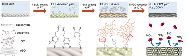
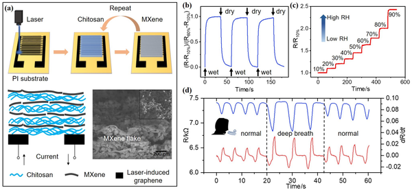
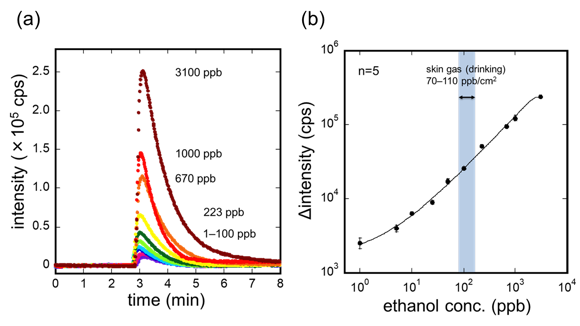
Figure 3. Selectivity and mechanical property of the DGY. (a) Normalized responses of the DGY under various gases exposure. (b) Responses of the DGY exposed to relative humidity (20-99 %). Each data point represents triplicate measurements. (c) Reponses of DGY upon repeated bending cycles up to 1000 times. The inset schematic shows a process of one cycle of bending test.

Uncaptioned visual Figure 4. Pratctical application test using DGY. The DGY was exposed to car exhaust; (a) gasoline and (b) diesel. (c) The response of DGY was changed after exposed to each car exhaust.

  1. Discussion

We demonstrated a wearable, disposable, cheap, and facile method e-textile gas sensor with improved sensing properties. The DGY seemed to have a possibility of being applicable for practical use by car exhaust test.


P4.113 A biocatalyst containing chitosan and embedded dye mediator adopted for promoting oxidation reactions and its utilization in biofuel cells

K. Hyun1, S. Kang1, Y. Chung2, Y. Kwon1
1Seoul National University of Science and Technology, Republic of Korea. 2Korea National University of Transportation, Republic of Korea

Abstract

Biofuel cells can be operated without membranes thanks to the glucose selectivity of enzymes. Since biofuel cells run on glucose, which is an abundant resource, they can be used as a new alternative to devices that use fossil fuels. However, enzymes have low reactivity and can't shuttle electrons alone. In this study, in order to solve the aforementioned disadvantages, a mediator (Methyl Red, MR) is adopted and affixed to an electrode to help electron shuttling. Not only does MR contain one azo and one carboxylic acid group which make the MR possible to be combined with chemicals per repeated unit, but also this MR is cheaper than other candidates, such as the osmium complex. Since the theoretical onset potential of the anodic reaction using MR is near 0 V vs. Ag/AgCl, it is expected that the sacrificed potential (overpotential) will be also comparable to that of other competitors.

In order to prevent leaching of the enzyme and MR, we used chitosan to entrap the GOx and mediators. As amino group of chitosan has interacted well with the hydroxyl groups of GOx, we predict chitosan could prevent leaching out of the enzyme complex. Through electrochemical analysis, we verified that not only does chitosan help attach MR onto the electrode, but excellent current density is also produced.


P4.114 The sensitivity improvement of bead-based sandwich assay sensor by charge confinement effect for the detection of Alzheimer’s disease

S. Roy1,2, K.S. Shin1, J.Y. Kang1,2
1Korea Institute of Science and Technology, Republic of Korea. 2University of Science and Technology, Republic of Korea

Abstract

Alzheimer’s Disease (AD) is the most prevalent neurodegenerative disorder, characterized by amyloid-beta (Aβ42) [1]ELISA is most popular immunoassay, but has detection limit above few tens of pg/ml. Since Ab is present in low concentration in blood, it lacks early detection of AD [2]To overcome this limitations, we propose a highly sensitive sandwich assay sensor consisted of small microwells (ø = 8 µm). Each microwell has an electrode pair located at the bottom (Fig. 1). The distance between each electrode pair is 2 µm, 500 times smaller than commercial Screen Printed Electrode (SPE). The implementation of microwell array (20 x 20) enhances the sensitivity by localizing the electric field on the captured magnetic beads between electrode pairs.


Surface-functionalized magnetic beads (2.8 µm) with anti-Aβ antibody and recombinant Aβ42 were mixed and incubated, followed by the reaction with detection antibody tagged by horseradish peroxidase (HRP). After washing with buffer, the bead solution was dropped onto the platform and external magnetic force is used to capture beads between the electrodes. A commercialized substrate (TMB/H2O2) solution was injected and incubated on the device before measurement(Fig.2).


The current responses for different concentrations of Aβ42 were observed by Cyclic Voltammetry (CV) and Linear Sweep Voltammetry (LSV) measurement (Fig.3). The CV data is compared with CV obtained from SPE. The current change is modulated by the change in concentrations. The signal of 1 pg/mL has large margin from negative control and predicted predicted to detect sub pg/mL with high signal to noise ratio, 10,000 time lower than that of SPE (LOD: 1ng/ml). Similar LOD was obtained from LSV measurement on our proposed device. The sensitivity is believed to be enhanced by reducing the diffusion length between the electrode pairs and charge confinement within the microwell.


 [1] "The Amyloid Beta Peptide: A Chemist's Perspective. Role in Alzheimer's and Fibrillization". Hamley IW, Chemical Reviews, 10, 112 (2012)

[2] “Amyloid Beta and Tau as Alzheimer’s Disease Blood Biomarkers: Promise From New Technologies.” Lih-Fen Lue, Andre Guerra & Douglas G. Walker, Neurology and Therapy volume 6, pages25–36(2017)


Uncaptioned visual


Fig. 1. (a) Commercial Screen printed electrode (WE = 1.6mm). The distance between WE and CE is 1mm (b) Scheme and setup of Bead-based Electrochemical Sensor with filed-focused microwell. The distance between each electrode pair is 2µm.


Uncaptioned visualUncaptioned visual

Fig. 2. (a) sandwich-type assay preparation of magnetic beads with capture antibody, oligomer Aβ as target antigen and HRP-detection antibody  (b) location of beads on the sensor device in the microwell 

Uncaptioned visual


Fig. 3. (a) CV response on commercialised SPE (LOD: 1ng/ml). (b) CV response on proposed bead-based sensor. The change in the CV graph shape was caused by the microwells. LOD is expected to be below 1 pg/ml. The highly sensitive detection was caused by faster diffusion between microelectrodes (c) LSV response on bead-based sensor also showed similar LOD.


P4.115 Advanced loop-mediated isothermal amplification actuated by a single self-priming phosphorothioated hairpin probe

J. Song, H.Y. Kim, Y. Jung, H.G. Park
KAIST, Republic of Korea

Abstract

We herein describe an ultra-simplified advanced version of Loop-mediated isothermal amplification (LAMP) actuated by only a single Self-priming Phosphorothioated hairpin probe (SPH-LAMP) for target nucleic acid detection. The hairpin probe (HP) employed in this strategy was designed to be opened through binding to target nucleic acid. Upon opening of HP, the self-priming domain (SP domain) within HP stem region is exposed and rearranged to serve as a primer. The following extension by DNA polymerase produces the extended HP (EP1) and displaces the bound target nucleic acid, which is then recycled to open another HP. The phosphorothioate (PS) modification at 5’ overhang and some loop part of HP should reduce the stability at the terminus of EP1 and consequently greatly enhance the formation of foldback hairpin, allowing the foldback extension. Since the PS modifications are always located at the same positions in the further extended hairpin probes (EP2, EP3, etc.), the foldback extension would be continuously repeated, consequently producing long hairpin concatamers. Based on this unique design principle, we successfully detected target DNA down to 115 zM under isothermal condition by employing only a single hairpin probe and eliminating the requirement for the complicated multiple primers in the conventional LAMP.


P4.116 Deep learning based lens-free shadow imaging platform (Cellytics)

S. Shin1, Y. Chae2, D. Seo3, H. Yang1, S. Myeong1, S. Oh3, M. Lee3, S. Seo1
1Korea university, Republic of Korea. 2Korea Institute of Science and Technology information, Republic of Korea. 3Korea Research Institute of Ships & Ocean Engineering, Republic of Korea

Abstract

Introduction: Recently, lens-free shadow imaging technology (LSIT) has been widely studied and applied to many cellular applications including cell counting, differentiation, size and morphology measurement, proliferation and viability testing, and etc. Since LSIT does not require any conventional optical lenses, it has many advantages such as cost-effectiveness, compactness, high-throughput, and focusing-free over microscopy based imaging. However, extraction of a specific shadow parameter corresponding to a specific cell’s property from cell shadow patterns, i.e., diffraction patterns, completely leans on human’s final decision with comparative experimental data. In this study, we demonstrate a deep-learning based lens-free shadow imaging platform, Cellytics, which can differentiate various sizes of microbeads and phenotypes of leukocytes.

Methods: We developed a deep learning based LSIT decision algorithm on the Alexnet model, a convolutional neural network, and applied to the Grad-CAM, a visual explanation algorithm via gradient-based localization (Fig. 1). And we also developed LSIT platform (Cellytics) along with dedicated cell chip, enabling automatic collection of shadow images of various micro particles and cells (Fig. 2). Shadow images of different sizes of polystyrene micro-beads (5, 10, and 20 um) and human leukocytes (CD4, CD8) were recorded with Cellytics and set to test the decision algorithm.

Results: Decision results of deep learning based LSIT platform for micro-beads and leukocytes are shown in Fig. 3. Automatically counted and cropped shadow images of polystyrene microbeads (5, 10, 20um, N=16,848) and leukocytes (CD4, CD8, N=3,736) were trained with the developed algorithm and the results showed 95.09% and 72.55% of detection accuracy for the micro-beads and leukocytes, respectively, in heterogeneous condition (Fig. 3).

Discussion: Deep learning based lens-free shadow imaging platform, Cellytics, holds a great promise in rare cell detection, e.g., circulating tumor cells, as well as in cell based diagnostics.

Acknowledgments:

This study was supported by the Basic Science Research Programs (Grant#: 2014R1A6A1030732, Grant#: 2017R1A2B4005499) through the National Research Foundation (NRF) of Korea. The authors also acknowledge the support of the projects titled ‘Development of Management Technology for HNS Accident’ and ‘Development of Technology to Support the Rapid Search and Rescue of Marine Accidents (Grant#: PES3130)’, of KRISO funded by KIMST.

Uncaptioned visual

Fig. 1 Decision algorithm of deep learning based lens-free shadow imaging platform

Fig. 2 Developed lens-free shadow imaging platform (Cellytics) (a) Cellytics and dedicated cell chip, (b) generation principle of lens-free shadow image, and (c) developed UIs for Cellytics

Uncaptioned visual

Fig. 3 Decision results of deep learning based LSIT platform for various micro-objects. (a) Cropped shadow images of polystyrene microbeads (5, 10, 20um) and leukocytes (CD4, CD8), (b) automatically extracted characteristics of shadow images after training with Alexnet and Grad-CAM, and (c) detection results for microbeads and leukocytes.


P4.117 Advances of Gold Nanoparticles for Bioimaging: Strategies and Future Outlooks

Ruth Stephanie, Su Min Kim, Sung Hyun Kim, Dae Yeon Lee, Min Woo Kim, Chan Yeong Park, Tae Jung Park
Chung-Ang University, Republic of Korea

Abstract

Gold nanoparticles (AuNP) are one of the most highly investigated, hence well-established types of nanomaterial. Its versatility has found its prominent role in diverse fields, including in bioimaging field. Their attractiveness is thanks to their tunable optical properties derived from the surface plasmon resonance (SPR) phenomenon, which enables them to have a wide-ranging variety of characteristics. In the context of bioimaging purposes, tissue transparency is often being the requirement for the operation. However, this characteristic is one of the constantly challenging requirements for AuNP to fulfill, as investigations in this topic remain continuing. For that purpose, this review is aimed to summarize (1) the basic information regarding gold nanoparticles and their importance to have tissue transparency properties; (2) recent advances in approaches and synthesis strategies to acquire AuNP with tissue transparency features; and (3) summarize the current progress, challenge, and future outlooks of AuNP with tissue transparent features for various imaging modalities.


P4.119 Development of graphene oxide nanoribbon-based organic electrochemical transistors

C-L. Sun1,2, C-H. Lin1, H-Y. Chen1, C-H. Kuo1, B-S. Lin1
1Chang Gung University, Taiwan. 2Linkou Chang Gung Memorial Hospital, Taiwan

Abstract

Since 2011 we started to study graphene oxide nanoribbons (GONRs) towards numerous applications. [1-13] In this presentation, we synthesized graphene oxide nanoribbons (GONRs) unzipped from multiwalled carbon nanotube (MWCNT) under different microwave powers. We used the MWCNT and various GONRs to modify the polystyrenesulfonate-doped poly(3,4-ethylenedioxythiophene) (PEDOT:PSS) thin film as the hybrid semiconductor layer of organic electrochemical transistors (OECTs). For the device fabrication, non-vacuum processes are adopted to prepare these OECTs and their device performances are compared. It was found that only the P-GONR20-100W device is better than the P device in terms of Id-Vd measurement. Furthermore, for the detection of dopamine (DA) all nanocarbon-modified devices are better than the original unmodified one. These results implied that our GONRs are holding great potential for developing advanced OECTs for biosensing.

References

[1] C. L. Sun et al. ACS Nano 5 (2011), 7788.

[2] L. Y. Lin et al. J. Mater. Chem. A 1 (2013), 11237.

[3] Y. J. Lu et al. Carbon 74 (2014), 83.

[4] M. H. Yeh et al. J. Phys. Chem. C 118 (30) (2014), 16626.

[5] C. L. Sun et al. Biosens. Bioelectron. 67 (2015), 327.

[6] X. Lu et al. J. Mater. Chem. A 3 (2015), 13371.

[7] R. Zhang et al. Anal. Chem. 87 (2015), 12262.

[8] Y. W. Lan et al. Nano Energy 27 (2016), 114.

[9] H. Y. Chen et al. Adv. Mater. Interfaces (2016), 1600357.

[10] C. H. Su et al. ACS Omega 2(8), (2017), 4245.

[11] T. E. Lin et al. Angew. Chem. Int. Ed. 56 (2017), 16498.

[12] R. Zhang et al. Carbon 126 (2018), 328.

[13] C. H. Su et al. J. Taiwan. Inst. Chem. Eng. 95 (2019), 48.


P4.120 High-performance polymer microneedle biosensor for continuous lactate monitoring

S. Seonu, H. Yoon, A. Zahed, Y. Shin, D. Kim, J. Park
Kwangwoon University, Republic of Korea

Abstract

Since lactate is a significant analyte for clinical care, it is widely used as an indicator of some pathological conditions and treatments. In addition, subtle changes may cause several sudden fatal effects such as lactic acidosis, endotoxic shock, and cardiac diseases on patient conditions. Thus, the continuous monitoring of lactate in interstitial fluid is highly effective to diagnose and manage critical patients suffering from the diseases above mentioned[1-2]. Specifically, this lactate control disorder results in a rapid increase to more than 5 mM[3]. However, as shown in Table1, previous studies of lactate sensors were not enough to measure this range. 

 In this work, we developed a microneedle-based biosensor with high sensitivity and a wide linear range for continuous lactate monitoring. The biosensor was fabricated by microelectromechanical system(MEMS) fabrication technologies and a femtosecond laser. Reduced graphene oxide(rGO) was drop-casted on the working electrode. After the electrodeposition of platinum nanoparticles(PtNPs), It was soaked in 3-mercaptopropionic acid(3-MPA) to make the carboxyl functional group. Finally, lactate oxidase was immobilized and nafion was coated on the electrode.

 Fig.1(a) illustrates the fabrication process of the microneedle biosensor. Fig.1(b) is the functionalization process of the electrode. Fig.2(a) and (b) depict a schematic and an optical image of the biosensor, respectively. Fig.3(a) and (b) show the amperometric responses of high and low lactate concentrations. Fig.3(c) indicates the linear regression analysis of the current responses. The as-fabricated biosensor exhibited an outstanding linear range of 0 to 6mM and a sensitivity of up to 18.21µA/mM·cm2. The detection limit and the response time were calculated to 8.3µM and 7sec, respectively (Fig.3(d)). Fig.3(e) shows the current response to several interferences and Fig.3(f) shows the stability test for 5 days, where the sensitivity variation is less than 8%.


Table 1. Performance comparison of state-of-the-art lactate biosensors

Electrode Materials

Limit of detection

(µM)

Sensitivity

(µA/mM·cm2)

Linear range

(mM)

Ref.

PPD-LOx/PB/PET

50

0.553 (µA/mM)

0.1 - 1

[4]

LOx/PBnc/SPCE

10

6.379

0.01 - 0.5

[5]

LOx-FSM8.0/Nafion/CoPC-SPCE

18

4.54

0.02-1.5

[6]

Nafion/LOx/3-MPA/PtNPs/rGO/Au

8.3

18.21

0.1 - 6

This work

* PPD: poly-orthophenylenediamine, PB: prussian blue, PET: polyethylene terephthalate, PBnc: prussian blue nanocubes, FSM8.0: mesoporous silica, CoPC-SPCE: screen-printed carbon electrode containing cobalt phthalocyanine, CNT: carbon nanotube, 4-ATP: 4-aminothiophenol


Uncaptioned visual

Figure 1. (a) MEMS fabrication process of the microneedle biosensor. (b) The functionalization process of the working electrode for making a lactate biosensor.


Uncaptioned visual

Figure 2. (a) A scheme of the microneelde biosensor to monitor lactate in interstitial fluid and (b) its optical image.


Uncaptioned visual

Figure 3. Electrochemical analysis of the fabricated lactate biosensor. (a) and (b) Amperometric responses of high and low concentration change of lactate, respectively. (c) Linear regression analysis of the current response in the range of 0.1 mM to 6 mM. (d) Amperometric response for confirming the LOD and response time of the biosensor. (e) Selectivity test of the lactate biosensor for 500mM lactate in presence of 10 µM of ascorbic acid (AA); 50 µM of uric acid (UA); 50 µM of Glucose (Glu); 5 µM of acetaminophen (AP). (f) Long term stability test by comparing the sensitivities for 5 days. 


Acknowledgments: 

This research was funded and conducted under ‘the Competency Development Program For Industry Specialists’ of the Korean Ministry of Trade, Industry, and Energy (MOTIE), operated by the Korea Institute for Advancement of Technology(KIAT). (No. P0002397, HRD program for Industrial Convergence of Wearable Smart Devices) and the Technology Innovation Program (20000773, Development of nanomultisensors based on wearable patch for nonhematological monitoring of metabolic syndrome) funded by the Ministry of Trade, Industry & Energy (MI, Korea).


Reference: 

[1] Rober D. Crapnell, et. al., MDPI sensors 2020, 21(3), 879

[2] Paolo Bollella, et. al., Biosens. Bioelectron. 2018, 123, 152-159

[3] M. Adeva-Andany, et. al., Mitochondrion 2014, 17, 76-100

[4] Jayoung Kim, et. al., Royal Society of Chemistry 2014, 139, 1632-1636

[5] Danfeng Jiang, et. al., Sensors and Actuators B: Chemical 2016, 228, 679-687

[6] Takeshi Shimomura, et. al., Analytica Chimica Acta 2012, 714, 114-120


P4.122 Detection of Escherichia coli based on FRET by graphene oxide and fluorescence-tagged aptamer

H.K. Kim1,2, H.R. Kim2,3, J.B. Kim1, B.C. Kim2,3
1Korea University, Republic of Korea. 2Korea Institution of Science and Technology, Republic of Korea. 3University of Science and Technology, Republic of Korea

Abstract

Bacterial contamination in drinking water and food is a serious problem for public health safety. Escherichia coli (E. coli) is an indicator of contamination in water and food, although not all E. coli harm the human. Therefore, rapid and accurate detection of coliform/E. coli is important for environmental and food safety. The FRET (Fluorescence Resonance Energy Transfer)-based biosensor can deal with these requirements. We used graphene oxide (GO) as an energy acceptor in this study. In the absence of the target cells, the intensity of fluorophore 5-Carboxyfluorescein (FAM) tagged to aptamers (FAM-aptamer) is weak when FAM-aptamers bind to GO because of FRET. If the FAM-aptamers bind to the target cells instead of GO because of high affinity and specificity, this leads to the recovery of the fluorescent intensity of FAM. We used a specific aptamer that targeted E. coli. We found the optimal concentration of GO and FAM-aptamer pairs to address FRET. Under the optimal condition, fluorescence intensity shows the increment as increasing the number of E. coli from 103 to 107 CFU·mL−1. The GO/FAM-aptamer based FRET sensor could be an effective way to measure E. coli concentration in foods or drinking water and, applicable to the detection of other bacteria.


P4.123 A Single Tube Assay using Azobenzene Functionalized Magnetic Nanoparticle for Rapid and Ultrasensitive Pathogenic Bacteria Detection

Feixiong Chen, TAEYOON LEE
Chungnam National University, Republic of Korea

Abstract

A single tube assay was developed to address the current issues of pathogenic bacteria diagnosis, limited by insensitive, labour-intensive and time-consuming nature. We present two strategies in a single tube assay to allow bacteria enrichment by hydrophobic effect and DNA extraction via electrostatic interaction, respectively. Thus, we engineered magnetic nanoparticles (MNPs) with a novel probe to display hydrophobicity that drives azobenzene-functionalized MNPs (Azo@MNPs) adsorbing onto hydrophobic bacterial surface. Next, after bacteria lysis, Azo@MNPs allowed the binding of DNAs by electrostatic interaction under a pH control, which performed DNA recovery serving to direct nucleic acid amplification test. We demonstrate the detection of S. Typhimurium, B. ovis, E. coli, with the detection limit of 5CFU/ml, 10CFU/ml and 5CFU/10mL urine by using our assay, respectively. Furthermore, via UV light trigger, the molecular structure of azobezene can be changed from trans-Azo@MNPs to cis-Azo@MNPs, so that these cis-Azo@MNPs with the enhanced surface hydrophobicity exhibited a unique biological performance of higher bacteria binding and lower DNA adsorption level, compared to that of trans-Azo@MNPs in our work, which further improve the diagnostic behavior of our assay. This assay can be performed in less than 90 mins and is robust to clinical urine and it combines dual-strategies of Azo@MNPs in bacteria enrichment and DNA extraction with nucleic acid amplification test, to achieve the simple, sensitive, and versatile diagnosis of bacterial species.


P4.124 Isolation and selection of aptamers specific for cell surface molecules of foodborne pathogens using whole cell-SELEX

T.T.Q. Nguyen, N.E. Lee, E.R. Kim, M.B. Gu
Korea University, Republic of Korea

Abstract

Food poisoning caused by the ingestion of food contaminated with harmful pathogens is one of the most important public health issues, affecting both developing and developed countries. Hence the control of foodborne pathogen outbreaks has been receiving global attention, developing a rapid and point-of-care (POC) method for pathogen detection which is proposed as alternative and innovative sensors over traditional methods is needed. To deal with this issue, biosensors using DNA/RNA aptamers as a sensing element known as an aptasensor have been rapidly developed for detecting foodborne pathogens. Aptamers are single-stranded DNA or RNA which can be obtained from Systematic Evolution of Ligands by Exponential enrichment (SELEX) and bind to their targets with high affinity and specificity. In this research, an improved SELEX method of using live bacteria cells has been applied successfully in selecting specific aptamers against cell surface molecules of whole-cell bacteria. A total of ten rounds of cell-based SELEX with target and three rounds of negative selection with counter targets mixture was performed to obtain specific aptamers. FAM-labeled aptamer pools of selection were tested for their binding to the target cells using fluorescence spectroscopic analysis for each round and flow cytometry in the later stage of selection. After 10 rounds of selection, the selected aptamer pools were cloned, sequenced and then divided into different families according to their sequence homology and secondary structure. For characterization of aptamer candidates, fluorescence spectroscopic assay and flow cytometry analysis was conducted to evaluate the binding affinity and specificity. These aptamers could be further implemented to the foodborne pathogens biosensing application which enables simple, rapid, and robust for detection.


P4.125 Sandwich-type electrochemical aptasensor for detecting avian influenza virus

T.T.T. Dang, B.H. Lee, S.H. Kim, M.B. Gu
Korea University, Republic of Korea

Abstract

In this study, we report a sandwich-type electrochemical aptasensor using a cognate pair of aptamers for avian influenza virus detection. Outbreaks of bird flu by avian influenza virus cause severe economic losses in the poultry industry every year. To deal with this problem, the development of sensitive biosensor for avian influenza virus is needed. Aptamer has been attracted great interest because of its high affinity and specificity. Moreover, cognate pair of aptamers, binding to target simultaneously, enable sandwich-type detection which could provide high sensitivity and stability to the sensor. Therefore, we developed a cognate pair of aptamer-based electrochemical aptasensor for highly sensitive detection of avian influenza virus.

The cognate pair of aptamer-based electrochemical aptasensor was fabricated by immobilizing the primary aptamer on the screen-printed gold electrode surface. Then 6-Mercapto-1-hexanol and human serum albumin were used as blocking reagents. For the detection of influenza virus, secondary aptamer conjugated with horseradish peroxidase was mixed with the target virus, then incubated on the gold electrode. 3,3’,5,5’-tetramethylbenzidine, as a redox mediator and electrochemical signal was measured using chronoamperometry technique. 

For the detection of influenza virus, the secondary aptamer conjugated with horseradish peroxidase and target mixture was incubated with primary aptamer immobilized electrode. The fabrication of the sandwich-type aptasensor was electrochemically characterized using cyclic voltammetry. The decrease of the peak current along with increase of peak-to-peak separation was observed, which showed the successful working of this biosensor. The analytical performance of this aptasensor was tested with different virus concentrations. By measuring chronoamperometry, the peak current was gradually increased with the increase in target concentration. The sandwich-type aptasensor platform showed a significant response to the target virus.

This work provides a promising sandwich-type aptasensor for detecting influenza virus with high sensitivity.

Uncaptioned visual


P4.126 The design and model of nanoscale-gap electrode for electrochemical sensing

Q-Z. Xie, H-T. Hsueh, P-H. Chen, C-T. Lin
National Taiwan University, Taiwan

Abstract

Introduction: In this work, a co-planar nano-gap electrode design/model was proposed and validated. Utilizing surface modifications on the nano-gap surface, a side-wall electric double layer (EDL) model was experimentally demonstrated. Based on this work, the sensitivity of electro-impedance measurement can be improved by nano-scale electrode designs.

Methods: To validate the proposed design of nano-gap electrode, the nano-gap electrode was fabricated by e-beam lithography with evaporation deposition of 50 nm Cr/Au as shown in Fig. 1. The gap surface was sequentially modified by aminopropyldimethylmethoxysilane (APDMMS), glutaraldehyde, and 3-aminobenzoic acid, as shown in Fig. 1. In other words, the gap surface was positively charged, neutral, and negatively charged, respectively. At each modification stage, the electro-impedance spectroscopy was employed to elucidate the relationship between measured impedance and gap-surface charges.

Results: The experimental electro-impedance spectrum can be shown as Fig. 2. The experimental result indicates that there are two main frequency domains (domain 1 and domain 3) which correspond to two different type of capacitive, e.g. double layer capacitor (EDLC) on electrode surfaces (domain 1) and PBS buffer solution capacitance (domain 3). These experimental results show gap-surface potential mainly modulates the EDL region. Since the modification is on the gap surface, this indicates the gap-surface potential modulates the EDLC of electrode surfaces. As a consequence, the effects of side-wall EDL model can be established.

Discussion: The surface-potential change induces the ion re-distribution on the top of the gap surface between two nearby electrodes. The phenomena of ion re-distribution modulate the EDL of electrode sidewall and changes its EDLC. Based on this, a two-path circuit model (Fig. 3) can be established to illustrate the gap-surface potential effect based on different gap-width experiments. Therefore, narrow-gap devices have better incremental capacitive response than wide-gap device do. And devices with nano-scale gap width have better sensing responses.

Uncaptioned visual

Figure 1

Uncaptioned visual

Figure 2

Uncaptioned visual

Figure 3


P4.127 Laser carbonization graphene and MXene-polyaniline composite based wearable patch sensor for personal skin pH real time monitoring

S. Yoon, H. Yoon, Md. Sharifuzzaman, J. Nah, J. Park
Kwangwoon University, Republic of Korea

Abstract

In this study, we have successfully developed flexible and stretchable sensor for skin pH real time monitoring without sweat. The pH-sensitive electrode was fabricated by laser carbonization graphene (LCG) that are subsequently permeated with MXene-Ti3C2Tx and polyaniline(PANI) as the conductive filler, binding material, and pH-sensitive membrane [1].

Since pH is a leading indicator of skin health, it is very important to measure personal skin pH levels and create customized product regimens to better care for skin. When pH balance is compromised, whether through environmental factors and underlying conditions, it can trigger inflammatory responses resulting in dryness, eczema, and atopic dermatitis.

Figure 1 shows the conceptual schematic of the proposed sensor. MXene-Ti3C2Tx offers flexibility and metallic conductivity [2]. PANI is highly selective for H+ ions against major interfering ions (Li+, Na+, K+). H+ ions present in the skin are selectively passed through the PANI membrane [3]. These H+ ions generate the redox potential on the working electrode. Figure 2 shows schematic diagram of fabrication process. LCG was formed through laser scribing and Laser cutting was carried out by aligning the polydimethylsiloxane(PDMS) onto the LCG. MXene-Ti3C2Tx was drop-casted on the working electrode and Ag/AgCl paste was applied as reference electrode. The sensor fabrication was then completed by electroplating PANI using cyclic voltammetry. Figures 3(a) and (b) show the fabricated sensor and its attachment on real skin. The sensor exhibited 135% stretching ability as shown in Figure 3(c). Figure 4(a) shows electroplating condition of PANI. Figure 4(b) shows CV for different electrodes in 2 mM K3[Fe (CN)6] with 0.1 M KCl under scan rate of 50 mV/s. Figure 4(c) shows calibration curve of the sensor. It exhibited the sensitivity of 52.5 mV/pH. Figure 4(d) shows a real time test result. In near future, wearable and battery-free skin pH monitoring system with NFC will be implemented.

REFERENCES :

[1] Rahim Rahimi, Manuel Ochoa, Ali Tamayol, Shahla Khalili, Ali Khademhosseini and Babak Ziaie, “Highly Stretchable Potentiometric pH Sensor Fabricated via Laser Carbonization and Machining of Carbon−Polyaniline Composite”, ACS Appl. Mater. Interfaces 2017, 9, 9015−9023

[2] Zheng Ling, Chang E. Ren, Meng-Qiang Zhao, Jian Yang, James M. Giammarco, Jieshan Qiu, Michel W. Barsoum, and Yury Gogotsi, “Flexible and conductive MXene films and nanocomposites with high capacitance”, PNAS, 111(47), 16676-16681

[3] M. Kaempgen, S. Roth, “Transparent and flexible carbon nanotube/polyaniline pH sensors”, Journal of Electroanalytical Chemistry, 586(2006), 72–76

Uncaptioned visual

Figure 2. Conceptual schematic of skin pH monitoring sensor.

Uncaptioned visual

Figure 1. Schematic diagram of fabrication process; (a) CO2 laser scribing, (b) Pouring and detach Polydimethylsiloxane, (c) CO2 laser cutting, (d) Skin pH sensor patch formation, (e) MXene drop casting and Ag/AgCl pasting, (f) PANI electroplating to complete the flexible skin pH sensor.

Figure 3. Optical image of the fabricated skin pH sensor; (a) Fabricated pH sensor, (b) Sensor attached on real skin, (c) Reliable sensor under 135% stretching condition.

Uncaptioned visual

Figure 4. (a) PANI electroplating using CV, (b) CV for different electrodes in 2 mM K3[Fe(CN)6] with 0.1 M KCl @ Scan rate: 50 mV/s, (c) Open circuit potential with pH of 3, 4, 5, 6, 7, respectively, (d) Real time test for personal skin pH monitoring.

Acknowledgements:

This research was supported by the Technology Innovation Program (20000773) by the Ministry of Trade, Industry & Energy (MI, Korea) and the Bio & Medical Technology Development Program (NRF- 2017M3A9F1031270) by the Korean government (MSIT).


P4.128 Aptamer-based electrochemical biosensing of dopamine using gold LTCC chips

S-H. Park1,2, D. Muaz3,4, J. Lee1,2, B. Natalia3, J. Opitz3, J-S. Yeo1,2
1School of Integrated Technology, Republic of Korea. 2Yonsei Institute of Convergence Technology, Republic of Korea. 3Fraunhofer Institute for Ceramic Technologies and Systems IKTS, Germany. 4École polytechnique fédérale de Lausanne-EPFL, Switzerland

Abstract

Dopamine (DA) is a neurotransmitter molecule with a variety of functions in the neuronal signal transduction and several critical illness. DA levels are related to the severity and progression of neurological disorders such as Parkinson's disease, attention deficit hyperactivity disorder, and Huntington's disease [1]. Furthemore, electrochemical sensing requires simple and low-cost set up and also spends few seconds time scale so that it shows great advantages for POCT (Point-of care testing). In this work, we demonstrated methylene blue (MB) reported DNA-aptamer as an electrochemical indicator to detect with label-free and regenerative characteristics. Aptamer formed by oligonucleotide acids may lose its thermally stable functionality irreversibly under electrochemical reaction and moreover are able to recover its function by self-refolding.
We developed aptamer based electrochemical biosensing of dopamine with enhanced signal using LTCC chip and functionalized MB. Aptamer probe containing 54 bases was synthesized with MB at the end of the 3' of the sequences, and functionalized with thiol at the end of 5' of the sequences. MB-anchored aptamer probe can be forced to associate from the interface after structure switching of the aptamer by combining with DA. Aptamer has a smaller molecular size than conventional receptors, therefore, the captured DA molecules are closer to the substrate of LTCC chip during the sensing, resulting in a stronger electrochemical field exerted from the MB which gives detection signals.

We use 6-MCH for passivation layer to make optimal concentration of the aptamer for its structural folding. The figure shows CV (Cyclic voltammetry) and DPV (Differential pulse voltammetry) response of LTCC chips. The 5' terminal of aptamer strand is modified with thiol, which enables chemisorption to the surface via SAM chemistry. Current peak shift is the largest at 10 𝝁M passivated, because optimized passivation concentration lead proper distance between aptamers so that it guarantees structural deformation.
The chips passivated with 6-MCH, detect the dopamine concentration 1, 5, 10, 20, 50 𝝁M respectively. The current change at redox peak shows a linear region at 1 to 20 𝝁M concentration and is saturated at 50 𝝁M. They show significant decrease in current with a relative signal change of 3, 6.1, 8.2, 10.2, 12.2 %. We demonstrate the electrochemical dopamine sensing platform based on MB reported aptamer with < 1𝝁M limit of detection.


P4.129 Stem cell-laden hydrogel microelectrode arrays fabricated by enzyme-mediated transfer printing for neuromodulation and cell therapy

S-H. Li, Y-X. Lin, W-C. Huang
National Yang Ming Chiao Tung University, Taiwan

Abstract

Implantable neural prosthetics offering neural signals recording and electrical stimulation have been successful in the treatment of multiple neurological diseases. The device enables a real-time neuromodulation, while the neuroreplacement or neuroregeneration is a critical goal for rehabilitation in nervous injury. Recent research is targeted in the development of hydrogel-based microelectrode arrays (MEA) due to the ultracompliant mechanical properties with that of local nerve tissue to reduce the adverse tissue responses and enhance neural signal transduction. Herein, we developed a biodegradable adhesive hydrogel microelectrode array (MEA) composed of extracellular matrix (ECM) component that can not only permit the neural signal tracking and neuromodulation, but also can provide a microenvironment that can ladening neural stem cells for promoting local tissue regeneration. Copolymerization of gelatin methacrylate and poly(3,4-ethylenedioxythiophene) led to the formation of a conductive hydrogel, called PDGA, which demonstrates both tissue-mimicked properties and hydroresponsive enhancement of electrical conductivity. By photopolymerization, the PDGA was directly micropatterned into a 7-channel electrode array with an electrode size of 50 μm. Furthermore, a receiver substrate was constructed with Transglutaminase (mTG)-crosslinked gelatin which exhibits temporally-controlled sol-gel phase transition and adhesion, enabling transfer printing prefabricated PDGA microelectrodes. Covered with PLGA as the passivation layer, the integrated final device demonstrated tissue-like structural and mechanical properties, showing a low impedance value compatible for neural stimulation. The adhesive nature permits the device to be conformably immobilized on the curvilinear tissues, whereas the tissue-mimicked microenvironment enables the in-situ adhesion, growth, and successfully differentiation of neuron stem cells into neuron cells. This adhesive tissue-mimicked cell-incorporated MEA can not only be immobilized well on the hydrated curvilinear nerve tissue surface, but can also permit electrical stimulation and recording to promote in-situ nerve activation and stem cell delivery, which was expected to be a next-generation smart neural implant to promote nerve regeneration.


P4.130 An instrument-free quantitative immunoanalysis kit with a specially patterned lateral flow membrane

K.W. Lee, K.Y. Jeong, K.R. Kim, H.J. Chun, D. Lee, E.K. Yang, H.C. Yoon
Ajou University, Republic of Korea

Abstract

In this study, we developed a membrane-based instrument-free optical immunosensor for the detection of microalbumin in the urine. In traditional colorimetric lateral flow immunoassay (LFI) using gold nanoparticles (AuNPs) as a probe, additional optics are required to quantify the colored signal of the test line because it presents as a single red line. Since the necessity of an additional optical transducer is one of the factors that makes it difficult to diagnose the disease in the field of resource-limited conditions, the device that can quantify the biomarker without optical instrument is essential. To exclude the external optical transducers, the signal on the test line should be quantifiable by naked eyes without the involvement of optical instruments. Given this objective, the test line of conventional LFI that prepared as a single line was modified to several herringbone-patterned spots for the construction of semi-quantitative lateral flow immunoassay (SQ-LFI). When the immunoassay was performed on the SQ-LFI by using AuNP as an optical probe, spots were colorized and the numbers of colored-spots were increased according to the increment of analyte concentrations. By counting the number of colored spots, the analyte concentration can be estimated with the naked eye. For the demonstration of the applicability of developed sensor, microalbumin which is a diagnostic marker for renal failure was analyzed by using the microalbumin-spiked artificial urine samples. Using the developed SQ-LFI immunosensor, the calibration results for microalbumin were obtained ranging from 0 to 500 μg/mL that cover the clinically required detection range. As this result, we expect that a membrane-based SQ-LFI would be an efficient diagnostic device in the resource-limited conditions.

Uncaptioned visual


P4.131 RNA aptamer as bio-amplifier for detection of amyloid-beta in human serum by field-effect transistor immunosensors

C.A. Vu, W.Y. Chen
National Central University, Taiwan

Abstract

Sensing proteins by Silicon Nanowire Field-Effect Transistor (SiNW-FET) immunosensors produces inconsistent signal due to non-uniformity of the surface chemistry after modification and detection. R18, an RNA aptamer targeting rabbit Immunoglobulin G (IgG), is not only able to stabilize current variations but also capable of enhancing electrical signal recorded from SiNW-FET immunosensors. In this report, we present an application of aforementioned strategy by designing SiNW-FET immunosensors to detect Amyloid β 1-42 (Aβ 1-42) in human serum (HS). Herein, mouse Immunoglobulin G1 (IgG1) was immobilized on the surface of SiNW-FET to capture Aβ 1-42 in HS before incubating with rabbit IgG and R18 for signal stabilization and amplification. Statistical data reveal that the developed nanosensors succeeded in recognizing Aβ 1-42 in HS at low concentration. The proposed method is potentially applicable for assays which diluting analytes is impossible and operation in serum samples is mandatory.


P4.132 Immunomagnetic microfluidic integrated system for potency-based multiple sorting of heterogeneous stem cells with high throughput abilities

B. Kang1, S. Han2, S. Haam2, E-K. Lim1
1KRIBB, Republic of Korea. 2Yonsei University, Republic of Korea

Abstract

Multipotent adult stem cells (MASCs) derived from Pluripotent stem cells (PSCs) have found widespread use in various applications, including regenerative therapy and drug screening. For these applications, highly pluripotent stem cells need to be selectively separated from those that show low pluripotency, and adult stem cells need to be collected for further application. Although fluorescent activated cell sorting (FACS) and magnetic activated cell sorting (MACS) are commonly used, these methods suffer from limitations namely low throughput efficiency and low purity, respectively. In this study, we developed immunomagnetic microfluidic integrated system (IM-MIS) for separation of stem cells depending on potency level. A mixture of PSCs and MSCs were used to demonstrate separation efficacy of IM-MIS. Compared to FACS, IM-MIS was found to be more efficient and rapid method with better throughput capabilities. Moreover, separation using IM-MIS was more accurate than that done using MACS. Furthermore, IM-MIS did not affect the key characteristics of stem cells including its potency, phenotype, genotype, and karyotype. IM-MIS may offer a new platform for the development of multi-separation systems for diverse stem cell applications.


P4.135 Dynamic characteristic analysis on the coupling of smart glasses and finger sensor for continuous dietary monitoring

W.K. Hong, W.G. Lee
Kyung Hee University, Republic of Korea

Abstract

Continuous dietary monitoring is a significant factor and its needs have been increasing, since knowing both intake food type and mass plays an essential role in revitalizing human healthcare and physical conditions. Here, we report a hybrid study that can combine smart glasses technologies (to analyze intake patterns) with finger sensors (to detects intake mass) for continuous dietary monitoring. To demonstrate our proof-of-concept validation, we built up a homemade smart glasses device that can detect facial activities (e.g., wink and mastication) moved by temporalis muscle and then amplify the force driven by their minute movements. We built a load cell onto the glasses hinges for force amplification based on the leverage mechanism (called Type 2 in our case) and placed an inertial measurement unit(IMU) onto the temple for head motion analysis. We then analyzed the patterns for buccal movement via a support vector machine(SVM) classifier. The sensor inputs can be mainly categorized into two parts: i) intake pattern from the smart glasses and ii) intake mass from finger sensor (by flexible force sensor). The results showed that after the process of classification and extraction of feature vectors for SVM, the accuracy of classification for the facial activities finally came to 0.982 in terms of testing the average F1-score (Figure 1A). The results also showed great potential for hybrid feasibility that the correlation of analog-to-digital converter(ADC) was proportional to the food mass delivered in spoon by R2=0.988 (Figure 1B). Therefore, we believe that this approach is potentially beneficial for developing a continuous dietary monitoring platform for personalized prevention of insulin resistance-related metabolic risks or diseases through continuous monitoring and automated management of food intake and mass in a quantitatively network-controlled manner.

Uncaptioned visual

Figure 1. A representative classification results data and measurement plot (A) Feature vector correlations of several facial activities, (B) ADC correlation of intake food mass in spoon measures by finger sensor


P4.136 Indirect quantitation of Aspergillus niger by electrochemical micro biosensors

J. Lee, H. Choi, J. Rye, A. Kang, S. Kim, W. Yun
Sungkyunkwan University, Republic of Korea

Abstract

We report on the electrochemical biosensor for detecting fungi, Aspergillus niger (A. niger), not directly from the fungi sample but indirectly from its filtrate. The redox signal from the interdigitated electrodes (IDEs) with a micrometer-scale gap where the antibody was immobilized to capture a fungi-derived antigen in the sample solution, was used for this study. It decreased upon antigen immobilization and showed a dependence on the initial concentration of A. niger. To our surprise, when applied in cyclic voltammetry (CV), electrochemical impedance spectroscopy (EIS), and differential pulse voltammetry (DPV), the indirect quantitation on the filtrate showed an improved quantitative behavior compared with the pristine one. Furthermore, the filter pore size dependence was also observed. It was found that too fine pored filter seems to remove too much from the original fungi solution, leading to the loss of concentration dependence in the detected signals. The result was discussed with the data from culturing the fungi, Infrared (IR) spectroscopy, biolayer interferometry (BLI), scanning electron microscopy (SEM) and matrix assisted laser desorption/ionization (MALDI) measurements.


P4.137 Hydrogel-based multiple SNP genotyping with toehold switch-mediated fluorescence turn on

W. Choi1,2, E. Park1,3, J. Choi2, H. Lee1, N. Choi1
1KIST, Republic of Korea. 2Korea University, Republic of Korea. 3Seoul National University, Republic of Korea

Abstract

Introduction: Single nucleotide polymorphisms (SNPs) are DNA sequence variations that occur when a single nucleotide is altered. The most widely used method for SNP genotyping is allele-specific PCR. However, this approach is relatively time-consuming due to many thermal cycles and exhibits limited applicability for point-of-care because of the typical size of PCR instruments. Here, we present a hydrogel-based multiplex SNP genotyping with the combination of the toehold switch-mediated fluorescence turn on and the sandwich assay. By immobilizing toehold probes within a hydrogel, we successfully discriminated against multiple SNPs simultaneously. Moreover, we utilized an isothermal sandwich assay for sample preparation, which does not require complicated DNA purification and bulk apparatuses.

Methods: Hydrogel microparticles were fabricated by the stop flow lithography (SFL) with polyethylene glycol diacrylate (PEGDA). The sandwich assay was performed with both 2.8 µm magnetic microparticles, and 50 nm gold nanoparticles (AuNPs) functionalized with invader DNA oligomers to cooperatively capture DNAs containing SNP sites. Reporter DNA probes conjugated with a fluorophore were immobilized within PEGDA microparticles, followed by hybridizing quencher DNAs to mute fluorescence. A significant number of invader DNAs were recovered from the AuNPs after the sandwich assay and then incubated with the hydrogel microparticles. Finally, fluorescence images from the microparticles were acquired.

Uncaptioned visual
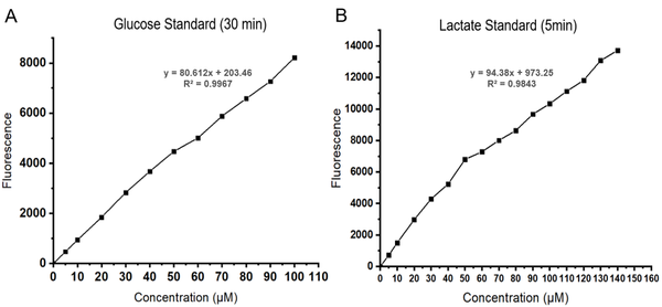
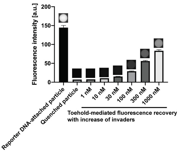
Results: After performing the sandwich assay isothermally at room temperature to capture SNP-containing DNA targets, resultant invader DNAs served effectively as amplified targets. We confirmed that the quenching efficiency between the quencher DNAs and the reporter DNAs immobilized within the hydrogel was over 97%. The recovery of fluorescence from the microparticles increased proportionally with the concentration of the invaders (Figure 1). We also verified the capability of multiplex SNP genotyping without false-positive (non-specific) signals.

Discussion: We have presented a hydrogel-based multiplex SNP genotyping with versatile and straightforward sample preparation. Our work can pave the new method for faster and more accurate SNP genotyping.

Figure 1. Fluorescence changes of hydrogel microparticles


P4.138 Electrochemical substrate for active profiling of cellular surface leucine aminopeptidase activity and drug resistance in cancer cells

S.T. Huang1, T.S.T. Balamurugan1, G.Z. Chen1, C.M. Lin2
1National Taipei University of Technology, Taiwan. 2Taipei Medical University, Taiwan

Abstract

Leucine aminopeptidase (LAP) is an essential proteolytic enzyme, and potential biomarker for liver malignancy. Overexpression of LAP is directly linked with some fatal physiological, and pathological disorders. Rapid and accurate detection of LAP is an effective way to diagnose a range of chronical liver diseases to the earliest and save the patients’ lives via timely medical treatment. In this regard, we have designed an activity based electrochemical substrate leucine-benzyl ferrocene carbamate (Leu-FC) for selective profiling of LAP activity in live cells. The Leu-FC consist of an amino ferrocene (AF) unit tag on to a self-immolative linker (amino benzyl alcohol); which is connected to the leucine trigger through an amide linkage (Scheme 1). LAP on interaction with Leu-FC (1) cleave the leucine amide linkage of the probe which triggers the self immolative cascade of amino benzyl alcohol linker via 1,6-elemination to expels the unmasked AF (2) reporter with its signature redox profile “turned on” at -0.15 V, vs Ag/AgCl. The substrate Leu-FC is distinctly specific towards LAP over other common biological species such, enzymes, amino acids, sugar, and electroactive bio species. The probe displayed broad working conditions with wide dynamic range and low detection limits in quick assay time. On top of this, the electrochemical substrate Leu-FC was employed in real time active profiling of cellular LAP activity in HepG2 cells and effect of LAP inhibitor. In extent, the substrate Leu-FC can effectively monitor cisplatin induced overexpression of LAP activity in HepG2 cells in presence and absence of bestatin. Our study indicates electrochemical molecular probes are emerging as a promising replacement for electrochemical immunoassays as they possess high storage stability, amiable signal reliability and good selectivity towards target analytes.

Uncaptioned visual

Scheme 1: Working principle of the electrochemical substrate Leu-FC towards LAP sensing


P4.140 Inhibition of DNA hybridization by magnesium ion and its mitigation using micro corona discharge technology

H. Jin1, B. Chua2, A. Son1
1Ewha Womans University, Republic of Korea. 2Korea University, Republic of Korea

Abstract

The NanoGene assay is a bacterial quantification assay that employs magnetic beads, fluorescent quantum dots, and DNA hybridization. It has been specifically developed for use on environmental samples and it has so far been resistant to most inhibitors except for Mg2+. In this study, we addressed its vulnerability to Mg2+ by first investigating inhibition due to the binding between Mg2+ and DNA. Circular dichroism (CD) and zeta potential analyses suggested that the presence of Mg2+ could result in a conformational change and the subsequent aggregation of DNA. This resulted in the inhibition of DNA hybridization and hence the NanoGene assay. Using direct micro corona discharge over the sample surface with a submerged anode, the inhibitory effects by Mg2+ were significantly reduced. In particular, these effects were observed as changes in the CD band at 280 nm, zeta potentials, and normalized fluorescence (NanoGene assay). On the other hand, the use of EDTA chelation did not yield a satisfactory result in terms of mitigation. Therefore, direct micro corona discharge could be a mitigation technique for DNA hybridization inhibition by Mg2+.

Uncaptioned visual


P4.141 Biomarker analysis of NSCLC in liqiud biopsy using IPS-Chip based biosensor

Y. Kang1, E. Shin1, N. Beshchasna2, R. Sivakumar2, S. Tanriverdi2, J. Opitz2, G. Penner3, I. Kang1
1Innopharmascreen Inc., Republic of Korea. 2Fraunhofer Institute for Ceramic Technologies and Systems IKTS, Germany. 3NeoVentures Biotechnology Inc., Canada

Abstract

An IPS-Chip coated with IPSLinker has been developed as a high-sensitive biosensor for detection of biomarkers in biological samples. In this work, we have investigated the potential for sensitive detection of a biomarker protein in biological samples using an IPS Linker-coated biosensor system. Quantitative analysis of a protein marker was successfully performed in lung cancer cells with an IPS-Chip. In order to develop a commercial ready biosensor with the necessary sensitivity, a polydimethylsiloxane (PDMS)-based microfluidics system was established. The creation of microfluidic channels in a PDMS-package was achieved with functionalized solid state substrate lithographic technologies (laser, UV and soft lithography).  The compatibility of these technological steps with sensitive aptamers was also tested. The fluorescent-microscopic images show no disturbance of aptamer immobilization on the sensor surface after oxygen plasma treatment (40 Watt, 1 minute, 1 mbar). In order to separate the target molecules from other blood components which can influence negatively the sensor measurements, the principle of deterministic lateral displacement (DLD), a method for the passive cell sorting was applied. Different sensor designs have been developed and the sensor-chips fabricated accordingly have been tested with polystyrene particles of relevant size (10 µm and 25 µm). The results confirm the dependency of sorting function on the geometry of microfluidic channels, particle size, flow rate and viscosity. Current works are focusing on optimization of sensor design, further development of aptamers sensitive to a protein marker and performance of sensor tests with human patient blood.

Uncaptioned visual


P4.142 Fabrication of liquid biopsy platform for sweat analysis using silver nanostar based surface enhanced Raman scattering

H.S. Kim, Y. Hong
Korea Institute of Machinery and Materials, Republic of Korea

Abstract

Traditional tissue biopsy reflects only a single snapshot of a small region, and therefore is inadequate for comprehensive characterization and monitoring for patients’ tumors. Recently, detection of tumor-derived components in body fluids (blood, urine, saliva, sweat, etc.), commonly referred to as “liquid biopsy”, has been acquiring enormous attention in both biomedical research and clinical applications. Among various approaches developed for liquid biopsy analysis, surface enhanced Raman spectroscopy (SERS) has emerged as one of the most powerful techniques due to its high sensitivity, specificity, tremendous spectral multiplexing capacity for simultaneous target detection, as well as its unique capability for obtaining intrinsic fingerprint spectra of biomolecules. In this research, we developed a liquid biopsy platform for sweat analysis based on SERS effect, which was utilized for detecting creatinine and cortisol in sweat.

The developed platform was fabricated in paper-based SERS substrates, comprising of silver nanostars. The silver nanostars were selected as a plasmonic material due to its morphological property that can enhance the Raman signals as well as characteristic of material itself that allows for precise measurement. A portable and hand-held Raman spectrometer, customized and developed for on-site diagnosis, was used for the measurement of SERS signal from silver nanostars in the substrates. Experimental results showed that the developed SERS-based sensor system can detect creatinine and cortisol in pico-mole level, and also successfully distinguished between normal and abnormal groups.

In conclusion, we have developed paper-based SERS substrates using silver nanostars with simple fabrication method. Moreover, the SERS substrates were used for examining creatinine and cortisol in sweat samples using the hand-held Raman spectrometer. Our next goal is to measure the biomarkers in various body fluids using paper-based sensor substrates we have developed and hand-held Raman spectrometer.


P4.143 High-throughput toxicity evaluation method using multi-object tracking and behaviour analysis of brine shrimp

M. Kim, J. Park, W. Kim, H. Park, S. Jo, W. Kim, C. Kim, W. Lee, J. Park
Korea university, Republic of Korea

Abstract

Heavy metal ion poses toxicity to ecology and human health. Studies have been conducted to assess heavy metal ion risks to ecology using bioassay. Lethality analysis has been major endpoint for bioassay, however recent studies show great potential of behavior analysis. Since facile and sensitive toxicity assessment is needed to effectively prevent the aquatic environment pollution, the behavior analysis has more advantages than lethality.

Development of image processing technology allowed behavior analysis of living organism. Swimming speed alteration is generally used as endpoint in behavior analysis. Automatic or manual tracking is conducted to observe speed. Several research teams have developed own automatic tracking system or used commercial tracking system. We have developed and optimized our own automatic tracking system with parallel computing algorithm using matlab to improve efficiency and accuracy.

In this paper, we have conducted behavior analysis of Artemia franciscana with the automatic tracking system. New endpoints were suggested, and general endpoints such as speed alteration has been studied.

Uncaptioned visual


P4.144 Palindromic primer-utilized rolling circle amplification enabling ultrasensitive microRNA detection

J. Song, Y. Jung, Y. Park, H.G. Park
KAIST, Republic of Korea

Abstract

We herein developed a palindromic primer-utilized rolling circle amplification (PP-RCA) reaction for microRNA detection. In this strategy, target microRNAs serve as primers to initiate RCA reactions by converting the dumbbell structure to the circular form. The palindromic primers also bind to the dissociated palindromic regions within the probe and promote the RCA reactions at the two separate sites. As a consequence of the RCA reactions promoted by both target microRNAs and palindromic primers, long concatenated DNA strands would be produced. Very importantly, the palindromic primers can also bind to multiple palindromic regions within the long concatenated DNA strands, allowing multiple simultaneous extension reactions. As a result, a large number of final double-stranded concatenated DNA products are produced under an isothermal condition, which can be monitored in real-time by detecting the fluorescence signal resulting from SYBR Green I staining. Based on this unique designed principle, we successfully detected target microRNA with excellent specificity. The PP-RCA technique developed in this work would greatly advance the conventional RCA reaction by significantly enhancing the sensitivity and enabling much simpler signaling by the duplex DNA-specific dyes.


P4.145 On-site optical biosensor by assembling portable electronics for sensitive analysis of c-peptide

Y.M. Park, T.J. Lee, N.H. Bae, Kyoung G. Lee, Moon-Keun Lee, S.J. Lee
National NanoFab Center, Republic of Korea

Abstract

Here, we established the portable optical biosensing system by employing the low power compact electronics and transparent polymer substrate. To effectively utilize an optical sensor as a point-of-care testing (POCT) device, we use the MODI which is conventional manufactures and consist of various type of connectable electronics such as rechargeable power supplier, power controller, LED light source, light detector, and display. By assembling the light source with detector, we could simply minimize the complex optical sensing system such as plate reader. Locating the chromogen-induced biosensing layer between light source and detector, the intensity of light passed through the biosensing layer could be changed in accordance with chromophore concentration. To this, we fabricated the target-specific recognition layer on the transparent polymer, and the polydimethylsiloxane (PDMS) was employed as a substrate. For modification of target-specific surface, O2 plasma was firstly treated to the PDMS to expose hydroxyl group, and then (3-aminopropyl) triethoxysilane (APTES) with succinimide-linked chemical were applied. The functionalized substrate could be stably modified with amine-contained biomolecules such as antibody or antigen. By using prepared biosensing system, the C-peptide was selected and analysed as a biomarker to monitoring of the diabetic disease. The C-peptide analogue molecule was immobilized on the PDMS surface, and mixture solution of target C-peptide with horseradish peroxidase (HRP)-conjugated antibody was loaded. After treat with tetramethyl benzidine (TMB), the chromogen-induced PMDS chip was located on the light detector on the MODI device, and the altered light intensity was immediately measured by displaying the optical signal. The 0 to 1.0 ng/mL of C-peptide in plasma samples were investigated, and high sensitivity (0.05 ng/mL) and reproducibility (<10%) results were obtained. Based on the findings, we successfully demonstrate on-site optical sensing system, and developed platform could be applied in the clinical application as a POCT device.

Uncaptioned visual


P4.146 Sensitive detection of hepatitis B surface antigen in real samples using gold nanoparticles via an immunoassay

J. Yoo, J. Kim
Gachon university, Republic of Korea

Abstract

According to the accounts of the world health organization (WHO), Hepatitis B is a potentially life-threatening liver infection caused by the hepatitis B virus (HBV) and it has been drawn major global health problems. Sensitive, selective, safer and point of care detection methodologies are always necessary despite having great advancements in nanotechnology. Thus, we have developed a simple and environmentally benign Hepatitis B antibody functionalized gold nanoparticles for the detection of Hepatitis B surface antigens (HBsAg) via point of care chip-based immunoassay approach. In this procedure, gold nanoparticles were synthesized at room temperature and surface functionalization was carried with the dihydrolipoic acid (AuNPS-DHLA) through soft acid Au3+ and soft base thiol functional groups of DHLA. Afterward, Hepatitis B antibody was also immobilized on the surface of AuNPS-DHLA with covalent interactions (AuNPS-DHLA@HBAb) in the presence of EDC and sulfo-NHS coupling reaction. Different size-dependent AuNPs were prepared by adjusting various time intervals. As-prepared AuNPS-DHLA@HBAb solution was placed on the nitrocellulose chip as a thin band. Soon after the brick red color band generation has been identified when the test sample with HBsAg was placed on the chip due to antigen and antibody interactions. In addition, several biological real samples have been examined to evaluate the potentiality of the developed protocol. Finally, as developed immunoassay was able to detect the HBsAg even at low concentrations (20 ng/mL). Based on the results, the proposed chip-based immunoassay can have the potential ability for onsite detection under hygiene environmental conditions.


P4.148 A simple and sensitive colorimetric dipstick assay for detection of salivary pepsin using a peptide

Y.J. Lee1, Y.G. Eun2, G.J. Lee1
1Kyung Hee University, Republic of Korea. 2Kyung Hee University Medical Center, Republic of Korea

Abstract

Laryngopharyngeal reflux (LPR) is a disease that causes gastric acid reflux up to the laryngopharynx. The gold standard method for the diagnosis of LPR - 24-hour double-probe pH monitoring - has some disadvantages including invasiveness, high cost, and discomfort. It is necessary to develop an accurate and non-invasive diagnostic method for LPR. Since pepsin is a proteolytic enzyme produced only in the stomach, pepsin in saliva can be utilized as a potential biomarker for LPR. Therefore, we developed a simple and sensitive colorimetric dipstick assay for detection of salivary pepsin using the proteolytic activity of pepsin. 

First, we designed a pepsin-specific peptide sequence including specific amino acids, which was cleaved preferentially by pepsin, and optimized the reaction conditions such as temperature and time for the proteolytic activity of pepsin. And the pepsin-specific peptide was modified with FITC and biotin for the colorimetric detection on nitrocellulose membrane. Using this peptide and a dipstick which consisted of gold nanoparticle-Anti-FITC antibody conjugates, we evaluated the analytical performance including sensitivity, specificity, and reproducibility of the colorimetric dipstick assay for detection of pepsin. Finally, we measured the concentration of pepsin in real saliva from healthy control and LPR patients using this colorimetric dipstick assay.

As a result, this assay could detect pepsin in the concentration range from 4 to 500 ng/ml with a good linearity (R2 = 1, 4-parameter logistic regression). And it showed an excellent specificity on pepsin and gold reproducibility (relative standard deviation 8.5%). Finally, the sensitivity and specificity of this assay using saliva samples (n= 52 for control, n=53 for LPR patients) were 91.11% sensitivity and 78.83% specificity, respectively (vs. pepsin ELISA). 

Therefore, we expect that this colorimetric dipstick assay using pepsin-specific peptide can be utilized as a simple, sensitive, and non-invasive method for detection of salivary pepsin in LPR patients.

Uncaptioned visual


P4.149 Development of sensitive electrochemical sensor for detection of PHB2 as a biomarker of hematologic cancer

Y-R. Yun1, S.Y. Lee2, B. Seo3, H. Kim4, M.G. Shin5, S. Yang1
1Gwangju Institute of Science and Technology (GIST), Republic of Korea. 2Jeonbuk National University Medical School and Hospital, Republic of Korea. 3Korea Advanced Institute of Science and Technology (KAIST), Republic of Korea. 4Dongseo University, Republic of Korea. 5Chonnam National University Medical School and Chonnam National University Hwasun Hospital, Republic of Korea

Abstract

Hematologic cancer is challenging to determine the cause, diagnosis, and prognosis. Thus, poor prognosis has been reported frequently. To this end, an analysis of biomarkers correlated with cancer cells has been proposed. Prohibitin2 (PHB2) is known as a potential biomarker that is overexpressed in patients with hematologic cancer. Sandwich ELISA can be referenced for quantitative PHB2 analysis. However, this method requires many sample volumes, technical expertise, complex procedures, and high cost. Hence, there is a limit to providing an analysis method for early diagnosis and continuous monitoring of hematologic cancer. On the other hand, an electrochemical immunosensor pays attention to a diagnostic method for analyzing biomarkers because of its high sensitivity, small sample volume, simplicity, and low-cost. 

Therefore, this study proposed an electrochemical immunosensor to detect PHB2, a biomarker of hematologic cancer. We used a screen printing electrode with gold nanostructures and a SWV with an HRP label to enhance the sensitivity. The developed sensor was capable of measuring the concentration of PHB2 with the range of 0-50ng/mL in PBS buffer. It showed a correlation of 0.998 between the PHB2 concentration and the electrochemical signal obtained from the sensor with the LoD of 0.04ng/mL. Similar experiments have been conducted using a WBC lysate, showing a little lower performance (LoD=0.63ng/mL, R2=0.999). However, the sensor satisfied the recovery rate in WBC lysate of 89.1~104.7%. Finally, the validity was evaluated using our sensor the concentration of PHB2 in WBC lysates from the hematologic cancer patients and healthy groups. It showed a calibration curve of R2=0.993 compared to the ELISA. This means that the developed sensor is a sensitive- and accurate-enough method for detecting the PHB2, a potential biomarker of blood cancer. We expect this study could be utilized for a sensitive, precise, and inexpensive analysis to diagnose hematological cancer in the future.


P4.151 Optimization of glucose and lactate assay in blood plasma for health monitoring

Y.S. Heo, S. Thapa
Keimyung University, Republic of Korea

Abstract

Glucose and lactate are not only related in diabetes but they both acts as an indicator in critically ill patients, as the hyperglycemia is also related to the mortality rate in critically ill patients in hospital especially in patients with undiagnosed diabetes. Even though glucose and lactate are related to each other in many aspects, they are still studied separately.

In this study, first, we optimized the protocol which allows us to measure glucose and lactate level with one substrate (Amplex Red) as shown in Figure1. Using this optimized protocol, we measured glucose and lactate level in 15 hours fasted C57Bl/6j normal and high fat diet (HFD) chow fed mice simultaneously. The blood sample used for the measurement were venous blood obtained from submandibular vein of the mice used.

Glucose level was higher in HFD fed mice than normal chow fed mice. Similarly, the lactate level showed the same pattern. With increase in mice body weight, lactate level showed a gradual increase. Our study found relationship between lactate and glucose when different data were plotted. Among which, the plot of glucose with L/G ratio showed a significance to find out the transitional period in between normal and obese mice. Especially, when L/G ratio was plotted against glucose it showed a distinctive transitional plot between normal and HFD fed specimen. This transitional period falls exactly between 2.155-3.17 (L/G) in our study. So, our study recommends that value of L/G <2.155 indicated that the mice might pose a health threat in coming future if this ratio continues to decrease. Whereas, the value >3.17 signify healthy mice group. This proves that our protocol can measure glucose and lactate at the same time using single kit which ultimately makes the measurement process fast, easy and economic.

Uncaptioned visual

Figure 1: Schematic diagram of Glucose (A) and Lactate (B) Assay principle.

Uncaptioned visual

Figure 2: Glucose and Lactate assay standard plot. A. The slope intercept form of fluorescence of glucose measured at 30 minutes to find out the unknown concentrations of blood glucose. B. The slope intercept form of fluorescence of lactic acid measured at 5 minutes to find out the unknown concentrations of blood plasma lactate.


P4.152 Self-responsive carbon nanotube yarn sensor for gastric electronics

Y. Jang, B-J. Kim, J.W. Park, E. Kim, L. Tao, S.J. Kim
Hanyang University, Republic of Korea

Abstract

The powerful peristaltic contraction of the stomach facilitates mixing and emptying of ingested food, occurring rhythmically 3 cycle per minute in human. However, its impairment leads to severe digestive disorders including gastroparesis. The patients with gastroparesis generally show gastric electrical dysrhythmia that is abnormal electrical pacing patterns in gastric motility. For treatment of the dysrhythmia, in vivo electrical impulses to lower stomach via implanted gastric stimulator have known to restore an altered motility. Nevertheless, current gastric stimulators are still deficient in sensing system to monitor gastric abnormality. Recently, a coiled carbon nanotube (CNT) yarn has revealed to convert tensile or torsional mechanical energy into electrical energy. For its application to gastric system, we attempted to characterize an electrochemical performance for the strain percent and low frequency in various simulated body fluids. In those electrolytes, a coiled CNT yarn showed stable open-circuit voltage (OCV) responding to the deformation by the frequency (0.02~0.1 Hz) and the strain (10~30%). To test performance of CNT yarn to volumetric change like gastric motility, we established in vitro artificial gastric system that applied CNT yarn sensor on the surface of the rubber balloon. Consequently, a coiled CNT yarn sensor generated a self-responsive OCV as an output voltage for volumetric change without no need for external input voltage. The electrical signals from CNT yarn represented the cycle and amplitude of volume’s change with a linear correlation, and importantly discriminated the disturbed cycle per minute of gastric motility shown in the patients with gastroparesis. Taken together, the present study show that a coiled CNT yarn can monitor the frequency and amplitude of volumetric change in a self-responsive manner. These findings suggest that the self-responsive detection can utilize as a biosensor to monitor gastric motility that is applicable to gastric electronics including gastric stimulator.


P4.153 Amine-functionalized reduced graphene oxide-based Pt@Pd bimetallic nanowires with high aspect ratio for selective human immunoglobulin G detection

M.A. Zahed, S.C. Barman, M. Sharifuzzaman, H.S. Yoon, J.S. Nah, J.Y. Park
Kwangwoon University, Republic of Korea

Abstract

The imbalance of human Immunoglobin (IgG) levels causes inflammatory bowel disease, rheumatoid arthritis, liver diseases, Alzheimer's disease, infectious and metabolic diseases [1][2]. Electrochemical immunosensor based on the highly specific recognition of analytes by bio-receptors has drawn much attention in recent decades owing to its high sensitivity, low detection limit, and feasible analysis without massive pre-treatment [3][4]. In this study, we synthesized a novel, scalable, and ex-situ hybridized Pt@Pd NWs/NH2-rGO nanocomposite as the matrix for anti-IgG loading to fabricate an efficient immunosensing platform to selectively detect the human IgG.

Figure 1 shows the nanocomposite synthesis process. The immaculate morphologies of the Pt@Pd NWs, NH2-rGO, and the final nanocomposite were confirmed from TEM images shown in Figure 2A. The existence of each element is proved by the EDS mapping (Figure 2B). Under optimized conditions, the electrocatalytic behaviors and interfacial properties of the Anti-IgG conjugated Pt@Pd NWs/NH2-rGO/GCE immunosensing platform were analyzed by Cyclic Voltammetry (CV) and Electrochemical Impedance Spectroscopy (EIS), as shown in Figure 2C and Figure 2D. The detection of IgG was investigated by differential pulse voltammetry illustrated in Figure 2E. The fabricated sensor exhibited a wide linear range of 1 – 610 ng/mL as shown in the inset and low detection limit (LOD) of 0.01 ng/mL. Figure 2F shows the data for the specificity of the immunosensor.

The above results show that the newly developed Pt@Pd NWs/NH2-rGO modified electrode is a promising platform for the electrochemical IgG immunosensing applications. We can also confirm that the proposed immunosensing platform is excellent as shown in Table 1.

It can be concluded that the NH2-rGO facilitated dispersion as well as obstructed aggregation of Pt@Pd nanowires, and the high aspect ratio nanowires combined with NH2-rGO knitted a robust network which provides plentiful active sites to load more anti-IgGs.

Uncaptioned visual

Figure 1. Pt@Pd NWs/NH2-rGO nanocomposite synthesis process where OA, DDAC, DCC, NH2-rGO, Pt@Pd NWs stand for oleylamine, Dimethyldioctadecylammonium Chloride, N, N′-dicyclohexylcarbodiimide, Amine terminated reduced graphene oxide, Platinum and Palladium nanowires, respectively.

Uncaptioned visual

Figure 2. (A) TEM (Transmission Electron Microscopy) images of (a) Pt@Pd NWs (b) NH2-rGO (c) Pt@Pd/NH2-rGO (d) A Pt@Pd nanowire displaying coherent lattice spacing consistent with the {111} facets (e) Magnified images of the nanowires. (B) EDS (Energy-dispersive X-ray spectroscopy) mapping of the Pt@Pd NWs/NH2-rGO nanocomposite. (C) CV and (D) EIS curves of (a) GCE, (b) Pt@Pd NWs/GCE(c) NH2-rGO/GCE (d) Pt@Pd NWs/NH2-rGO/GCE, (e), Anti-IgG/Pt@Pd NWs/NH2-rGO/GCE (f) BSA/Anti-IgG/Pt@Pd NWs/NH2-rGO/GCE (g) IgG/BSA/Anti-IgG/Pt@Pd NWs/NH2-rGO/GCE. (E) DPV response of the immunosensor at different concentrations of IgG, from 0.01 ng mL−1 to 610 ng mL−1 and the inset is the calibration plot. (F) Specificity of the immunosensor response to 1 ng/mL IgG (1), 1 ng/mL IgG + 5 g/mL Glu (2), 1 ng/mL IgG + 5 g/mL AA (3), 1 ng/mL IgG + 5 g/mL DA (4), 1 ng/mL IgG + 5 g/mL Thrombin (5). All electrochemical characterizations were accomplished in 0.1M KCl electrolyte containing 5mM Fe (CN)64−/3−.

Table 1. Performance comparison of state-of-the-art IgG detection based on various materials.

Sub-strate

Electrode materials

Detection Method

Linear Range
(ng/mL)

LOD (ng/mL)

Year

Ref.

GCE

CNT/Fc-IL-CHO/Ab2

DPV

0.05-30

0.01

2018

[5]

SPCE

Poly(2-aminobenzylamne)

DPV

1-50

0.15

2017

[6]

GCE

GNC/LFSB

-

0.5-50

0.10

2017

[7]

GCE

G2 FC /anti-IgG

CV

5-50

2

2016

[8]

GCE

Anti-IgG/Pt@Pd NWs/NH2-rGO

DPV

1 – 610

0.01

2019

This work

[SPCE = Screen-printed carbon electrode, GCE = Glassy carbon electrode, DPV = differential pulse voltammetry]

References:

[1]C. Thunkhamrak, P. Reanpang, K. Ounnunkad, J. Jakmunee, Talanta 171 (2017) 53–60.

[2]H. Zhang, L. Ma, P. Li, J. Zheng, Biosens. Bioelectron. 85 (2016) 343–350.

[3]N. Wang, C. Gao, Y. Han, X. Huang, et. al., J. Mater. Chem. B 3 (2015) 3254–3259.

[4]S. Zhang, N. Huang, Q. Lu, M. Liu et. al., Biosens. Bioelectron. 77 (2016) 1078–1085.

[5]Y. Shen, G. Shen, Y. Zhang, Int. J. Electrochem. Sci. 13 (2018) 8905–8914.

[6]T. Putnin, W. Jumpathong, et. al., Nanomedicine Biotechnol. 46 (2018) 1042–1051.

[7]Y. Yang, M. Ozsoz, G. Liu, Microchim. Acta 184 (2017) 2023–2029.

[8]S. Chandra, C. Gäbler, C. Schliebe, H. Lang et.al., New J. Chem. 40 (2016) 9046–9053.

Acknowledgments:

This research was supported by the Technology Innovation Program (20000773) by the Ministry of Trade, Industry & Energy (MI, Korea) and the Bio & Medical Technology Development Program (NRF- 2017M3A9F1031270) by the Korean government (MSIT).


P4.154 Microfluidic-based sensor with 3D-nanostructured hydrogels for one-step detection of exosomal mRNA in cancer liquid biopsy

J. Lim, B. Kang, E-K. Lim
KRIBB, Republic of Korea

Abstract

Metastasis is responsible for approximately 90% of cancer-related deaths. Thus, the detection of metastatic tumor–derived components in the blood assists in determining cancer recurrence and patient survival. Microfluidic–based sensors facilitate analysis of small fluid volumes and represent an accurate, rapid, and user-friendly method of field diagnoses. In this study, we develop a microfluidic chip-based exosomal mRNA sensor (exoNA-sensing chip) for the one-step detection of exosomal ERBB2 in the blood by integrating a microfluidic chip and 3D-nanostructured hydrogels. The exoNA-sensing chip is a vacuum-driven power-free microfluidic chip that can accurately control the flow of trace fluids (< 100 µL). The sensing part of the exoNA-sensing chip includes 3D-nanostructured hydrogels capable of detecting ERBB2 and a reference gene by amplifying a fluorescent signal by an enzyme-free catalytic hairpin assembly reaction at room temperature. This hydrogel offers a detection limit of 58.3 fM with good selectivity for target sequences. The performance of the exoNA-sensing chip was evaluated by testing in vitro/in vivo samples, and was successfully proven to be effective for cancer diagnosis and liquid biopsies. 


P4.155 Chemical modulation of graphene oxide fluorescence sensors for the selective detection of dopamine in neuron cells

C.H. Choi, S.J. Jeon, T.W. Kang, J.H. Lee, J.H. Kim
Hanyang University, Republic of Korea

Abstract

Designing nanomaterials capable of detecting the target molecules of interest selectively without the use antibodies is of great interest in biosensing and nanomedicine because of the limitations of the antibodies, including high cost, easy denaturation, and the multistep procedure of antibody-based assays. Herein, we demonstrate an approach for the chemical modulation of graphene oxide (GO) fluorescence with a combination of amino acids (AA) and metal ions (MI), producing to various types of GO-organometallic complexes (GO-AA-MI) for the selective fluorescent detection of catecholamine neurotransmitters. To this end, GO was coupled with six different amino acids, in which eight different metal ions were chelated in an aqueous solution. The synthesized GO-AA-MI complexes exhibited unique and selective fluorescence responses to catecholamine neurotransmitters, including dopamine. Particularly, the GO-Y-Fe hybrid showed selective quenching response in its fluorescence in the presence of dopamine, but the GO-K-Au hybrid exhibited a selective increase in its fluorescence upon addition of norepinephrine. GO-Y-Fe hybrid sensor was able to differentiate dopamine from other catecholamine neurotransmitters such as norepinephrine and epinephrine at nanomolar concentration and blood abundant ascorbic acid and uric acid without the use of antibodies. Finally, the GO-Y-Fe hybrid sensor successfully detected dopamine secreted from living PC12 cells through its fluorescence response. This chemical approach for the modulation of GO fluorescence can be extended to a variety of fluorescent nanomaterials to design optical biosensors for the selective detection of targets without the use of antibodies.


P4.156 Meta-surface AAO membrane coated with silver nanoparticles or localized surface plasmon resonance biosensor platform

B.H. Zhu1, E.S. Kim1, S. Mishra1, Y. Li2, N.Y. Kim1
1Kwangwoon University, Republic of Korea. 2University of Jinan, China

Abstract

Meta-surface anodic aluminum oxide (AAO) nanostructure with a highly ordered and tunable nanopore array is widely utilized in sensor applications, resulting from the merit of the facile fabrication process and high aspect ratio. And silver nanoparticles (AgNPs) with a diameter lower than 100 nm features extraordinary optical properties comparing with a traditional silver layer or bulk silver material. In this study, an localized surface plasmon resonance (LSPR)-based biosensor platform is developed based on a 3D nanostructure formed by AgNPs deposited on a meta-surface AAO membrane. The optical spectrum of the proposed device is measured in real-time to characterize the refractive index sensitivity induced by LSPR of noble metal nanoparticles, based on the confinement of light with a certain wavelength by an electromagnetic field resulting from the collective oscillation of electrons with the light-electrons coupling. AAO membrane can be easily fabricated using the two-step anodization process, and the parameters of the membrane, such as the diameter of pores and thickness of the membrane, are controllable via the tuning of processing parameters. AgNPs are covalently bonded on the prepared AAO/glass substrate with a highly ordered nanopore structure with a diameter of 80 nm, featuring an LSPR located at 406 nm. To verify the sensing performance of the developed sensor platform, the phosphate buffer solution (PBS) with a refractive index of 1.335 is utilized to test the refractive index resolution. The biosensor performance to selectively detect biomolecular is testified after functionalizing AgNPs with the antibody of coronavirus. The merit of rapid detection, label-free, real-time detection of the proposed biosensor platform shows a potential application in clinical diagnose after the Ag particles functionalized with specific bio-materials, such as antibodies and DNA probes.


P4.157 Reduced graphene oxide based biosensor for various aptamer detection methods

S.J. Lee, S.M. Roh, Y. Park, Y. Jung, H. Kim, E. Lee, J. Kim
Dongguk University, Republic of Korea

Abstract

Reduced graphene oxide (rGO) is a novel candidate to utilize graphene in low cost with utilzing the great properties of graphene such as high electrical conductivity, mechanical strength and etc. The rGO has more reaction site than graphene to be sufficient to be utilized as biosensor. The reaction sites can be the location to immoblize or absorb receptor on sensing zone to detect speicific biomolecules. And the robust rGO biosensor which has richful potential reaction sites are sufficient to apply various methods of detection.

From the above, we introduced the simple field effect based rGO biosensor to applicate widely. The rGO biosensor was fabricated by surface micromachining and aqueous solution based fabrication with graphene-solution and meniscus dragging deposition (MDD). MEMS techniques were utilized to fabricate robust, accurate and high sensitive rGO biosensor.

High sensitive property of aptamer is easily denatured by level of pH, existence of DNAase, temperature or etc. Therefore, stabilized condition of aptamer was required in order to measure or detect aptamer by certain biosensors. Sensitive rGO biosensor is relevant device to detect certain biomolecules by aptamer on the area of rGO formatted. The interaction between aptamer and rGO by particular binding affinity such as π-π bond. Specially, immobilized aptamer on sensing zone in rGO biosensor has specific ability to detect or bind target biomolecule such as thrombin, it was detached adsored aptamer on rGO biosensor. Accordingly, formation of specific bind between rGO and aptamer was changed resistance of rGO biosensor. Moreover, different concentration of aptamer was changed resistance of graphene biosensor in each point of concentration of aptamer. Thus, resistance change of rGO biosensor by interaction between rGO and aptamer is utilized for quantitative analysis of aptamer on rGO biosensor can be practicably approach in various fields of biosensor.


P4.158 An aptamer based microfluidic chip for impedimetric detection of ranibizumab in a bioreactor

T. Bhardwaj, P. Dalal, A.S. Rathore, S. Kumar Jha
Indian Institute of Technology Delhi, India

Abstract

Introduction: Ranibizumab is a therapeutic drug injected into eyes of the patient suffering from neovascular wet age-related macular degeneration. In absence of a suitable inline monitoring tool for direct measurement of ranibizumab in bioreactor, manufacturers resort to use tedious and expensive offline analytical techniques like HPLC. This not only contributes to the cost of testing but also lowers the productivity. Therefore, an aptamer based microfluidic chip is developed for the first time for inline monitoring of ranibizumab in bioreactors.

Methods: Aptamers for ranibizumab were generated using 10 rounds of SELEX process. Their binding affinity was tested using docking programs and thermofluorimetric analysis. The aptamer with highest binding affinity was immobilized on gold micro-electrodes on a microfluidic chip which was fabricated with glass and PDMS using conventional photolithographic technique. Immobilization steps were characterized using FTIR and EIS. Non-faradic EIS measurement was used for label-free detection of ranibizumab. The proposed detection system was validated using real samples of culture broths.

Results: Best aptamers for ranibizumab had dissociation constant in 20-30nM range. The linear range of detection and LOD were found to be 1-100nM and 1nM respectively which was significantly better than HPLC technique (about 240nM). Statistical analysis confirmed the correlation between the microfluidic chip and HPLC based methods for real sample analysis.

Discussion: Low dissociation constant represented higher binding affinity of aptamer for ranibizumab. The microfluidic chip had low LOD without the need of sample pretreatment and preconcentration in comparison to traditional HPLC and showed excellent selectivity and specificity for ranibizumab. The label free microfluidic chip reduced detection time to half an hour for detection of ranibizumab. Hence, the proposed microfluidic chip could be quite useful in biopharmaceutical production, including clone selection, process optimization, and inline monitoring during commercial manufacturing. Additionally, as the selected aptamer is highly specific for ranibizumab, it can be used for purification of ranibizumab in affinity columns and process analytical technique (PAT) application in bioproduction stage.

Uncaptioned visual


P4.159 Evaluating the role of Orco in in-vitro insect odorant receptor sensors with graphene field effect transistors.

M. Thanihaichelvan1,2, C. Hamiaux3, D. Colbert3, A.V. Kralicek4, N.V.O. Plank1,5, C. Carraher3
1Victoria University of Wellington, New Zealand. 2University of Jaffna, Sri Lanka. 3The New Zealand Institute for Plant & Food Research Ltd, New Zealand. 4Scentian Bio Limited, New Zealand. 5The MacDiarmid Institute for Advanced Materials and Nanotechnology, New Zealand

Abstract

Insect odorant receptors (ORs) are an excellent primary receptor for biosensors due to their ability to detect thousands of odorants at extremely low concentrations, and the fact that insects can do so with only a small number of receptors. Insect ORs are seven-transmembrane proteins that uniquely function as ligand-gated cation channels, made up of at least two subunits: the odorant specific receptor OR and the co-receptor Orco. The exact stoichiometry of the complex is unknown, however both subunits are needed for sensing in vivo. We have previously demonstrated that liposomes and nanodiscs containing only the OR subunit can be used for artificial olfaction on both carbon nanotube field effect transistors CNT-FET and electrical impedance spectroscopy platforms. However, so far, no studies have been carried out on evaluating the role of Orco in in-vitro sensing.

Here we have evaluated the insect OR expression system and the role of Orco co-expression in in-vitro sensing. Monolayer graphene field-effect transistors were used to fabricate the sensors. Two insect ORs (DmelOR10a and DmelOR22a) were reconstituted into liposomes with and without the Orco subunit. Biosensors were fabricated by immobilizing the OR±Orco liposomes onto the graphene channel. The biosensors showed a selective electrical response to their respective positive ligands, methyl salicylate, and methyl hexanoate. Interestingly, we found that the Orco co-expressed liposome sensors showed an enhanced sensitivity when compared to the OR liposome sensors. Our experimental results are the first to report the increased in-vitro sensitivity when the Orco subunit is co-expressed.