Introduction
Brugada Syndrome (BrS) is a rare arrhythmogenic entity that poses a risk of sudden cardiac death (SCD) due to ventricular arrhythmias. Sinus node dysfunction (SND) is a less recognized and acknowledged conduction abnormality associated with BrS, but nevertheless an important potential manifestation that can alter management.
Case description
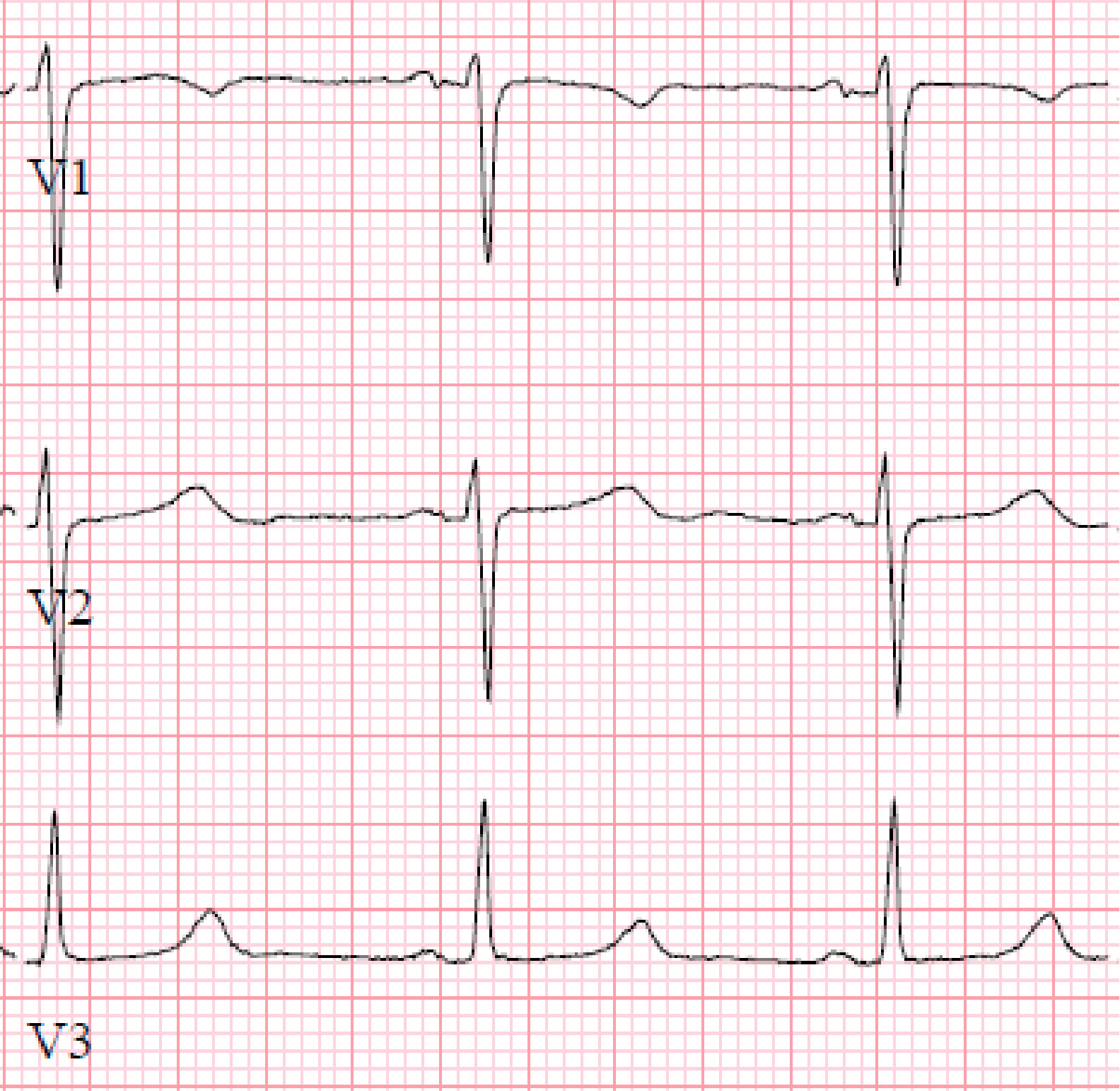
This is a case of a 31-year-old male presenting with syncope and an ECG consistent with type-1 Brugada pattern (Figure-1-A). His workup including an echocardiogram was unremarkable. Due to the concern for Brugada associated ventricular tachyarrhythmias as the culprit for his syncopal event, the placement of a defibrillator was recommended. The patient elected to have a subcutaneous defibrillator (S-ICD) due to its cosmetic advantage, lack of vascular injury risk and low systemic infection risk. Unfortunately, after placement he had another inpatient syncopal episode associated with dizziness secondary to a 12 second sinus arrest documented on telemetry monitoring (Figure-1-B). Continuous monitoring revealed multiple more episodes of sinus pauses of different durations. Given the concern that his presenting syncope was a result of sinus arrest rather than the presumed ventricular arrythmia that is classically associated with Brugada, the patient was presented with different management options including the placement of a pacemaker or a dual chamber ICD and the future removal of the S-ICD. He deferred further procedures and elected to pursue outpatient cardiac event monitoring.
Discussion
Syncope or SCD in patients with BrS is classically attributed to ventricular arrhythmias.1 SND in patients with BrS has been reported in the literature; but remains clinically a less recognized presentation. Fatty tissue deposition, atrophy and fibrosis of the sinus node was illustrated on autopsy of a young patient with BrS who died suddenly.2 In a study of 59 asymptomatic patients with Brugada, 17% demonstrated SND induced during electrophysiology study.3 Clinically documented SND in patients with BrS was estimated to be 1.1-1.7% in large population studies.4,5 A possible bradycardic mode of death was suggested with a carrier of the a mutant SCN5A gene, who suffered from sinus arrest > 9 seconds.6
A mutation in the cardiac predominant sodium channel α-subunit SCN5A which accounts for 20- 30% of patients with Brugada, has been linked to SND.7, 8, 9, 10 The abnormal sodium channel is thought to prolong the action potential of sinus node cells and slow depolarization.6 As a result, an overlap syndrome of cardiac sodium channelopathy including BrS, conduction disease and long QT syndrome has been proposed.11,12 Unfortunately, SND in our patient was apparent after his S-ICD was already placed for a presumed ventricular arrhythmogenic etiology of his presenting syncope.
Conclusion
It is imperative that providers and cardiologists are aware of the association of SND with BrS which is likely related to common genetic mutations leading to an overlap syndrome. We recommend avoiding the placement of S-ICD which may necessitate future procedures such as the placement of a pacemaker or transvenous ICD and the removal of S-ICD in the event that SND is to develop or is revealed as the etiology of a syncopal event.
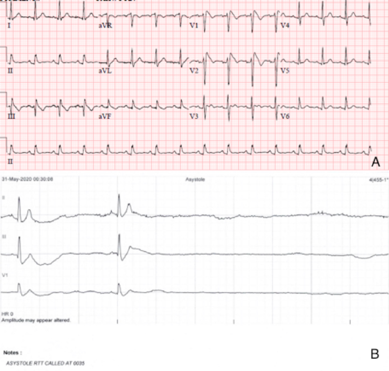
Figure1-A-ECG shows Brugada pattern; B-Telemetry strip reveals sinus arrest.
Introduction / Objective:
Esophageal perforation is an uncommon entity that most often occurs after penetrating injury or blunt trauma. Other causes include catheter ablation, ingestion of caustic/erosive agents, pill esophagitis, Crohn’s disease, peptic ulcers, dilatory injury, amongst others. Esophageal perforation can be managed conservatively in select patients, where the perforation is contained and the patient is without signs of systemic sepsis. Although conservative management has less morbidity and mortality compared to surgical management, life-threatening complications can still arise. Here, we report a case of pyopericarditis, a rare and life-threatening process that occurred in the setting of an esophageal perforation.
Case Description:
A 24-year-old man with Arnold-Chiari malformation, myelomeningocele, and cerebral palsy presents with one day of chest pain. Notably, patient was recently admitted for contained distal esophageal perforation related to NSAID use, which was managed conservatively with a proton-pump inhibitor. He now re-presents with constant retrosternal chest pain radiating to the back. Vital signs revealed pulse of 166 beats per minute, blood pressure of 138/84 mmHg and pulsus paradoxus of 14 mmHg. Physical exam demonstrated muffled heart sounds, a pericardial rub and knock, and jugular venous distention. Laboratory testing revealed a white-cell count of 13.6 K/uL (normal, 4.0 - 10.0 K/uL). Chest X-Ray showed lucencies in the left hemithorax with CT Chest demonstrating a pyopneumopericardium with tamponade effect, concerning for esophagopericardial fistula. ECG demonstrated PR elevation and ST depression in aVR (“knuckle sign”), along with diffuse ST elevations and PR depressions. The patient was started on Vancomycin, Cefepime and Metronidazole and subsequently underwent a pericardiocentesis, esophageal stenting, and thorascopic pericardial window with chest tube placement. Pericardial cultures ultimately grew Streptococcus anginosus and Streptococcus salivarius groups – consistent with pyopericarditis likely from esophageal perforation. Patient’s postoperative course was complicated by provoked deep venous thrombosis, pulmonary embolism, gastrointestinal bleeding, and esophageal stricturing several months later requiring repeated esophageal dilations.
Discussion / Conclusion:
Pyopericarditis is a rare and life-threatening complication due to 1) direct hematogenous spread, 2) extension from subdiaphragmatic focus, or in our case -- 3) direct inoculation. Gram-positive organisms cause upwards of 45% of pyopericarditis cases, with S. aureus being the most common, followed by S. pneumonia. Polymicrobial infections are uncommon – however when present, they generally arise from gastrointestinal sources. Mortality rate for pyopericarditis is upwards of 30%, with about 4% developing constrictive pericarditis. Prompt diagnosis and treatment with source control and broad-spectrum antibiotics is imperative to reduce risk for future complications. Invasive management with a pericardial window allows for more complete drainage and manual lysis of adhesions and loculations. Although diffuse ST elevations is the classical sign of pericardial inflammation, the presence of PR segment elevation with ST segment depression in aVR (“knuckle” sign) can help further support the diagnosis.
Introduction: Dextrocardia associated with situs inversus is a very rare form of adult congenital heart disease. Isolated dextrocardia is an even more rare finding. It is unclear whether or not isolated dextrocardia is associated with an increased risk of developing cardiovascular disease. It is also unclear if there is a difference in terms of cardiovascular risk between dextrocardia with or without situs inversus. Such cases, specifically coronary artery disease, have been described in both instances, however there are no large trials to investigate the pre-existing risk.
Case Description: Patient is a 56 year old female with a past medical history of hypertension who presents to the hospital with complaints of persistent chest pain on exertion. Patient takes no medications and has not seen a physician in many years. Vital signs on arrival were within normal limits. Heart sounds on auscultation were more prominent on the right side of the chest and apical impulse was felt on the right mid-axillary line at the 5th intercostal space. Labs were significant for two negative troponin values that were separated by 3 hours. EKG revealed normal sinus rhythm, extreme axis deviation, poor R wave progression, and no significant ST changes. CXR revealed dextrocardia without radiographic evidence of situs inversus. Patient was admitted to the hospital and underwent myocardial perfusion imaging which revealed no evidence of reversible ischemia and a left ventricular ejection fraction of 66%. Prior to discharge, the patient was still having significant chest pain on exertion. Therefore, the patient was taken for a coronary angiogram. Angiography via right radial arterial access was obtained and revealed 40% stenosis of the proximal right coronary artery (RCA) and 30% stenosis of the mid right coronary artery. There was no significant disease in the left main coronary artery (LMCA), left anterior descending (LAD) artery, and left circumflex (LCX) artery. However, there was TIMI 2 flow noted in both the right and left systems. Therefore, there was a strong consideration for diffuse microvascular disease. Patient was ultimately started on goal directed medical therapy for coronary artery disease, optimized on antianginal therapy, and discharged home.
Discussion: This was a peculiar case of newly diagnosed isolated dextrocardia in a patient presenting with anginal type chest pain. Though this patient had a normal stress test, her symptoms and her newly diagnosed medical history warranted further evaluation which revealed abnormal results. This begs the question whether or not isolated dextrocardia itself increases the risk of developing cardiovascular disease.
Conclusion: Isolated dextrocardia is a very rare form of adult congenital heart disease. It is unclear whether or not this finding alone, or if associated with situs inversus, imposes an increased risk of cardiovascular disease. Further investigation needs to take place to better understand if there is a true association.
Figure 1: Films of RCA (RAO CRANIAL), LMCA (LAO CAUDAL), and LMCA (RAO CRANIAL)
*RAO (Right Anterior Oblique), LAO (Left Anterior Oblique)
Introduction:
Although a known complication, high output heart failure from an arteriovenous fistula is often overlooked, especially in the setting of multi-organ dysfunction.
Case Description:
A thirty year old male presents with worsening shortness of breath and multi-organ failure. He has a past medical history of renal failure secondary to membranoproliferative glomerulonephritis on hemodialysis for the past eight years with a failed renal transplant, and severe aortic stenosis. Physical exam was notable for volume overload, left upper extremity fistula, and induration of his right hand. A transthoracic echocardiogram was obtained showing normal left ventricular systolic function and severely dilated right sided heart chambers with reduced RV systolic function, RVSP 100 mmHg, and severe aortic stenosis. His last TTE report from the year prior showed normal RV function with RVSP 27 mmHg.
He was emergently transferred to a tertiary care center where he underwent extensive workup for pulmonary hypertension. A right heart catheterization was performed showing elevated right sided filling pressures, normal left sided filling pressures, and an elevated pulmonary artery pressure 88/48 mmHg with mean 61 mmHg. His cardiac output and index by thermodilution were elevated at 10 L/min and 5.4 L/min/m2. His calculated pulmonary vascular resistance was 5 Wood units. Blood sampling showed an oxygen saturation 88% in the innominate vein in the setting of a large left arm AV fistula. A fistulogram was performed showing a dilated arteriovenous anastomosis with a high flow state at 7-10 L/min for which he underwent banding of the left brachiobasilic fistula. Since discharge, he has improved significantly. He is now able to do all activities of daily living and his RSVP has reduced to 49 mmHg.
Discussion:
High output heart failure is defined as elevated cardiac output >8 L/min or resting cardiac index >2.5-4 L/min/m2 with reduced systemic vascular resistance. Causes of high output heart failure range from obesity, liver disease, arteriovenous shunts, severe anemia, lung disease, hyperthyroidism, or myeloproliferative disease. As a result, high output heart failure is due to an underlying etiology with a normal functioning heart that is unable to keep up with the body's demand for increased blood and oxygen. Obtaining a clinical history from patients that present with high output heart failure is essential to diagnosis as symptoms can range depending on the etiology along with the standard heart failure like symptoms of shortness of breath, hypervolemia, orthopnea, and fatigue. In our patient, a broad differential diagnosis led to a quick identification for the cause of his high output heart failure resulting in a favorable outcome.
Conclusion:
It is essential to identify high output heart failure and obtain a thorough clinical history to identify a possible etiology as many cases are reversible. An arteriovenous fistula is an uncommon but known cause of high output heart failure and early recognition can lead to better outcomes. As seen in our patient who presented with multi-organ dysfunction and hypervolemia, by receiving AV fistula banding he was eventually able to return to his daily life.
Case presentation.
76 Years old male with past medical history of Tetralogy of Fallot (TOF), He presented with severe low flow-low gradient Aortic stenosis.
Our patient had an interesting history of TOF, underwent Blalock-Taussing shunt at age of 3, that was followed by definitive surgery which included shunt removal, patch augmentation of RVOT and VSD patch repair. The patient had uneventful cardiovascular complications away from atrial fibrillation until he presented complaining of fatigue and decreased exercise tolerance of 6 months duration.
EKG showed Atrial fibrillation with left anterior fascicular block and right bundle branch block. Echo-cardiogram revealed decreased EF of 20-25% with severely calcified aortic valve and mean pressure gradient cross aortic valve of 30 mm Hg, LVOT VTI 13 cm, DVI ratio 0.17 and Mild AI.
Patient subsequently underwent LHC that showed mean pressure gradient across aortic valve of 45 mm Hg and group 2 pulmonary hypertension.
Patient underwent trans-catheter Aortic valve placement using 29 mm Sapien-3 bioprosthetic valve, with excellent results and mild paravalvular leak. Patient subsequently developed high grade AV block that required Dual chamber pacemaker placement. LVEF recovered back to 55-60%.
Discussion and conclusion:
Long-term survival is excellent following TOF repair, particularly for patients who are operated on at a young age, among whom survival is >90 percent 25 years after repair. Survival has improved considerably with advances in surgical techniques.
Patients who have undergone TOF repair are at risk for chronic postoperative complications, which may require reintervention. These include Pulmonary regurgitation with associated RV enlargement, Residual RVOT obstruction and RV dysfunction, Aortic root dilation and aortic valve insufficiency, Arrhythmias, including atrial and ventricular tachycardia. Arrhythmia and heart failure leading to sudden cardiac death are the most common causes of late death following surgical repair.
In less than 15 years, transcatheter aortic valve replacement (TAVR) has progressed from a procedure of last resort in patients at prohibitively high perioperative risk for major morbidity and mortality from surgical valve replacement to a viable alternative option to surgery in most patients with native aortic valve stenosis.
Background
Vascular complications from trans-radial cardiac catheterization are uncommon. Mediastinal hematoma is a rare complication with life threatening potential. We present a case of a patient who underwent cardiac catheterization and subsequently experienced odynophagia from a perforated anomalous subclavian artery that led to a mediastinal hematoma.
Case Report
A 59 year old female with past medical history of coronary artery disease presented with complaints of angina and underwent a trans-radial cardiac catheterization. Immediately after the procedure, the patient complained of chest pain and odynophagia. EKG and echocardiogram were unremarkable and a CT scan of the chest demonstrated an ill-defined fluid collection present in the superior mediastinum and an aberrant right subclavian artery. The patient was closely monitored in the Intensive Care Unit and the patient remained hemodynamically stable throughout the admission. Patient was subsequently discharged home in good condition and did well on outpatient follow up.
Conclusion
Vascular perforations associated with delivery of standard radial catheters in the subclavian artery are rare, with very few cases reported in the literature. We presented the case of a patient who had a previously unidentified right aberrant subclavian artery with a retroesophageal course which precipitated the hematoma and subsequently resulted in odynophagia despite an uncomplicated catheterization. This rare complication of a commonplace procedure necessitates prompt recognition, appropriate hemodynamic management, and possible repair of the injured vessel to appropriately manage a potentially life-threatening condition.
Learning Objectives: Anticoagulation in patients with atrial fibrillation and CHA₂DS₂-VASc score of zero.
Keywords: Atrial Fibrillation (AF), CHA₂DS₂-VASc Score, Coronary Embolism (CE) & Thromboembolism,
Introduction:
Coronary embolism (CE) is a rare cause of acute coronary syndrome (ACS) and accounts of 3% of cases [1]. Atrial fibrillation is the commonest cause of CE. Diagnosing CE in the setting of atrial fibrillation is of paramount importance as the indications for anticoagulation in these patients may not correlate with the CHA2DS2VASc score.
Case Description:
We report the case of a 53-year-old obese gentleman with a history of paroxysmal atrial fibrillation off anticoagulation because of a CHA2DS2-VASc score 0 who presented with acute sudden onset pressure-like chest pain associated with lightheadedness and was subsequently diagnosed with an NSTEMI and hyperthyroidism. The patient was placed on a baby aspirin, high dose statin and a beta blocker along with a heparin drip. A left heart catheterization was performed and revealed evidence of thrombotic occlusion of the distal LAD without any evidence of epicardial atherosclerotic coronary disease. A 2.5 mm X 16 mm balloon was used to dilate the lesion with marginal benefit likely due to organized thrombus. The patient was then placed on anticoagulation and dual antiplatelet therapy.
Discussion:
Coronary embolism (CE) is a rare and important cause of non-atherosclerotic acute coronary syndrome. Previous studies estimated that non-atherosclerotic coronary artery disease accounts for 4-7% and 13% of all ACS cases at coronary angiography or autopsy respectively [2-3]. The largest retrospective study by Shibata et al demonstrated a 2.9% of patients with an MI was due to a CE in their study population. Coronary embolism can be from a cardiac source (left atrium, left ventricle, mitral valve or cardiac tumors), paradoxical from a peripheral vein through a patent foramen ovale (PFO) or iatrogenic (sequelae to a cardiac procedure) [1]. Non-valvular AF is the commonest cause for CE with a higher prevalence in chronic atrial fibrillation than paroxysmal atrial fibrillation [4].
Conclusion:
This case clearly illustrates the imperfection of the assumptions underlying a “low” CHA2DS2-VASc score and the importance of clinic judgement to guide the decision to anticoagulate such patients.
References:
1. Claire E. Raphael. Coronary Embolus J Am Coll Cardiol Intv. 2018 Jan, 11 (2) 172-180.
2. Waller BF. Atherosclerotic and nonatherosclerotic coronary artery factors in acute myocardial infarction. Cardiovasc Clin. 1989;20(1):29‐104.
3. K. R. Prizel,“Coronary artery embolism and myocardial infarction: a clinicopathologic study of 55 patients,”Annals of Internal Medicine, vol. 88, no. 2, pp. 155–161, 1978
4. Shibata,. Prevalence, Clinical Features, and Prognosis of Acute Myocardial Infarction Attributable to Coronary Artery Embolism. Circulation.doi:10.1161/circulationaha.114.015134
Introduction: Coronary anomalies affect ~1% of the population. Congenital coronary artery anomalies are usually found incidentally. Their presentation is variable-from being asymptomatic to causing angina, syncope, or sudden cardiac death (SCD). In 14-40 year olds, coronary anomalies are involved in 12% of sports-related SCD versus 1.2% of non-sports deaths. Case Description: 27 y/o male with Kikuchi Nectrotizing Lymphadenitis and an extra-muscle myopathy who presented with chest pain in the setting of influenza B infection. Labs showed elevated troponin (hs-trop peak of 51), CPK (237), and BNP (332) and his ECG showed T-wave inversions V4-V6. Echocardiogram demonstrated reduced ejection fraction of 30-35% with hypokinesis of the mid-distal apical, periodical, septal and inferior walls. He was diagnosed with viral myocarditis, and treated for his heart failure. Cardiac MRI and Coronary CT Angiograhy (CCTA) were ordered. Cardiac MRI confirmed myocarditis while the patient was still hospitalized. Outpatient CCTA showed left dominance with normal left main and left anterior descending artery. Left circumflex was normal. However, the distal circumflex was noted to continue in the right AV groove and course superiorly in the expected location of the right coronary artery. No right coronary osmium or proximal RCA was present. Discussion: A single coronary is defined as an isolated artery originating from a single osmium and supplying the whole heart. Single coronary arteries are very rare, with an autopsy incidence of 0.29%. There are many classifications of coronary anomalies. The system described by Dr. Martin Lipton, M.D. is the most popular. With the growing use of CCTA for diagnosing coronary artery disease, CCTA is also identifying coronary anomalies with a precision and descriptiveness that is unmatched by coronary angiography. CCTA is also important in treatment therapies for coronary anomalies. There are some coronary anomalies that require surgical correction such as anomalous RCA arising from the left aortic sinus because of an increase risk of SCD.The significance of an absent RCA is controversial. One study of 21 patients with this anomaly identified a cardiac cause of death in 10 patients, of which 5 were sudden. Given the normal nature of the patient's LM, LAD, and dominant LCx, we feel confident his troponin, ecg and chest pain were related to viral myocarditis, which was supported by cardiac MRI. His systolic heart failure was treated with GDMT. Forty percent of single coronary anomalies occur in association with other congenital heart abnormalities. Our patient had no structural heart disease identified on cardiac MRI or CCTA. We also found no association with coronary anomalies with Kikuchi Histiocytic Necrotizing Lymphadenitis. Extra muscle myopathies (another co-condition in our patient) are typically associated with conduction disturbances not anomalous arteries. Conclusion: Anomalous coronary arteries are rare congenital cardiac defects with varying symptomatology and controversial clinical significance. Single coronary arteries such as absent RCA are the most rare of the described defects. Advanced cardiac imaging including CCTA is an essential tool in modern cardiology and will hopefully help increase our knowledge and understanding of coronary anomalies including single coronary anomalies.
Introduction: coronary artery fistulas have been described as the most common hemodynamically significant congenital coronary anomaly. These fistulas arise from the right and left coronary trees with similar frequency, but infrequently they may be bilateral. Many patients with congenital coronary artery fistulas are asymptomatic at the times of diagnosis, but the occurrence of symptoms increases with increasing age. The most common symptoms are congestive heart failure and angina due to left to right shunting and coronary steal respectively, but rarely can be associated with myocardial infarction.
Case description: an 82-year-old female with chronic dyspnea presented with atypical chest pain and was found to have a non-ST elevation myocardial infarction. Coronary angiography demonstrated a large coronary to pulmonary artery fistula with feeding vessels originating from the left anterior descending and right coronary arteries. There was no significant coronary artery disease present. Right heart catheterization demonstrated pulmonary to systemic flow ratio of 0.8:1. Given the patient’s symptoms and the concern for these fistulas causing myocardial ischemia the decision was made to proceed with fistula closure. Taking into consideration her advanced age and absence of other indication for cardiac surgery an endovascular approach was attempted. Coiling of both branches of the fistula was ultimately successful.
Discussion: the 2018 American Heart Association guideline for the management of adults with congenital heart disease recommends the presence of coronary artery fistulas requires review by a knowledgeable team that may include congenital or non-congenital cardiologists and surgeons to determine the role of medical therapy and/or percutaneous or surgical closure. Symptoms, complications, and significant shunt are the main indication for the treatment of CAFs. Given the patient’s chronic shortness of breath, presentation with an acute myocardial infarction in the setting of non-obstructive coronary artery disease, and no other cardiac disease necessitating cardiac surgery the decision was made to pursue percutaneous closure of both feeding vessels of the fistula.
Conclusion: Coronary artery fistulas are rare congenital anomalies which are more commonly asymptomatic. They rarely present bilaterally and even less often with acute myocardial infarction. They present a unique therapeutic challenge in part due to the infrequency with which it is encountered. The decision for closure should be based on symptoms, degree of shunting, or complications associated with the fistulas.
Introduction:
Takotsubo (stress-induced) cardiomyopathy is a clinical syndrome that is commonly triggered by an emotional or a physical stressor. The syndrome is most common in post-menopausal women and can often mimic acute coronary syndrome. This entity was first described in Japan in 1990 and has since been further characterized. We present a case of a healthcare worker who presented with stress-induced cardiomyopathy during the height of the COVID-19 pandemic in Detroit, illustrating the tremendous mental and emotional toll on healthcare personnel that was caused by this public health emergency.
Case Description:
A 60-year-old female healthcare worker with a medical history of hypertension, hyperlipidemia, and sickle cell trait presented to the hospital with left-sided chest pain and diaphoresis. The patient reported that her work duty hours and demands had been increased during the height of the COVID-19 pandemic. In the months leading up to her illness, the psychological stress at work had led to insomnia, poor eating habits, and irritability. While in the emergency department, her vital signs indicated blood pressure of 188/82 mmHg, and she was tachycardic to 115 bpm. Laboratory evaluation showed brain natriuretic peptide elevation at 318 pg/mL and mildly elevated high sensitivity troponin I of 24, 29, and 27 ng/L. Her EKG showed a new left bundle branch block, not meeting Sgarbossa criteria for STEMI. A previous EKG performed 9 months before her presentation showed normal sinus rhythm with an incomplete left bundle branch pattern and non-specific ST-T wave changes. She was given aspirin and started on a heparin infusion in the emergency department. An echocardiogram was performed, which revealed a reduced ejection fraction (EF) of 20-25% and moderate basal and apical left ventricular hypokinesis. The findings were concerning for either Takotsubo cardiomyopathy or proximal LAD or left main stenosis. Given her chest pain, decreased EF, and wall motion abnormalities, the patient underwent left heart catheterization which revealed no coronary disease. She was subsequently placed on guideline-directed medical therapy and discharged home with a LifeVest.
Discussion:
Takotsubo cardiomyopathy is often precipitated by emotional or physical stress. Many proposed mechanisms to explain the pathophysiology of this clinical entity have been postulated, including adrenergic surge and direct cardiac myocyte injury; however, the exact mechanism remains unclear. This case highlights the consequences of psychological stress for healthcare workers during the height of the COVID-19 pandemic in one of the most heavily affected cities. Our case is important because it describes the extent of the psychological toll that the pandemic had on healthcare workers, thus raising awareness about this issue. Also, this case highlights that understanding the patient as a whole and paying close attention to patients’ life circumstances and emotional health is crucial and can aid greatly in diagnosis.
Conclusion:
Healthcare workers have been exposed to extreme stress during the COVID-19 pandemic, and Takotsubo cardiomyopathy is an organic manifestation of acute psychological stressors that may be seen in this population. Healthcare workers endure significant emotional duress, the awareness of which is critical and must be greatly emphasized.
Introduction
Unicuspid aortic valve (UAV) is an extremely rare congenital abnormality. Prevalence of UAV is noted to be .02% in the general population; however, it is recognized as a cause of severe aortic stenosis in surgically excised aortic valves at around 5%. The most common congenital abnormalities of the aortic valve include bicuspid, quadricuspid, or a unicuspid aortic valve. UAV exists in two forms, as unicuspid acommisural and unicuspid unicommisural aortic valve. Unicuspid acommisural aortic valve presents in early infancy or childhood as severe aortic stenosis, while unicuspid unicommisural aortic valve presents between the 3rd and 5th decade.
Case Description
A 58 year old gentleman with hypertension and hyperlipidemia presented to urgent care for worsening shortness of breath at rest. Patient had been experiencing exertional dyspnea and leg swelling ongoing for a year. On evaluation patient was noted to have an ejection fraction (EF) of 10-15% per transthoracic echocardiography with additional findings of a bicuspid aortic valve, a significantly dilated aortic root, and moderate-severe aortic stenosis. Subsequent studies revealed a 6.5cm ascending aortic aneurysm (AAA) with extensive calcification of the aortic valve. Patient underwent surgical aortic valve replacement with a mechanical aortic valve, aortic root replacement, and repair of AAA. Intraoperatively patient was noted to have a unicuspid aortic valve with extensive calcification. Patient's postoperative course was complicated by hemorrhagic shock requiring numerous transfusion products. Patient additionally developed global cerebral and cerebellar infarcts along with upper and lower extremity deep venous thrombosis. Patient was eventually discharged after a prolonged hospital stay to rehab without associated neurological deficits with hemodynamic stability and an improved EF of 30-35%.
Discussion
Unicommisural unicuspid aortic valve is an extremely uncommon congenital abnormality that presents between the 3rd and 5th decade and is usually associated with moderate to severe regurgitation or stenosis at the time of presentation. Additional abnormalities adding to the morbidity and mortality include aortic aneurysms, aortic regurgitation, aortic dissection, coarctation of the aorta, coronary anomalies, patent ductus arteriosus, and aortic root dilatation. It remains under diagnosed with current imaging modalities with a definitive diagnosis made either intraoperatively or at autopsy. The definitive treatment for UAV remains surgical replacement.
Conclusion
With our case we intend to highlight the importance of early recognition and inclusion of UAV in the differential diagnosis of a young patient presenting with heart failure symptoms, murmurs and physical examination suggestive of aortic stenosis. Patients with UAV develop symptoms 20-30 years before patients with bicuspid aortic valve along with associated congenital anomalies. Early diagnosis and evaluation with a multidisciplinary team approach is vital to improve the morbidity and mortality in patients with UAV due to the complexity and under diagnosis of this entity.
Introduction
A variant form of angina pectoris that was first described in 1959 by Prinzmetal et al still remains as a mimicry of a type 1 ST-elevation myocardial infarction. Generally thought to have good prognosis, those with refractory cases of vasospastic angina (VSA) can lead to myocardial infarction, arrhythmia, or death.
Case Description
A 62-year-old female presented post cardiac arrest. She has a history of VSA in the left anterior descending artery (LAD) and medial ramus intermedius (RI) branch, limited systemic sclerosis, Raynaud phenomenon, and hypertension.
Earlier in the morning, the patient complained of chest pain and took a sublingual nitroglycerin. The spouse incidentally found her unresponsive several hours later. In route to the hospital, the patient was in ventricular fibrillation, necessitating several defibrillation. Her electrocardiogram (ECG) showed Wenckebach with anterior and inferior ST-elevation. Her emergent cardiac catheterization showed a complete occlusion of her proximal LAD. All of the left system vessels as well as posterior left ventricular branches of the right system were significantly small in caliber. The estimated left ventricular ejection fraction was 25% with anterior hypokinesis/akinesis and inferior hypokinesis. The proximal LAD was cannulated and a balloon inflation was performed. After intracoronary nitroglycerin, all of the caliber of vessels increased.
About 6 hours later on targeted temperature management (TTM), patient suddenly became hypotensive and bradycardic. Her ECG showed anterolateral ST-elevation. On repeat angiogram, the same proximal LAD was totally occluded and the other vasospastic vessels again appeared small. With intracoronary nitroglycerin, the flow in the LAD remained occluded. The LAD lesion was crossed and an undeployed balloon was placed in the artery for support. Another intracoronary nitroglycerin was given with no change to obstruction. Unfortunately, the patient rapidly declined with bradyarrhythmia, hypotension, and ventricular fibrillation with no recovery.
Discussion
Acute management of refractory focal and diffuse vasospasm is a challenging scenario. There was one focal stenosis of the same location as the prior angiogram and evidence of diffuse spasm in multiple vessels that improved with subsequent intracoronary nitroglycerin. However, the possibility of organic stenosis cannot be ruled out as the proximal LAD lesion had to be cannulated after the lack of initial nitroglycerin response. Historically percutaneous coronary balloon angioplasty yielded mixed outcomes with some recurrence of coronary restenosis, though optimal medical therapy reduced the incidence. Coronary stenting has been successfully utilized in refractory cases with nonobstructive or obstructive organic stenosis. There are still risks of in-stent restenosis or stenosis development in the stent surrounding area or in a different location(s). Finally, the use of intravenous calcium channel blocker and/or nitroglycerin is unconventional for conducting the TTM for post cardiac arrest patients.
Conclusion
This case demonstrated the complexity of the acute management of severe VSA and cardiac arrest. Refractory VSA to oral, intravenous, and intracoronary medical therapies was complicated by persistent hypotension, incessant arrhythmias, and multiple ischemic insults. Halting this rapid decline proved to be difficult. Therefore, clinicians should strive for aggressive medical therapy in the forefront to prevent any escalation of angina.
Introduction
Normal coronary arteries on two-dimensional (2-D) coronary angiogram were one of the principles of Takotsubo Cardiomyopathy (TTC) diagnosis based on the Mayo Clinic Diagnostic Criteria. However, recent cohorts have increasingly shown the concomitant cases of TTC and obstructive coronary artery disease (CAD) with higher morbidity and mortality rates compared with TTC alone. Additionally, optimal treatment for concomitant cases is not well described in the literature due to limited data. Therefore, a high index of suspicion is required to promote more accurate prevalence of coexistence and treatment optimization.
Case Description
An 86-year old Caucasian female with no hypertension, dyslipidemia, diabetes mellitus or history of tobacco use presented with acute chest discomfort. Physical examination was only notable for tachycardia without evidence of congestive heart failure or major valvular heart disease. Presenting EKG showed ST segment elevations (STE) in leads V2–V6 with no clear reciprocal changes in the inferior leads. Her blood work showed an elevated troponin- I level of 5.09 ng/mL and a BNPEP level of 3951 pg/mL. A cardiac team alert was called for acute anterior STEMI. Angiography showed 90% hazy stenosis in the mid portion of the left anterior descending artery (LAD) concerning for vasoactive plaque rupture (Picture A). Primary percutaneous coronary intervention was performed with a drug eluting stent (DES) implantation to the mid LAD without any complications. Left Ventriculography (LVG) demonstrated anteroapical akinesis with hyperkinetic basal segments along with moderately low LVEF, which was consistent with classic TTC (Picture B).
Discussion
Presenting EKG showed ST elevations without clear reciprocal changes; however, were consistent with an anterior infarction, which was supported by the vasoactive lesion in the mid LAD. Although the culprit lesion was not a total occlusion, the presence of TIMI II flow was possibly due to spontaneous lysis of an occlusive thrombus. Interestingly, wall motion abnormalities on the LVG did not correlate the area of distribution of the LAD and in fact showed apical ballooning sparing the basal segments. Overall, the findings indicated a coexistent obstructive CAD in a patient with TTC.
Conclusion
TTC and obstructive CAD co-exist more frequently than it was previously thought based on the current studies. Moreover, the mortality rates were reported much higher in the literature in the presence of concomitant cases than isolated TTCs. Our case highlights the importance of being aware of coexistence in order to provide more accurate prevalence and treatment optimization.
Introduction/Objective
Deglutition syncope is an uncommon cause of syncope. Although its pathophysiology is not clear, there are several postulated mechanisms that are described in case reports. It’s thought be mediated by the vagus reflex in the setting of swallowing that causes inhibition of the cardiac conduction system. It has been described to be associated with various esophageal and gastric disorders including stricture, hiatal hernia and achalasia. However, many casese report no association with structural abnormalities. Deglutition syncope was first reported in 1793 by Thomas Spens and there have been numerous case reports describing this phenomenon.
Case Discussion
SC is a 60-year-old male with a past medical history of hypertension, diabetes, COPD, and small vessel brain aneurysm, who initially presented with altered mental status and dizziness. He reported of consuming 4 alcoholic beverage the night prior to his admission and his family reported of his abnormal behavior the following morning. After a comprehensive work up including CT of head, initial diagnosis was metabolic encephalopathy. However, the next day, continuous telemetry demonstrated an 8 second sinus pause when he was eating his lunch. The patient did feel mildly dizzy, but reported no loss of consciousness. Subsequently,he demonstrated multiple episodes of sinus pause ranging from 4 to 7 seconds only associated during meal time.
Upon further questioning, he stated having multiple episodes of transient loss of consciousness in the past that had been worked up with a holter monitor. Unfortunately, his holter monitor was unable to capture any arrhythmia. His last syncopal episode was approximately 1 year ago when he was dining at a restaurant with his wife. After a few bites, the patient suddenly lost consciousness for 10 seconds and came back to his baseline. He reported of having a few more episodes of having similar episodes that were associated with food consumption.
Carotid sinus massage was performed bilaterally at bedside following negative auscultation failed to induce significant sinus slowing or pauses. Bedside evaluation of left lateral recumbent and sleeping demonstrated apnea not associated with sinus pauses. The patient also underwent Barium swallow study which revealed no stricture or a normal-appearing gastroesophageal junction into the stomach.
He was started on pantoprazole 40 mg daily and was discharged with an outpatient 30-day monitor. Holter monitor showed no recurrence of sinus pause and the patient had no recurrence of syncope or dizziness after initiation of pantoprazole.
Discussion/Conclusion
Deglutition syncope is a rare cause of syncope. Although the exact mechanism is unknown, it is likely mediated by external stimulation of left atrium. Diagnosis of deglutition syncope involves careful history taking as well as physician examination. It is critical to examine the need for pacemaker before committing the patients to a lifelong pacemaker implantation. Careful withdrawal of offending agents and initiation of GI treatment should be performed first.
In our patient, we were able to exclude any esophageal structural abnormalities and his symptoms improved with initiation of pantoprazole. Further investigation is warrnated to study the association between GERD and deglutition syncope.
Introduction
Indwelling central line catheters and hemodialysis predispose to bloodstream infection. This can lead to cardiac abscess formation either by dissemination or by direct extension of a pre-existing cardiac infective focus. The incidence of cardiac abscesses continues to be investigated and is of great importance when deciding the prognosis and management of patients. All these patients have increased risk of embolization, morbidity, and mortality.
Case Description
34-year-old women with diabetic nephropathy on home hemodialysis (HD) presented to the hospital for fever and lethargy. Two days prior to presentation she had mild chest pain managed with aspirin and nitroglycerin. She recorded a temperature of 103.8F at that time. On presentation she was found to have fever and leukocytosis. Computer Tomography (CT) scan showed two cavitary lung nodules. Echocardiogram demonstrated normal left ventricular function and a prominent, independently mobile echodensity attached to the left atrial wall. Differential diagnosis included endocarditis, thrombus, or mass. She was started on antibiotics and heparin infusion. Blood cultures subsequently grew Staphylococcus aureus and a transesophageal echocardiogram (TEE) was performed. This revealed a large mobile echodensity in the left atrium attached to the Coumadin ridge with small abscess formation. Anticoagulation was discontinued and she was evaluated by cardiothoracic surgery. Source of the infections was determined to be the HD catheter. She was managed with six weeks of intravenous antibiotics.
Discussion
When a patient on hemodialysis presents with fever endocarditis needs to be on the differential diagnosis. These patients are at increased risk of bloodstream infection with possible multiorgan dissemination.
In this case echocardiogram was performed early, however due to the unusual appearance and location of the lesion a broad differential diagnosis was maintained and TEE was needed to arrive at a more definitive answer and guide therapy.
Conclusion
Despite unusual appearance and location, when a high clinical suspicion is maintained for infective endocarditis, patients can be diagnosed quickly and effectively using echocardiographic interrogation of the heart so specific and appropriate therapy can be rendered.
Introduction
Stress cardiomyopathy, also known as Takotsubo cardiomyopathy, is a cardiac syndrome that involves a transient decrease in the ejection fraction (EF) in the absence of significant coronary artery disease with reversible left ventricular (LV) apical akinesis. This is often triggered by an intense emotional or physiological stressor; however, it uncommonly occurs without any identifiable causes. This is the case of a 66-year-old male who developed Takotsubo cardiomyopathy after an exercise treadmill stress test.
Case Description
A 66-year-old male was being seen in the electrophysiology clinic for bradycardia and dyspnea on exertion, thought to be secondary to sick sinus syndrome. A treadmill exercise stress test was ordered to assess for chronotropic incompetence and to rule out structural heart disease. His resting echocardiogram was normal with a LV EF of 55%. The test was stopped after seven minutes on the treadmill because of fatigue and muscle cramps. Post-exercise echocardiography showed significant LV chamber dilatation and development of severe global hypokinesis. With concern for multivessel ischemia, he underwent left heart catheterization and coronary angiogram the next day that showed no coronary artery disease. Left ventriculogram was performed and the LV EF was 35% with hyperdynamic basal function and normal apical function in a classic pattern of Takotsubo cardiomyopathy. The patient was continued on his Losartan. A beta blocker could not be initiated due to bradycardia. An echocardiogram was repeated one month later and showed recovery of the LV ejection fraction to 50% with mild hypokinesis of the apex.
Discussion
Takotsubo cardiomyopathy is a rare but well-known entity estimated to account for up to 0.02% of inpatient admissions. Almost 90% of cases occur in women, typically post-menopause. The typical presentation of Takotsubo cardiomyopathy is chest pain and dyspnea often with EKG changes and elevated cardiac biomarkers, mimicking acute coronary syndromes. Obstructive coronary artery disease must be excluded.
The pathologic etiology is not understood; there are a few hypotheses. The most commonly accepted hypothesis is that severe emotional or physiologic stress induces an endogenous catecholamine surge leading to myocardial stunning. Exercise intrinsically induces catecholamine release, which can subsequently lead to transient subendocardial microvascular ischemia. This, in conjunction with the hypothesis that a hypertensive response during exercise gives rise to altered myocardial contractility could be a possible mechanism for triggering Takotsubo cardiomyopathy in our patient. Additionally, a marked increase in afterload during exercise due to a hypertensive response can precipitate wall motion abnormalities.
The association of Takotsubo cardiomyopathy with exercise stress testing is exceedingly rare. There are very few case reports with similar presentations. Such case reports lend data to further understanding the mechanism by which exercise stress testing induces this cardiomyopathy. The prognosis in stress induced cardiomyopathy is very good, with most patients recovering completely within 4-8 weeks.
Conclusion
Takotsubo cardiomyopathy is an exceedingly rare but potential complication from a treadmill stress test and should be kept as a differential diagnosis in patients who go from normal LV systolic function at rest to severe dysfunction post-exercise.
Background:
Covid-19 related perimyocarditis has typically been associated with inflammatory markers such as elevated C-reactive protein, erythrocyte sedimentation rate and troponin elevation.This case highlights that perimyocarditis following Covid-19 may be present without elevation of these inflammatory markers. Further work up with cardiac magnetic resonance imaging (MRI), in patients under high suspicion, may assist in diagnosis and appropriate management.
Case:
A 21 year old female presented to the Cardiology Clinic with complaints of exertional shortness of breath, orthopnea, paroxysmal nocturnal dyspnea worsening over a 1 month duration. Patient reported Covid-19 symptoms such as fevers, chills, cough and shortness of breath 7 months prior to this presentation, though she had not been tested at the time, with subsequent improvement in symptoms. 3 weeks prior to cardiology clinic presentation, patient was admitted for right-sided abdominal pain, had liver congestion, inflammatory markers were negative, echocardiography showed ejection fraction of 45%, with global hypokinesis, mildly enlarged left ventricle, severely enlarged left atrium, moderately enlarged right atrium, right ventricular systolic pressure at 65-70 mmHg. Following discharge, patient was seen at the primary care physician’s office for complaints of shortness of breath. Covid-19 immunoglobulin G (Ig G) was positive, thyroid stimulating hormone, human immunodeficiency virus and hepatitis panel were unremarkable. In the Cardiology clinic physical exam showed stable vital signs with patient saturating well on room air, though demonstrating shortness of breath with exertion. Chest exam was positive for a pericardial friction rub. Patient was sent to the emergency department, where cardiac echocardiogram showed ejection fraction of 15-20% with severe global hypokinesis, grade 3 diastolic dysfunction, myocardial appearance suggestive on noncompaction, severely increased right ventricular cavity size with reduced systolic function, severely dilated left atrium, dilated right atrium, mild mitral regurgitation, severe tricuspid regurgitation with severe pulmonary hypertension of 76 mmHg, moderate pulmonary insufficiency, nonsignificant aortic regurgitation and small pericardial effusion. Hemoglobin was stable around 9.7, troponins less than 0.02, erythrocyte sedimentation rate 11, C-reactive protein 6.7, ferritin 97, fibrinogen level 213, elevated D-dimer level 1919, elevated pro-Brain Natriuretic Peptide 3070, elevated Lactate Dehydrogenase 487. Covid-19 nasal swab negative. Cardiac magnetic resonance imaging (MRI) showed findings suggestive of inflammatory myocarditis in the post viral setting. Telemetry during hospitalization was remarkable for intermittent episodes of non-sustained ventricular tachycardia. Patient was managed with steroids, lisinopril, low dose beta blocker, colchicine, furosemide 20 mg and discharged home on a Lifevest following significant improvement in clinical symptoms.
Discussion
The Covid-19 virus invades cells through angiotensin-converting enzyme 2 protein, which is found in high concentration in pneumocytes and myocytes, explaining why these organs are mostly involved in the disease process. Presence of positive Covid-19 Ig G in the above patient shows previous exposure to the virus, and likely progressive inflammation of the myocardium and worsening clinical status.
Conclusion:
This case highlights that Covid-19 related perimyocarditis and cardiomyopathy may be present without elevation of inflammatory markers such as ESR,CRP and troponins and further work up such as cardiac MRI may be appropriate to establish diagnosis.
Objective: We present a case in which excellent interdisciplinary communication during pericardiocentesis after PEA arrest resulted in an excellent patient outcome and augmented education.
Case Description: A 74 year-old male who presented to a community hospital ED with episodes of PEA arrest was found to have a large pericardial effusion with tamponade physiology. Despite lack of previous experience with emergent pericardiocentesis by the ED resident and attending physician, the cardiology fellow on call was able to provide immediate guidance at bedside, resulting in a successful aspiration. The patient subsequently underwent definitive treatment and survived to discharge in stable condition.
Discussion: Previous studies have demonstrated good outcomes when senior, experienced operators provided guidance and education to junior operators within a tertiary healthcare setting. This case demonstrates how inter-professional relationships and communication between the emergency and cardiology departments directly resulted in improved patient care and excellent outcome in an emergent situation, despite lack of resources available at tertiary centers.
Conclusion: This case emphasizes how physician-to-physician communication, notwithstanding training level or department, leads to improved patient care when time is of the essence.
Background
Congenital long QT syndrome (LQTS) is an inherited disease with a prevalence of ~1:2000 characterized by prolongation of the QT interval on electrocardiogram and the potential risk for ventricular arrythmias. The majority of congenital long QT cases can be explained by a mutation in one of 3 genes. KCNQ1 leading to the clinical phenotype of long QT type 1, KCNH2 causing long QT type 2, and SCN5A causing long QT type 3. Defining genotype in long QT carries particular importance as there exists distinct associations between genotype, triggers for arrhythmia, and response to beta blocker treatment. In long QT1 exercise is often a trigger due to excessive prolongation of the QT during this period. Startling auditory events, such as a loud alarm clock, sleep deprivation, and being post-partum can trigger events in long QT2. Events in long QT3 are less predictable. Here we present a case of congenital long QT type 1 with particularly interesting genotype.
Case Description
The patient is a 27-year-old woman who presented for pre-operative evaluation and was found to have a QTc of 515ms. Her history is notable for Beckwith-Wiedemann Syndrome (BWS) which led to macroglossia, hemihypertrophy, neonatal intraventricular hemorrhage, and omphalocele. Despite her complicated neonatal course, the patient was an active and normal functioning adult at the time of evaluation. Despite her prolonged QTc she did not report a history concerning for ventricular arrythmia events. A repeat QTc was 470ms, surface echocardiogram demonstrated a structurally normal heart and an event monitor showed no ventricular arrythmias. Genetic testing was sent and demonstrated a pathogenic splice variant in KCNQ1. This variant leads to haploinsufficiency of a potassium channel and her predisposition for a prolonged QT interval. In order to reduce her risk of ventricular arrythmias she was started on nadolol 20mg daily with a goal dose of 60mg daily.
Discussion
At first glance this case seems to be a relatively straightforward presentation of long QT type 1. However, her combination of inherited diseases is peculiar and are indeed related. KCNQ1 is located on chromosome 11, nearby an imprinting center. Certain regions of DNA are only expressed from the maternal or paternal allele, this is referred to as imprinting. BWS occurs when a non-functioning imprinting center on chromosome 11 is maternally inherited. This patient’s particular KCNQ1 variant was in an intronic region of KCNQ1 that is responsible for regulation of the nearby imprinting center. Thus, this particular variant disrupts both normal KCNQ1 expression and regulation of the imprinting center. Since imprinting at the BWS region is determined by maternal inheritance, we can conclude that the variant must have been inherited from her mother, whereas inheritance from her father would have resulted in isolated LQTS. Should this patient choose to have children they would each have a 50% risk of inheriting both long QT syndrome and BWS.
Conclusion
This case represents a rare connection between Beckwith-Wiedemann and congenital long QT syndrome elucidated by genetic testing.
Introduction:
MINOCA (Myocardial Infarction with no obstructive coronary atherosclerosis) responsible for about 5-6 % of all MI cases, which encompasses coronary microvascular dysfunction, thrombogenic disorders, embolism from other sources, etc. it is important to recognize and investigate these minority of patients for the underlying cause.
Case:
54 year old gentleman with hypertension and cannabis use disorder presented after having an episode of chest pain. Initial EKG revealed ST elevations in antero-lateral leads without reciprocal changes. The catheterization laboratory was activated and the subsequent angiogram revealed a large, organized, non-occlusive thrombus in the proximal LAD, occluding 50% of the vessel (TIMI III flow). There was significant thrombus burden in the apical portion of the LAD with 95% occlusion, which was thought to be secondary to embolization from the proximal LAD thrombus. Other epicardial coronary arteries were angiographically normal. Due to the risk of distal embolization, presence of TIMI III flow, and absence of active symptoms,PCI was not performed. He received aspirin, prasugrel, eptifibatide, and heparin infusion. There was no family history of thrombophilic disorders. He reported regular cannabis use, most recently on the night prior to his presentation. UDS however was negative for any substances. He was admitted to CVICU and a repeat angiography three days later revealed persistent proximal LAD organized thrombus with some improvement distally. Factor V leiden genetic mutation test was performed, which came back negative. Protein C and S assay could not be sent as the patient was on anticoagulation. He was discharged on aspirin, prasugrel and rivaroxaban with the plan for follow-up with cardiology and hematology.
Discussion:
Identifying an organized thrombus is vital in managing patients with STEMI, since the majority of ACS are due to atherosclerotic coronary artery disease causing plaque rupture. Studies have compared MINOCA patients to patients with obstructive coronary artery disease, and have found that the former have a lower in-hospital and 12-month mortality as compared to the latter (1.1 vs 3.2, and 3.5 vs 6.7). Nonetheless, since the management of patients with MINOCA needs to be individualized given a wide range of differentials, it is important to appropriately investigate and follow these patients closely to improve outcomes. Investigating the cause of MINOCA with an isolated thrombus usually begins in the hospital with thorough history taking and studies including genetic/functional assays for coagulopathies. Due to the broad differential, answers may not be immediately found and continued close follow up may be needed. It is also important to monitor these patients for worsening of clot burden in the immediate setting, particularly when a major coronary artery territory is affected and a PCI is not deemed to be an appropriate treatment strategy. Since these patients usually are initiated on both antiplatelet and anticoagulation medications, monitoring closely for potential signs of bleeding is essential.
Conclusion:
MINOCA is a rare etiology of STEMI worldwide and its management is often tailored to be patient specific. Our patient will need a continued ongoing investigation of potential etiologies as well as close monitoring for return of his symptoms.
Objective: To discuss a rare and potentially fatal risk of alcoholism and medical noncompliance leading to recurrent left ventricular (LV) thrombus formation and the burden of embolization to the brain. The literature and challenges of medical therapy in this unfortunate quagmire will also be reviewed.
Introduction: LV thrombus formation is a fearful pathologic anomaly that can lead to devastating complications from embolic phenomena, affecting both morbidity and mortality. It most often develops in areas of poor contractility within the LV leading to more turbulent and static blood flow setting up a predisposition to thrombus formation. Poor contractility can also result in left ventricular apical aneurysms with dyskinesia or akinesias further increasing the chances of thrombus formation. Embolism from such a thrombus can not only be devastating to the patient, but also challenging when it comes to therapeutic strategies.
Case Description: We present a rare case of a 47-year-old African-American male with recurrent LV thrombus formation years apart, however, suspected to be from the same etiology. The patient presented to our hospital due to left-sided weakness leading to admission for acute stroke outside the range for tissue plasminogen activator (tPA). Upon further questioning, it was discovered that the patient was on chronic anticoagulation therapy with warfarin due to multiple episodes of pulmonary embolism as well as a remote history of a LV thrombus three years prior. His first LV thrombus was discovered incidentally on a transthoracic echocardiography (TTE) and was attributed to alcoholic cardiomyopathy with an estimated ejection fraction (EF) of 35-40%. Shortly after initiation of anticoagulation therapy, the patient’s LV thrombus was no longer seen on repeat TTE or cardiac magnetic resonance imaging (MRI), indicating either resolution or embolization.
During admission at our facility, the patient admitted he was noncompliant with his warfarin therapy for 3 weeks due to hematuria, however, restarted taking his warfarin again for 1 week prior to arrival. Although his INR was therapeutic on admission, he did have multiple acute right hemispheric infarcts which were likely embolization from the newly discovered LV thrombus on TTE. It was also discovered that the patient did quit drinking alcohol after the incident with his first LV thrombus 3 years prior, however, began drinking again for the past 7 months which correlated with his cardiomyopathy revealing an EF <20%.
Discussion: Although the patient’s first incident of LV thrombus was successfully treated and should have been a warning sign, his decision to drink again and lack of follow-up resulted in a downward spiral that affected both his morbidity and mortality placing him in an unfavorable medical dilemma. Management for such a situation carries a high risk of mortality regardless of treatment modality. Without significant literature favoring one treatment over another, it becomes a difficult decision that ultimately falls on the patient or their caregivers.
Conclusion: This case highlights not only the detrimental effects of alcoholism, but also the importance of medical compliance.
Introduction: Apical hypertrophic cardiomyopathy (AHCM) is an uncommon variant of hypertrophic cardiomyopathy (HCM) and was initially described in Japan, accounting for up to 25% of HCM in Asian populations and 1 to 10% in non-Asians. It was initially described with a “spade-like” configuration of the LV cavity and “giant” negative precordial T waves on ECG. AHCM, however, is frequently missed by echocardiography and this case illustrates the importance of a multimodality imaging approach to identify this entity as it does have long term implications for the patient.
Case Presentation: A 57-year-old Asian male with no prior medical history was referred to the cardiology clinic for palpitations and ECG abnormalities. ECG indicated increased voltage, with deep T wave inversions throughout the precordium (Figure 1A). Recent echocardiography (Figure 1B) showed preserved systolic function and was otherwise unrevealing for any abnormalities. Cardiac monitor did not detect any arrhythmia. Given increased clinical suspicion for AHCM, cardiac MRI (Figure 1C) was completed revealing concentric wall thickening localized to the apex. The patient was started on a beta-blocker and improved symptomatically.
Discussion: Compared with classic HCM, AHCM is more sporadic, genetic mutations are detected less frequently, but atrial fibrillation is more common (1). The prognosis of AHCM is relatively benign, but up to 25% of individuals can develop significant late cardiovascular morbid events including heart failure, apical aneurysms, and strokes (2, 3). Detection of this variant is a challenge and using a multimodality approach should be the standard of care when clinical suspicion is high. ECG most commonly shows repolarization abnormalities and marked T wave inversions (>1mv) throughout the precordial leads. On TTE, apical HCM has been defined as an apical thickness of more than 15mm with a ratio of apical to basal LV wall thickness of more than 1.3. Though TTE remains the first line imaging modality, the inherent limitations imaging the apex may lead to a missed diagnosis. As a result, in patients with the above ECG abnormalities and normal echocardiography, advanced imaging such as cardiac MRI may be necessary. Cardiac MRI reveals obliteration of the LV cavity in the LV apex, often referred to as an “ace-of-spades” silhouette. Medical management of apical HCM with a normal ejection fraction includes beta-blockers and calcium channel blockers. If the systolic function is decreased, guideline medical therapy is recommended. In severe cases that are refractory to medical management, surgical intervention may be required. Overall, the risk of sudden cardiac death in apical HCM is lower compared to classic HCM, particularly that with left ventricular outflow obstruction. Some patients may require ICD implantation, with the indications being similar to that of classic HCM. Therefore, early detection is critical in patients with AHCM and periodic lifelong follow-up is key.
Conclusion: In conclusion, apical hypertrophic cardiomyopathy is frequently associated with marked T-wave inversions in the precordial leads. Routine echocardiography may miss the diagnosis, therefore, advanced imaging with cardiac MRI is often necessary.
A quadricusp aortic valve (QaV) is a rare congenital valvular anomaly that is often presented as an incidental finding. The reported incidence of QaV is <0.05%. Here, we report three cases where a QaV was found on transesophageal echocardiogram (TEE) at a small, 200 bed community hospital. A 20 year old male with syncope and palpitations, a 36 year old female with palpitations and a prominent eustachian valve concerning for a mass, and a 61 year old male who presented for an elective TEE and synchronized cardioversion for atrial fibrillation. Due to it's rarity, long term outcomes and general guidelines regarding surveillance and management are not well established. However, with further advancements in echocardiography and cardiac imaging, over the years we have started to see gradually increasing reports of the QaV, and it's associated characteristics. Many patients develop clinically significant manifestations; progressively worsening aortic regurgitation requiring surgical intervention being the most common. As such, patients with a QaV should have careful follow up and evaluation.
Introduction/Objective
Inappropriate sinus tachycardia (IST) is described as a largely benign condition in the literature, occurring in approximately 1% of the population. According to the Heart Rhythm Society, the syndrome of IST is described as a diagnosis of exclusion, specifically with sinus heart rate (HR) >100 beats per min (bpm) at rest with a mean HR >90 bpm over 24 hours, associated with activity limiting symptoms including palpitations, dizziness, syncope or weakness. Herein, we present the diagnosis, pathophysiology, and management of this rare entity.
Case Description
A 22-year-old, G2P1001 35-week pregnant female with a history of chronic UTI on prophylactic antibiotics presented with one-week history of progressive shortness of breath, dizziness, and palpitations. Symptoms were worse when ambulating, not fully alleviated when lying down and no improvement on the left lateral decubitus position. Vital signs on arrival included blood pressure 106/59 mmHg, heart rate 130/min, respiratory rate 17/min with no oxygen requirements. She had no cardiac issues in the past. Physical exam was remarkable for tachycardia with no appreciated murmurs or gallops. Routine labs did not reveal any electrolyte abnormalities, thyroid dysfunction or evidence of infection. Orthostatic vital signs were negative. ECG showed sinus tachycardia. Heart rate trends during hospitalization are shown in Figure 1. Computed tomography of chest revealed no evidence of pulmonary embolism. Transthoracic echocardiogram revealed normal left ventricular systolic function and diastolic function with no wall motion, valvular or other structural abnormalities. Patient received 500 cc fluid challenge with no appreciable change in heart rate and continued to have tachycardia despite liberal oral fluid intake.
Discussion
IST needs to be distinguished from other forms of tachycardia including supraventricular tachycardias and atrial arrhythmias. The etiology of IST is not fully elucidated but comprises several mechanisms which can be classified into two groups: intrinsic, implying an increase in sinus node automaticity or extrinsic, including beta-adrenergic hypersensitivity, decreased parasympathetic activity, and impaired neurohormonal activation. There is also evidence that in some patients, IgG anti-B receptor antibodies can continuously activate B-adrenergic receptors without desensitization, causing positive chronotropic response and resulting tachycardia. Numerous treatment strategies for IST have been described in the literature ranging from lifestyle modifications including adequate fluid intake to pharmacologic therapies comprising beta-blockers as first line therapy followed by non-dihydropyridine calcium channel blockers. Most recently, ivabradine, via inhibition of If current in the sinoatrial node, has also been described as a treatment option for IST. An advantage of ivabradine is lack of effect on blood pressure with primary effect on sinus node rate. Radiofrequency ablation can also be performed in the setting of drug-refractory IST.
Conclusion
It is important to recognize IST as a clinical entity to appropriately and effectively treat patients. A thorough medical history review and physical examination needs to be performed along with evaluation of other possible etiologies of tachycardia prior to diagnosing this condition.
Figure 1. Heart rate trends during hospitalization.
Introduction:
Coronary Artery Aneurysm(CAA) is defined as localized dilatation of the coronary artery exceeding 50% of the reference vessel diameter. CAA is relatively uncommon and is usually identified incidentally during angiography with incidence varying from 0.3%–4.9%. Although hemodynamically significant giant CAA is encountered infrequently, when present, it is associated with poor prognosis. At present, there are no guidelines on the management of CAA. By presenting our case, we acknowledge the scarcity of data on this topic and support the use of PCCS for clinical situations that may mimic our case.
Clinical Case:
71-year-old female with a history of hypertension, advanced COPD, and prior history of abdominal aortic aneurysm(AAA) repaired endovascularly, presented to the emergency department with acute substernal chest pain found to have NSTEMI. Subsequently, the patient underwent coronary angiogram which showed subtotal occlusion of proximal right coronary artery (RCA) with a large aneurysmal dilatation of mid-RCA post stenotic lesion. Due to multiple comorbidities, the patient was deemed a poor surgical candidate for bypass grafting. It was decided to pursue with PCI of proximal RCA stenosis and simultaneous repair of coronary aneurysm using a covered coronary stent. With femoral access, angiogram of RCA was performed. Proximal RCA had 95% stenosis with TIMI flow 3, with a large aneurysm distal to the stenotic segment. 0.014 mm guidewire was introduced across the occluded proximal RCA. Stenotic segment was dilated with 2mm balloon. Intravascular ultrasound (IVUS) was then performed which revealed a reference vessel of 4mm, with large aneurysm measuring 20mm. 3mmx20mm balloon was used to dilate the proximal RCA and segment distal to the aneurysmal neck. Post balloon angioplasty, residual stenosis was 70% with TIMI flow 3. Following this, a 4mmx26mm PCCS was deployed. Repeat IVUS of the stented segment revealed a well-sealed aneurysm. The stent was well opposed to the wall, without residual stenosis and no communication with the aneurysm. End results were excellent with TIMI flow 3. Patient was discharged on dual antiplatelet therapy and high-intensity statin for 1 year. At 6-month follow-up, the patient had stayed asymptomatic with no major adverse cardiovascular event.
Discussion:
At present, there is no consensus on preferred treatment modality for CAA. Treatment options vary from medical management, to PCI with covered coronary stents, to surgical repair. Coronary covered stents were originally approved for treating coronary leaks and perforations. Its use in treatment for CAA is not well studied. In our case, the patient had prohibited surgical risk. Clinical presentation was complicated with the presence of a significantly large aneurysm in conjunction with a clinically significant subtotal stenosis just proximal to it. Due to this, it was required to fix both the lesions simultaneously. We decided to intervene percutaneously and were eventually able to achieve favorable results treating both subtotal stenosis and the aneurysm using a PCCS with no short- and long-term postoperative complications.
Conclusion:
Giant CAA though occurs infrequently, It may have dreaded complications like rupture and death. PCCS could potentially be used to seal-off CAA, if it is hemodynamically indicated in poor surgical candidates.
Introduction:
A 41 year old male with a history of CABG presented complaining of exertional left chest wall pain associated with swelling. Despite coronary intervention the patient’s symptoms persisted. Selective angiography of the LIMA revealed inadequate antegrade flow of a proximal lateral branch of the LIMA with evidence of fistualization between patent distal LIMA graft and pulmonary venous vasculature. Steal phenomenon was suspected. Patient underwent successful embolization of distal LIMA graft with restoration of adequate antegrade flow to the lateral branch of the LIMA with complete resolution of symptoms.
Case Description:
A 41 year old male with past history of CABG presented complaining of exertional left chest wall pain associated with swelling in the mid-axillary region. Repeat heart catheterizations were performed to evaluate native and bypass vessels for progression of disease or occlusion following atypical ischemia on stress testing. Patient underwent stenting of the native LAD artery across an atretic LIMA-LAD anastomotic site which was expected to have shutdown flow in the distal LIMA graft. Despite intervention and complete coronary revascularization the patient experienced persistent left chest wall discomfort and swelling with exertional activity. CT with contrast demonstrated relative edema within the left mid-axillary musculature. Selective angiography of the LIMA revealed a large proximal lateral branch that demonstrated inadequate antegrade flow. Patent flow of the distal LIMA graft along with early enhancement of the left pulmonary vein and left atrium during arterial phase was also observed. Fistualization between distal LIMA and pulmonary venous vasculature was of concern. Exertional left chest wall pain was attributed to steal phenomenon occurring causing left chest wall claudication. Following heart team approach the patient was scheduled for elective embolization of the LIMA distal to the bifurcation of the lateral thoracic artery. Patient underwent successful embolization of distal LIMA graft with successful occlusion of flow to the fistula and restoration of adequate antegrade flow to the lateral branch of the LIMA with resolution of symptoms.
Conclusion:
Selective LIMA angiography from the LIMA ostium confirmed the source of symptoms were the result of steal phenomenon from the distally occluded but patent LIMA and pulmonary venous fistula from the lateral thoracic artery. Left chest wall pain was concluded to be attributed to steal phenomenon causing left chest wall claudication with exertion. The patient tolerated the procedure well without complication and had resolution of exertional chest pain and swelling following the procedure.
Introduction/Objective
Left atrial appendage (LAA) ostial stenosis is a rare occurrence that is usually found incidentally on transesophageal echocardiography. LAA is the site with the highest blood stasis in the heart, leading to an increased risk of transient ischemic attacks and cerebrovascular accidents. Although most cases occur as a complication of incomplete surgical closure of LAA, a few reports of idiopathic LAA ostial stenosis have been reported in literature.
Case Description
A 47-year-old female with a history of atrial septal defect (ASD) who initially underwent surgical repair of ASD at age of four. Her ASD repair eventually failed by the age of thirty-two. She subsequently underwent surgical closure of ASD, mitral valve repair, and LAA closure with running prolene sutures done at age of thirty-three. Preoperative transesophageal echocardiogram (TEE) showed a normally appearing LAA.
Fourteen years later, the patient started to have shortness of breath. A transthoracic echocardiogram revealed a normal left ventricular ejection fraction and severe mitral valve stenosis. The patient underwent a TEE. She was indeed found to have severe mitral stenosis, but no shunting was seen across interatrial septum. Additionally, she was found to have a stenotic LAA ostium with a diameter of 3 mm, turbulent inflow through LAA ostium with unusually high inflow velocity up to 220 cm/second (Figure 1), and otherwise normally appearing LAA with no thrombus formation.
Discussion
LAA ostial stenosis can be idiopathic or secondary to incomplete closure of LAA. Only 10 cases of idiopathic LAA ostial stenosis have been reported thus far, and their clinical sequelae are unknown. Surgical closure of LAA is done by either excision or exclusion.
Incomplete closure of LAA is thought to increase the risk of LAA thrombus formation and subsequent thromboembolic events due to stagnant blood flow within the appendage. Previous studies reported an increased incidence of thromboembolic events in patients with unsuccessful closure of LAA, with a stroke risk approaching 11-15%. In our case, however, it is unclear if the patient has an increased risk of LAA thrombus formation given the high velocity turbulent blood flow into LAA.
Suggested treatments include LAA excision or anticoagulation to prevent systemic embolization.
Conclusion
We are reporting a case of LAA ostial stenosis as a late complication of surgical closure of LAA, resulting in unusually high inflow velocity through LAA ostium up to 220 cm/second. LAA ostial stenosis is a rare occurrence that is generally associated with increased thromboembolic events.
Figure 1: Transesophageal echocardiogram with color and spectral doppler showing a high velocity turbulent flow into LAA through a stenotic LAA ostium.
Introduction
Sarcoidosis is a granulomatous disease that involves multiple organs. About 25% of patients with systemic sarcoidosis have cardiological involvement. Cardiac manifestation of sarcoidosis ranges from conduction abnormalities, congestive heart failure, pericarditis, atrial or ventricular arrhythmias (that can lead to sudden cardiac death), but it usually spares the valves.
Case Description
Herein, we present a 71 year old male, with history of coronary artery disease, atrial fibrillation, sick sinus syndrome, who presents with persistent and worsening shortness of breath. Twenty nine months ago he had a two vessel coronary artery bypass grafting along with cox maze ablation and left atrial appendage ligation. Echocardiogram, at that time, showed mild mitral regurgitation (MR) with preserved left ventricular function. Post operatively, he was incidentally diagnosed with cardiac sarcoidosis (CS) based on the left atrial appendage biopsy results. His chest computed tomography was consistent with pulmonary sarcoidosis. Due to his worsening symptoms, echocardiogram was repeated which now shows thickened mitral valve leaflets, mitral annular calcification, severe mitral regurgitation (figure 1), moderate mitral stenosis with preserved left ventricular function. His primary MR progressed from mild to severe in a span of twenty nine months with stable left ventricular function. Other causes of primary MR were systematically ruled out. Given his biopsy confirmed diagnosis of CS and rapidly deteriorating mitral valve, his MR is believed to be a manifestation of CS. In addition, he has a history of arrhythmia and sick sinus syndrome requiring pacemaker, which could be attributed to his then undiagnosed CS. He has been referred to get transcatheter mitral valve replacement at a tertiary medical center.
Discussion
The diagnosis of CS is challenging, as there is no clear gold standard testing at this time. An endomyocardial biopsy showing non caseating granuloma can be confirmatory, but due to the patchy nature of the disease the sensitivity is as low as 20%. Treatment for CS is mainly corticosteroids, however data is limited on its efficacy in cardiac involvement.
Conclusion
This is a very unique case of CS causing primary severe MR, which was diagnosed incidentally on a left atrial appendage biopsy. Though valves are not a common manifestation of CS, clinicians should consider it as a cause of mitral regurgitation valvular heart disease.
Figure 1: Color doppler across mitral valve that displays severe mitral regurgitation
Introduction: The differential diagnosis for intracardiac mass is broad and includes benign tumor, primary cardiac malignancy, metastasis, thrombus and endocarditis. Differentiating between these pathologies is challenging, but incorporating history, physical exam, and objective data including various cardiac imaging modalities provides key insights into the characteristics of an intracardiac mass .
Case Description: A 25-year-old previously healthy woman presented with acute hemoptysis and subacute decline in functional capacity over several weeks. She denied unexpected weight loss and night sweats. She denied intravenous drug use. Initial thoracic CT scan suggested embolic or metastatic disease to the lungs and a liver lesion of unclear etiology. Transthoracic echocardiography revealed a round, mobile, cystic mass containing free-floating hyperechoic debris. Transesophageal echocardiography confirmed the mass was affixed to the right atrial (RA) wall and prolapsed through the tricuspid valve into the right ventricle (RV) with each cardiac cycle (Figure 1A-1C). Cardiac MRI (CMR) revealed that the RA mass was non-enhancing and not infiltrating the endocardium. (Figure 1D-1F). Abdominal MRI revealed multiple hemangiomas, in addition to a large thrombus within the inferior vena cava (IVC).
Discussion: Echocardiography findings were initially concerning for complex abscess, with thrombus and malignancy lower on the differential. CMR findings described above were consistent with thrombus, though the large size, cystic nature, and prolapsing motion with cardiac cycle were atypical for thrombus. Given the lack of definitive diagnosis as well as risk of downstream embolization of the mass, cardiothoracic surgery was consulted and excised the mass. Pathology found that the mass was entirely thrombus, contiguous with the IVC thrombus noted on imaging. As the patient had minimal risk factors for thrombosis, she was referred to hematology for hypercoagulable evaluation.
Conclusion: Noninvasive imaging, including echocardiography and CMR, provided crucial clues to diagnosis and guided the ultimate treatment plan of this unusual presentation of RA mass.
Objective: We present a case where a rare complication of COVID-19 is manifested in a severe form due to underlying uremic condition.
Case Description: The patient is a 55-year-old male with a history of COVID-19 infection one month ago presenting initially to the emergency room with hypotension during dialysis. Upon admission the hospital the patient was initially in atrial fibrillation with rapid rate that was subsequently cardioverted to normal sinus rhythm. The patient had blood pressure recovery but was severely short of breath. CT scan was then performed showing a large pericardial effusion, with subsequent POCUS revealing tamponade physiology. Emergent pericardial drain was placed with drainage of 3L of serosanguinous fluid. The patient was then transferred to another facility where a pericardial window was placed and with multiple day with drains still in the chest cavity due to copious fluid drainage. The patient was then stabilized.
Discussion: COVID-19 has been a world-wide pandemic since the winter of 2019, including a multitude of extrapulmonary manifestations. Recently an article had been in the journal of the American college of cardiology describing multiple cases of cardiac tamponade as a result of COVID-19 infection. At this time there have been no clear link as to what would cause the severity of the effusion and chronicity. This case demonstrates that a possible underlying uremic state could be influential for the development of a large pericardial effusion with multiple days requiring a drain despite intervention.
Conclusion: This case emphasizes the need for correlation of underlying comorbid conditions to extra-pulmonary manifestations of COVID-19.
Introduction:
The COVID-19 infection, which emerged as a pneumonia outbreak in Wuhan city (China) in December 2019, has been implicated in multiple cardiovascular complications including myocarditis. Myocardial injury occurs in 7%-23% of the cases, and is associated with a higher rate of morbidity and mortality. Magnetic resonance features of myocarditis can persist for months after COVID-19 infection.
Case description:
A 41-year-old female with a previous history of hypertension, type 2 diabetes mellitus, and previous MINOCA three years prior to presentation, who was admitted with cough, chest pain, and a new atrial flutter. Laboratory work showed an elevated troponin-I of 0.92 ng/ml and elevated BNP of 907 pg/ml. Nasopharyngeal swab PCR was positive for COVID-19 infection. TTE revealed a new low LVEF of 35-40% (was 55% one year prior) and a granular appearance of LV myocardium. CMR showed extensive myocardial inflammation with a typical pattern of sub-epicardial and mid-wall delayed enhancement (Figure 1), confirming the diagnosis of COVID-19 myocarditis.
Discussion:
SARS-CoV-2 virus binds with high affinity to human angiotensin-converting enzyme 2 receptor which is abundant in the lung and heart tissues. COVID-19-related cardiac injury has shown disproportionate rates of mortality, with rates approaching 51% when cardiac involvement has been established. Pathophysiology of COVID-19–related myocarditis is thought to be a combination of direct viral injury as well as cardiac damage due to the host’s immune response, so called “cytokine storm”. In a previous series by Kariyanna et al, CMR revealed late gadolinium enhancement in all reported patients, while myocardial edema was identified in more than half of patients. CMR imaging is an integral test in the diagnosis of myocarditis. It offers major imaging advantages over echocardiography, but it is limited by availability, cost, requirement for some breath holding, and other technical requirements.
Conclusion:
This case report presents acute myocarditis as a complication of COVID-19 infection. Echocardiography and CMR are important diagnostic tools. Myocardial involvement is not only seen in the acute phase of COVID-19 infection, but can also be detected months after recovery. This further supports the need for CMR imaging in COVID-19 patients with symptoms suggestive of cardiac involvement.
Figure 1: extensive myocardial inflammation with a typical pattern of sub-epicardial and mid-wall delayed enhancement on CMR