To save this page as a PDF, click this button and choose the PDF destination.

Poster Session 3: Light sheet engineering and LSFM hardware

16:30 - 17:30 Friday, 25th September, 2020


11 The importance of the excitation light polarization state for the optimization of the signal levels in two-photon light-sheet microscopy

Giuseppe de Vito1,2, Pietro Ricci2, Lapo Turrini2,3, Vladislav Gavryusev2,3, Caroline Müllenbroich2,4,5, Natascia Tiso6, Francesco Vanzi2,7, Ludovico Silvestri2,3,5, Francesco Saverio Pavone2,3,5,8
1University of Florence, Department of Neuroscience, Psychology, Drug Research and Child Health, Viale Pieraccini 6, Florence, FI 50139, Italy. 2European Laboratory for Non-Linear Spectroscopy, Via Nello Carrara 1, Sesto Fiorentino, FI 50019, Italy. 3University of Florence, Department of Physics and Astronomy, Via Sansone 1, Sesto Fiorentino, FI 50019, Italy. 4School of Physics and Astronomy, Kelvin Building, University of Glasgow, Glasgow, G12 8QQ, United Kingdom. 5National Institute of Optics, National Research Council, Via Nello Carrara 1, Sesto Fiorentino, FI 50019, Italy. 6University of Padova, Department of Biology, Via Ugo Bassi 58/B, Padua, PD 35131, Italy. 7University of Florence, Department of Biology, Via Madonna del Piano 6, Sesto Fiorentino, FI 50019, Italy. 8francesco.pavone@unifi.it, Italy

Abstract Text

Two-photon (2P) light-sheet fluorescence microscopy (LSM) is a variant of traditional one-photon LSM that exploits the 2P absorption process to excite fluorescent dyes with infra-red light. Owing to the recent developments in scanning systems and fast cameras, in 2P LSM the volumetric acquisition frequency is limited by the signal-to-noise ratio and this is the reason why maximizing the signal levels is of paramount importance to achieve a high temporal resolution.

The polarization state of the excitation light plays an important, and often overlooked, role in the fluorescence excitation process. This is particularly true in 2P microscopy, due to the photoselection rules, and in LSM microscopy, due to the interplay between the directionality of the emitted fluorescent light and the orthogonality of the detection orientation; nevertheless, this role was not yet characterized for 2P LSM.

In this contribution, I will present our recent work [1] where we tested the observed 2P LSM signal levels for three different polarization states of the excitation light—circular polarization, linear polarization parallel to the detection axis, and linear polarization orthogonal to it—in five typical sample types characterized by spatially unordered dye populations: fluorescein solution and EGFP- or GCaMP6s-expressing live and fixed zebrafish (Danio rerio) larvae.

In all observations, we consistently detected the highest level of signal for the linear polarization perpendicular to the detection axis, while the circular polarization produced lower signal levels. The linear polarization parallel to the detection axis produced high signal levels in the fluorescein solution and low signal levels in zebrafish larvae. These observations are in agreement with our theoretical predictions that link the (2P) LSM levels with the environmental fluorophore rotational mobilities.

In conclusion, our results highlight the importance of controlling the polarization state in (2P) LSM and show that, by carefully orienting the polarization axis of linearly-polarized light, it is possible to significantly increase (e.g. even more than doubling, for the living GCaMP6s case) the signal levels with respect to the orthogonally-oriented polarization, thus potentially enabling higher-speed 2P LSM [2].


12 UC2 Light sheet microscope: From an education tool to a biological application

Barbora Marsikova1,2, Benedict Diederich1,3, René Lachmann1,2, Rainer Heintzmann1,2,3
1Leibniz Institute of Photonic Technology, Jena, Germany. 2Faculty of Physics and Astronomy, Friedrich Schiller University, Jena, Germany. 3Institute of Physical Chemistry, Friedrich Schiller University, Jena, Germany

Abstract Text

The open-source optical toolbox UC2 [YouSeeToo] [1] simplifies the process of building optical setups, by combining 3D-printed cubes, each holding a specific component (e.g. lens, mirror) on a magnetic square-grid baseplate. The use of widely available consumables and 3D printing, together with documentation and software, offers an extremely low-cost and accessible alternative for both education and research areas. In order to reduce the entry barrier, we provide a fully comprehensive toolbox, TheBOX, that allows the user to go from brightfield to light sheet microscope within a few minutes, by simply exchanging some components. This is highly beneficial in the field, where one wants to try several imaging modalities on the same sample and in educational environments where optical concepts can be explained easily. 

In the previous LSFM conference we presented our simplest selective plane illumination microscope, that is easily portable and reproducible, thanks to the use of of-the-shelf-components and with the price tag around 400 EUR. It proved itself to be a valuable tool for demonstrating the principles of this method to users, who might work with a light sheet system all the time but never looked inside the black box. 

In the next step, we move from the classroom to the lab and focus on the scientific application of our light sheet setup. We decided to change the configuration to the rather unusual open-top SPIM [2], where the optics is located underneath the sample. This makes the microscope ideal for classical samples mounted with a coverslip or, for example, for microfluidic chips. In the microfluidic chip, the cells grow in conditions that simulate the in-vivo situation. For observing these samples, it is crucial to not disturb the conditions and therefore a compact light sheet microscope that doesn’t have any special requirements for sample mounting seems to be the ideal solution. 

The project is in a work-in-progress state and aims to grow along with an active community of interested researchers, biologists, teachers but also other enthusiastic users and makes microscopy affordable, portable, understandable and generally accessible.


13 “Organ-on-chip” specific light sheet fluorescence microscope for vascularization investigation

Caroline Bissardon1, Xavier Mermet1, Clément Quintard1, Yves Fouillet1, Sophie Morales1, Frédéric Bottausci1, Marie Carrière2, Florence Rivera1, Pierre Blandin1
1Univ. Grenoble Alpes, CEA, LETI, DTBS, F-38000 Grenoble, France, France. 2Univ. Grenoble-Alpes, CEA, CNRS , IRIG, SyMMES, F-38000 Grenoble, France, France

Abstract Text

Introduction 

Nowadays, light sheet fluorescence microscopy (LSFM) is a trendy optical imaging technique used in biology from molecular biology to tissue analysis and is highly interesting in microdevice applications. This technique possesses several advantages such as high-speed volumetric acquisition rate and low photo-toxicity that are essential for rapid 3D and 4D imaging for biological applications. Based on a simple setup design, we developed an original LSFM around a microfluidic chip and its environment to allow 3D imaging of “Organ-on-chip”-like biostructures inside microfluidic chips whatever their types or designs. Initially, this microscope was built to respond to crucial “Organ-on-Chip” issues relative to the visualization and the understanding of the vascularization occurring within the organoid-like samples. To follow up the vascularization process, we use microfluidic chips with a specific microfluidic injection system, and we reject sample preparations that would cause a loss of the sample functionality, such as clarification or fixation. The optical design promotes a large field of view at the expense of high spatial resolution, and enables to study organoids and other tissues cultured in various kind of microfluidic chips.

Set-up & Characterization

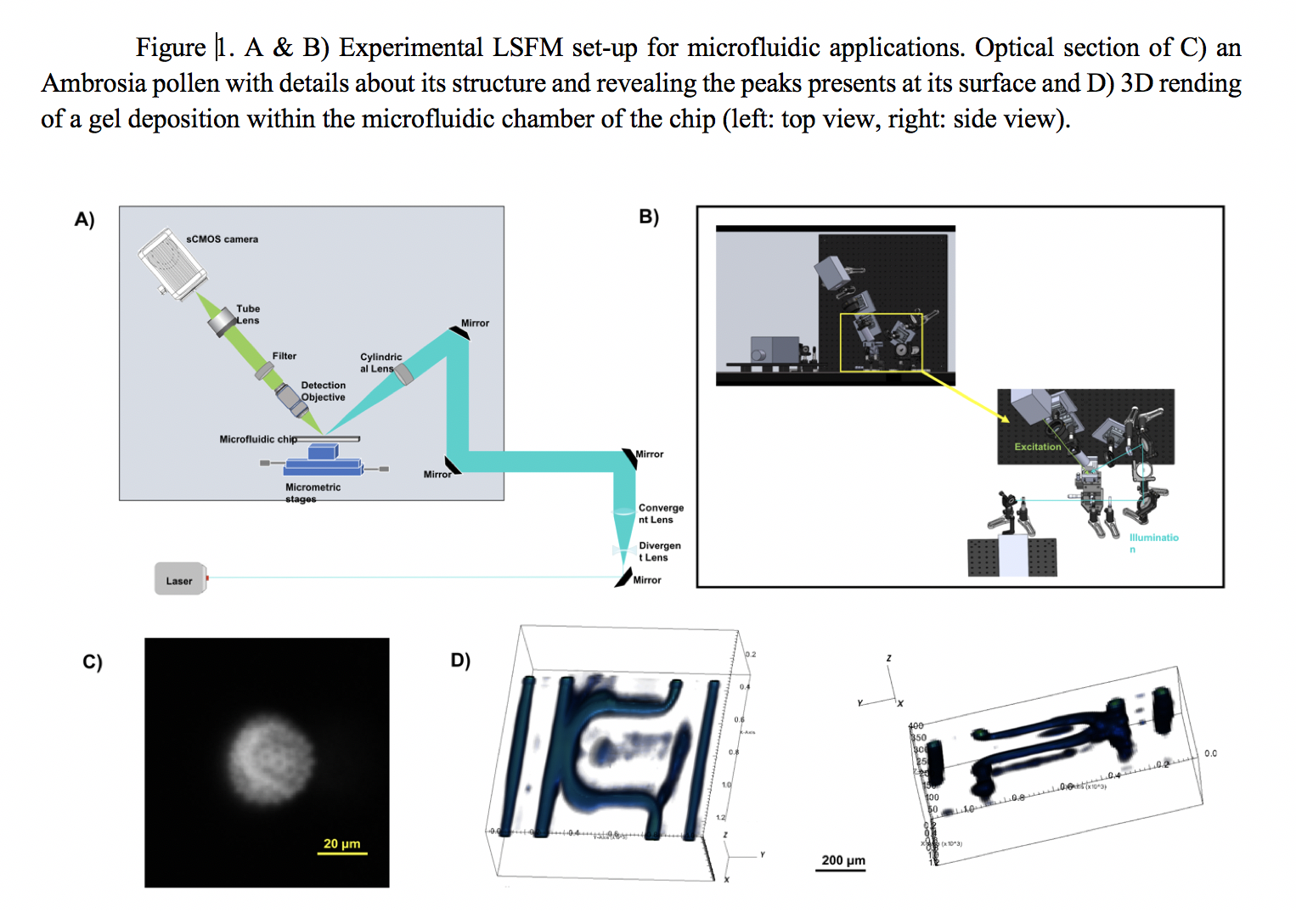
As shown in Fig.1(A&B), an expanded 488 nm Ar laser beam is shaped into a light sheet using a cylindrical lens. The propagation direction of this excitation beam creates a 45° angle with the microfluidic chip, which will necessary be maintained horizontally. The microfluidic chip can then move thanks to translation stages to scan the whole sample volume. The emitted fluorescence is collected by a long working distance 20x objective, associated with a long-pass filter and a tube lens. The wide-field image is acquired on a sCMOS camera . Illumination and detection arms are at 90° from each other. We characterized the optical performances of our system using fluorescent microspheres with a size lower than the expected resolution. Fitted Gaussian curves of the PSFs give for the lateral resolution measured at the FWHM a value of 6,5 ± 0.7μm inside the microfluidic chamber. We investigate the impact of the coverslip, the buffer solution and the induced optical aberrations on the image quality.  

 Results & Conclusion

To validate this system with more complex samples, we first observed diverse samples such as pollen (Fig.1.C). To demonstrate 3D imaging ability inside microfluidic chips, we imaged fluorescence gel deposits in channels of an original microfluidic design. Using a simple data stacking and a minimal data analysis, their 3D rending can then be obtained (Fig1.D). In our case, it is then possible to evaluate the thickness of the gel deposit, and then decide which working flows to use in our microfluidic experiment. We will also present the first images obtained on vascularized network.

This LSFM development leads to an innovative and relatively simple microscope dedicated to sample imaging in microfluidic chip. The system is currently under optimization to enhance the image quality and to reduce optical aberrations by modifying the incidence angles on the chips. The next step is to perform a real time 3D volume acquisition of a continuously perfused biological sample.

Uncaptioned visual


17 Miniaturized Light-Sheet Microscope with active control of optical paths

Spyridon Bakas, Deepak Uttamchandani, Ralf Bauer
University of Strathclyde, United Kingdom

Abstract Text

Light-sheet microscopy systems are starting to become more advanced in order to revise and improve aspects such as, light-sheet uniformity, axial resolution over wider field of view,  or imaging resolution and contrast [1]. Inevitably, the cost of the microscopes starts to increase, reaching levels that can be considered unaffordable for certain labs and settings. In this work, we present a miniaturized version of a LSM that aims to reduce the cost and size as well as provide active control of all the optical paths. Imaging of the whole 3D voxel takes therefore place with the sample at rest. Control of the optical paths through small-scale active optical elements are based on Microelectromechanical Systems (MEMS) technology, using a bimorph varifocal mirror, a piezoelectric 2D scanning mirror, and an electrical tunable lens (Optotune). In previous work, we have shown synchronised movement of the two optical paths in order to create a 3D image of the sample over a 200 μm depth and 500 μm x 280 um width and height [2]. In this work we introduce a custom 3D printed optical prism to reduce astigmatism in the image collection for flat bottomed sample holders. In this way we attempt to image both “thin” samples fixed on microscope slides as well as “thick” samples embedded in an agarose gel volume. We manage to achieve significant reduction of astigmatism, especially for “thick” samples whereas the imaging resultion is set to sub-micron levels.

Uncaptioned visual

Figure 1. a) 3D schematic of the Miniaturized LSM b)Schematic of the light-path intersection and sample mounting including 3D printted prism 


25 Video-rate remote refocusing through continuous oscillation of a deformable mirror.

Terry Wright, Hugh Sparks, Carl Paterson
Photonics Group, Dept of Physics, Imperial College, London, United Kingdom

Abstract Text

KEY WORDS: Light sheet fluorescence microscopy, high-NA imaging, remote refocusing, video frame-rate imaging.


There are numerous applications in microscopy where it is desirable to refocus a high numerical aperture objective lens rapidly [1]. In this work, an Alpao DM97-15 membrane deformable mirror was used to refocus a 40×/0.85 air objective and a 40×/0.80 NA water-immersion objective through a defocus range of -50 to 50 µm at 26.3 sawtooth sweeps per second (Figure 1). Such mirrors are known to exhibit viscoelastic creep and temperature dependent variations in the mirror response [2]. In this work, viscoelastic creep was avoided by ensuring that the temporal average of the surface applied to the mirror was flat over timescales comparable to the creep time constant. Optimisation of the mirror surface to achieve refocusing with a high-NA objective was performed with the mirror continuously refocusing at the desired refocus sweep rate. An initial warm-up period of 5 minutes of oscillation was used to allow thermal effects to stabilise prior to the start of the mirror optimisation procedure. This deformable-mirror-based refocusing system was incorporated in a light-sheet fluorescence microscope in order to perform video-rate volumetric imaging.

Uncaptioned visual

Figure 1. An array of 1 µm pinholes was illuminated with an LED and imaged in transmission by the system. The pinholes were on a motorised stage and were moved towards the microscope objective at 6 µm/s. The objective was refocused by the DM at a rate of 26.3 refocusing scans/s over a range of -50 to 50 µm. Each column shows the central 50×50 µm2 sub-region of the field of view of images acquired for DM refocus positions over the range -50 to 50 µm.


27 On-Chip high-throughput LSFM analysis of single cancer cells

Federico Sala1,2, Roberto Memeo1,2, Michele Castriotta1, Petra Paié2, Andrea Farina2, Roberto Osellame2,1, Francesca Bragheri2, Andrea Bassi1
1Politecnico di Milano, Italy. 2Centro Nazionale delle Ricerche - Istituto di Fotonica e Nanotecnologie, Italy

Abstract Text

The study of cellular populations heterogeneity is becoming a standard in biology, thus single-cell analysis techniques able to process tens or hundreds of cells per minute are required to collect statistically significant quantity of data in a reasonable amount of time. In this framework, fluorescence cytofluorimetry showed the ability to analyse large number of samples in a fast, automated and user-friendly fashion. However, the information that can be obtained from this technique are limited, like presence, absence or intensity of a specific fluorescence signal, or widefield 2D images. Light sheet fluorescence microscopy can play an important role in this field, by combining its fast imaging capability with fluidic continuous sample delivery.

We present an integrated all-glass device where an engineered microchannel is used to handle and deliver single cells, one after the other, in an automated fashion. The samples are flown in the channel through a prealigned light-sheet, allowing continuous acquisition.  A cylindrical microlens is embedded in the chip, in order to shape the light-sheet directly on the same device, with no need of external optical systems, guaranteeing a robustness and precise alignment. The chip, with a footprint smaller than a coin, is designed to be easily mounted on a standard inverted microscope as a functional add-on. A scheme of the device is presented in Figure 1.a.

Uncaptioned visual

The whole device is realized using femtosecond laser micromachining, a versatile technique that allows the fabrication of 3D structures in biocompatible and inert materials like fused silica glass. The precision of the technique allowed us to shape the microchannel in order to reduce to the minimum the optical aberration in signal detection and to optimize the microlens profile, to be compatible with different excitation wavelengths.

We demonstrated the performances of our device by acquiring dual-color 3D stacks of hundreds of single cancer cells with a totally automated system, driven by a custom software. Each cell can be analysed in less than one second, still ensuring a three-dimensional resolution that allows the identification of cellular structures and sub-nuclear vesicles. In Figure 1.b a sequence of acquired sections of the same cell is reported. We believe that our system could be a powerful tool for cellular population studies and inspection of cancer cell heterogeneity and epigenetic alterations.


31 Development and application of a flexible light sheet fluorescence microscope for high speed 2D and 3D imaging of calcium dynamics in cardiomyocytes.

Liuba Dvinskikh, Hugh Sparks, Alice Francis, Carl Paterson, Sian Harding, Ken MacLeod, Chris Dunsby
Imperial College London, United Kingdom

Abstract Text

The high prevalence and poor prognosis of heart failure are two key scientific drivers behind research into the electrophysiology of healthy and damaged cardiac tissue. Dyssynchronous calcium release and disorganization in the t-tubule structure within individual cardiomyocytes has been linked to poor contractile function and arrhythmia. Confocal line scan microscopy has been used widely for single dimensional analysis of calcium signals in cardiomyocytes. However, the physiological anisotropy of cardiomyocytes is fundamental to calcium signalling and impulse propagation through adjacent cells in heart tissue and the extension of confocal scanning to more spatial dimensions comes at the cost of reduced signal to noise ratio and temporal resolution. 

Correlative imaging of the calcium dynamics of cardiomyocytes and their microstructure calls for the use of imaging techniques capable of high-speed two-dimensional or real-time video-rate three-dimensional microscopy of cardiac tissue at subcellular resolution.  This work implements a custom-developed inverted light sheet fluorescence microscope for studying calcium dynamics in cardiomyocytes [1]. The system can operate using two different illumination modes: slower high-resolution imaging using scanned line illumination, and light sheet illumination for fast high-speed 2D and 3D imaging with lower phototoxicity and photodamage to the cells. Video rate volumetric acquisition is achieved by scanning the detection focal plane using folded remote refocusing [2], scanned synchronously with the axially-swept illumination light sheet, with minimal mechanical disturbance of the sample. 

The spatial and temporal resolution achieved allows multidimensional characterization of calcium dynamics across single cardiomyocytes in correlation with t-tubule microstructure. We present data obtained with this system demonstrating two-dimensional dual channel optically sectioned time-lapse imaging of calcium dynamics in isolated live rat cardiomyocytes at 150 fps and 0.5 μm lateral resolution. Electrical pacing is used to stimulate calcium transients with decoupled contraction. The fluorescence intensity trace of calcium indicator Fluo-4 is used to characterize the spatial variation in transient rise time across the cell (Fig. 1). Preliminary results indicate some variation in the time to half maximum within a cell, which was seen to be consistent across multiple transients within the same cell. 

Uncaptioned visual

Fig. 1 Calcium transient analysis in live cardiomyocytes (a) Spatially separated Cell Mask Orange (CMO) and Fluo-4 (F4) emission at peak of Ca2+ transient. Intensity traces of pixels at indicated positions (green dots) are shown in (b): Fluo-4 emission intensity cell average (bold) and example individual pixel traces with linearly interpolated half-maximum (HM) frames. (c) Variation of relative frame number (6.67ms/frame) corresponding to the half-maximum  across cell for an individual transient. Image dimensions: 85 μm x 170 μm.


39 Optimal Laser Pulse Repetition Rate for Fast In Vivo Imaging using Multiphoton Light-sheet Microscopy

Vincent Maioli, Antoine Boniface, Pierre Mahou, Júlia Ferrer Ortas, Lamiae Abdeladim, Emmanuel Beaurepaire, Willy Supatto
Laboratory for Optics and Biosciences, Ecole Polytechnique, CNRS, INSERM, Institut Polytechnique de Paris, France

Abstract Text

Light-sheet microscopy has demonstrated for many years its ability for fast in vivo imaging of delicate samples. The orthogonal arrangement of excitation and detection reduces the photodamage compared to collinear microscopy techniques such as confocal imaging by limiting out-of-focus excitation. This orthogonal geometry exhibits additional benefits in the case of two-photon excited fluorescence with pulsed lasers. Indeed, the low focusing used to generate the light sheet implies lower peak intensities at the sample compared to other fast multiphoton microscopy techniques, mitigating highly nonlinear photodamage without compromising signal, axial resolution nor optical sectioning. In addition, compared to multifocal multiphoton microscopy, the parallelization of the excitation is obtained without splitting the beam, which is critical in the case of a nonlinear optical process and results in lower illumination mean power required. Together, fast imaging is achieved using lower peak intensity and mean power compared to other strategies for fast multiphoton microscopy thanks to this orthogonal arrangement. Does it result in a different photodamage regime compared to point-scanning microscopy and a different strategy to balance fluorescence signal and photodamage? Is 80 MHz laser repetition rate commonly used multiphoton microscopy adapted to take full advantage of light-sheet microscopy? In this study, we investigated these unexplored questions. We aimed at quantifying the influence of laser parameters (pulse frequency, wavelength, and mean power) on both nonlinear signals and photoperturbations during in vivo imaging using multiphoton light-sheet microscopy. To this end, we developed a systematic experimental workflow using the heartbeat rate of zebrafish embryos as a reporter of linear and nonlinear disruptions. We then quantified linear effects such as heating and established the scaling laws of nonlinear photodamage. This study demonstrates thermal effects potentially due to water absorption dominates photoperturbation in current implementations of two-photon light-sheet microscopes using 80 MHz femtosecond laser sources, unlike in the case of other multiphoton techniques. As a result, we show an order-of-magnitude increase in signal can be obtained at constant heating effect and without reaching nonlinear photodamage threshold using lower laser pulse repetition (~10 MHz) [1]. Applying this new optimum to live imaging, we could capture cardiac valve motion at 500 frames per second using two-photon excited fluorescence. Such significant improvement using optimal laser duty cycle opens new opportunities for fast and deep in vivo imaging using multiphoton light-sheet microscopy.


8 Light Sheet Fluorescence Microscopy

Abdullahi Aborode1, Raheemat Rafiu2, Monsour Zakariyah3
1University of Ilorin, Nigeria. 2Missouri University of Science and Technology, USA. 3Federal University of Agriculture, Nigeria

Abstract Text

Light sheet fluorescence microscopy (LSFM) functions as a non-destructive microtome and microscope that uses a plane of light to optically section and view tissues with subcellular resolution. 

This method is well suited for imaging deep within transparent tissues or within whole organisms, and because tissues are exposed to only a thin plane of light, specimen photobleaching and phototoxicity are minimized compared to wide-field fluorescence, confocal, or multiphoton microscopy. 

LSFMs produce well-registered serial sections that are suitable for three-dimensional reconstruction of tissue structures. 

Because of a lack of a commercial LSFM microscope, numerous versions of light sheet microscopes have been constructed by different investigators. 

This review describes development of the technology, reviews existing devices, provides details of one LSFM device, and shows examples of images and three-dimensional reconstructions of tissues that were produced by LSFM. 


Keywords

OPFOS, TSLIM, light sheet microscopy, optical sectioning