Stream 4: Diamond Light Source Session 2

14:45 - 16:45 Thursday, 8th July, 2021

Sessions Diamond Light Source Conference Session

Session Organiser Peijun Zhang

Diamond Light Source is the UK’s national synchrotron user facility with 32 beamlines along with two state-of-the-art electron microscopy laboratories (eBIC for cryo-EM and ePSIC for aberrationcorrected transmission electron microscopy). Users at Diamond have access to a wide range of cutting edge experimental techniques in microscopy and spectroscopy across multiple length scales, including X-ray imaging, hard and soft X-ray microscopy, XANES and XRD mapping, tomography , and phase-retrieval techniques such as ptychography and coherent diffraction imaging. These have wide ranging applications in fields from biology, environmental, earth and planetary sciences, to materials science and engineering. As for cryo-EM, eBIC provides cryoEM single particle analysis, cryo-electron tomography, electron crystallography, and cryoFIB/SEM. Through this session we aim to showcase a selection of user studies conducted at Diamond, with an introductory staff presentation briefly discussing some of the capabilities and access routes. 


15:00 - 15:30

352 Looking inside the cell; molecular pathology using electron tomography

Professor James Naismith
Rosalind Franklin Institute, Harwell Campus, United Kingdom. Oxford University, Oxford, United Kingdom

Abstract Text

The abstract content is not included at the request of the author.


15:30 - 15:42

105 Cryo-EM and single-particle analysis reveals pore dynamics and asymmetric cargo loading in an encapsulin nanocompartment.

Dr Jennifer Ross1, Mr Zak McIver2, Dr Cecilia Piergentili2, Miss Jasmine Bird2, Dr Efraín Zarazúa Arvizu3, Dr Kevin Waldron2, Dr Marcus Wilson3, Dr Arnaud Basle2, Dr David Clarke3, Dr Jon Marles-Wright2
1University of Bristol, Bristol, United Kingdom. 2Newcastle University, Newcastle Upon-Tyne, United Kingdom. 3Edinburgh University, Edinburgh, United Kingdom

Abstract Text

The abstract content is not included at the request of the author.

Keywords

Cryo-EM, Single-Particle, Encapsulin, Ferritin, Iron, Ferroxidase, Nanocompartment


References


15:42 - 15:54

126 Multi-modal chemical imaging of catalysts – from micro to nano scale resolution

Dorota Matras1,2, Antonios Vamvakeros3,4, Simon Jacques3, Andrew Beale4,5,3, Paul Quinn1
1Diamond Light Source, Didcot, United Kingdom. 2The Faraday Institution, Didcot, United Kingdom. 3Finden Ltd, Abingdon, United Kingdom. 4Department of Chemistry, University College London, London, United Kingdom. 5Research Complex at Harwell, Didcot, United Kingdom

Abstract Text

Summary

Chemical imaging techniques were applied to investigate the complex solid-state chemistry in catalytic systems used for methane conversion processes. The results, obtained with high resolution diffraction and spectroscopic imaging, improved our understanding of the catalyst structure-function relationships.

Introduction

Heterogenous functional materials, such as solid catalysts and battery materials typically possess a non-uniform 3D structure and are known to change with time under operating conditions. High resolution chemical imaging techqniues have the potential to provide a unique insight into the complex structure-function relationships. For example, the combination of computed tomography (CT) with techniques such as X-ray diffraction (XRD), X-ray fluorescence (XRF) and X-ray absorption near edge spectroscopy (XANES) enables for the extraction of local chemical and physical state information within the interiors of intact materials 1,2 . The obtained spatially-resolved signals can reveal information that would otherwise be lost in bulk measurements. We show here how such chemical imaging techniques have been applied to study catalytic materials at microfocus (I18) and nanofocus (I14) beamlines of the Diamond Light Source.

Materials and methods

1. Materials

The 2 wt.% La - 2 wt.% Mn – 1.6 wt.% Na – 3.1 wt.% W/SiO2 and the 2 wt.% Mn – 1.6 wt.% Na – 3.1 wt.% W/SiOcatalysts were prepared by the sequential incipient wetness impregnation method. The 10% wt.% Ni – 0.2 wt.% Re/10 wt.% CeO2-ZrO2/Al2O3 catalyst was prepared by sequential impregnation method. All materials were kindly provided by the Boreskov Institute of Catalysis (BIC). 

2. Methods

Combined μ-XRF-CT and μ-XRD-CT measurements were performed at beamline I18 of the Diamond Light Source using a 13 keV monochromatic X-ray beam focused to a spot size of 2.3 μm (V) × 3.5 μm (H). XRF spectra were collected with the Standard Vortex Si Drifts detector and the XRD patterns were collected with a Photonic Sciences CMOS-based X-ray imaging detector with exposure time of 350 ms per point. Catalyst particles were placed inside a quartz capillary (0.5 mm outer Ø and 0.4 mm inner Ø) supported by glass wool. Tomographic measurements were performed with 125 translation steps (with 5 μm step size) and 55 angular steps (with 3.5° step size) covering angular range from 0 to 189°. The reaction mixture (50 % of CH4/Ar and 20 % of O2/He) with the molar ratio of CH4/O2 equal 4 was introduced to reactor during the measurements, while the reactor was heated to 800 °C by an infrared heater. Every 2D diffraction image was converted to a 1D powder diffraction pattern after applying a 30% trimmed mean filter to suppress the single-crystal diffraction artefacts using in-house developed MATLAB scripts 2

Combined 3D-XRF-CT and 3D-XRD-CT measurements were performed at beamline I14 of the Diamond Light Source using a 18 keV monochromatic X-ray beam focused to a spot size of ~ 50 nm (V) × 50 nm (H). XRF spectra were collected with the XRF SDD in the backscatter geometry and the XRD patterns were collected with the Excalibur detector in transmission geometry with exposure time of 200 ms per point. Catalyst particle was mounted on a standard tomography pin. Tomographic measurements were performed with 230 translation steps (with 800 nm step size), 37 vertical steps (with 800 nm step size) and 46 angular steps (with 4° step size) covering angular range from 0 to 180°. 

Results and discussion

1. Na2WO4-Mn2O3/SiO2 catalyst for oxidative coupling of methane

This study aimed to understand the role of La as a promoter in the Na2WO4-Mn2O3/SiO2 catalyst for the oxidative coupling of methane. Hence, both catalysts (i.e. the La promoted and the unpromoted) were measured under operating conditions with μ -XRF-CT and μ-XRD-CT. As revealed by XRF-CT (Figure 1), at high temperatures catalyst active W species become volatile and migrate from the catalyst particle towards the reactor vessel, a phenomenon that is observed to a lesser extent in the case of the La promoted catalyst. As shown with XRD-CT measurements, this could be explained by formation of NaLa(WO4)2 phase, which is more stable at high temperatures when compared to unpromoted Na2WO4 phase. In addition, the La promoter was found to act as a chemical promoter, enhancing the interaction between metallic species and support material through formation of La-Mn-Si-O phases 3.

Uncaptioned visual

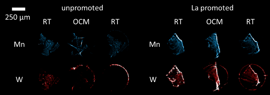
Figure 1. Elemental maps of Mn and W of the unpromoted and La promoted Na2WO4-Mn2O3/SiOcatalysts at room temperature, under OCM reaction and after OCM reaction at room temperature.

2. Ni-Re/CeO2-ZrO2/Al2Ocatalyst for methane reforming

In this study we aimed to understand the role of the noble metal promoter (Re) and the redox promoters (CeO2-ZrO2) by investigating changes, induced during the reforming reaction, in the solid-state chemistry of a multi-component Ni-Re/CeO2-ZrO2/Al2O3 catalyst 4. With 3D-XRF-CT we observed a significant agglomeration of Re species at the surface of the catalyst particle. At the same time, Ce and Zr showed a similar distribution suggesting the formation of a mixed CeO2-ZrOredox material, with Ce species being also present in high concentration without Zr species in only a small volume of catalyst particle. These heterogeneities are bound to have an impact on how coke formation and sintering of metallic active species, will occur during the methane reforming reaction.

Uncaptioned visual

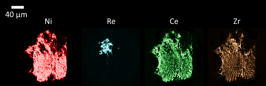
Figure 2. Elemental maps of Ni, Re, Ce and Zr of the Ni-Re/CeO2-ZrO2/Al2O3 catalyst at room temperature before reaction.

Conclusion

Chemical imaging methods allow us to investigate multi-component functional materials with micron and sub-micron resolution. Multi-modal imaging of catalyst particles with XRD-CT and XRF-CT provided a unique insight into the catalyst active site(s), the influence of each promoter on the catalyst performance and the reasons behind the catalyst deactivation.

Keywords

chemical imaging, tomography, catalysis, diffraction, spectroscopy

References


(1) Beale, A. M.; Jacques, S. D. M.; Weckhuysen, B. M. Chemical Imaging of Catalytic Solids with Synchrotron Radiation. Chem. Soc. Rev. 2010, 39 (12), 4656–4672. https://doi.org/10.1039/C0CS00089B.

(2) Matras, D.; Pritchard, J.; Vamvakeros, A.; Jacques, S. D. M.; Beale, A. M. Tomography in Catalyst Design. In Heterogeneous Catalysts; John Wiley & Sons, Ltd, 2021; pp 263–278. https://doi.org/10.1002/9783527813599.ch15.

(3) Vamvakeros, A.; Jacques, S. D. M.; Di Michiel, M.; Middelkoop, V.; Egan, C. K.; Cernik, R. J.; Beale, A. M. Removing Multiple Outliers and Single-Crystal Artefacts from X-Ray Diffraction Computed Tomography Data. J. Appl. Crystallogr. 2015, 48 (6), 1943–1955. https://doi.org/10.1107/S1600576715020701.

(4) Vamvakeros, A.; Matras, D.; Jacques, S. D. M.; di Michiel, M.; Price, S. W. T.; Senecal, P.; Aran, M. A.; Middelkoop, V.; Stenning, G. B. G.; Mosselmans, J. F. W.; Ismagilov, I. Z.; Beale, A. M. Real-Time Multi-Length Scale Chemical Tomography of Fixed Bed Reactors during the Oxidative Coupling of Methane Reaction. J. Catal. 2020, 386, 39–52. https://doi.org/10.1016/j.jcat.2020.03.027.

(5) Matus, E. V.; Ismagilov, I. Z.; Yashnik, S. A.; Ushakov, V. A.; Prosvirin, I. P.; Kerzhentsev, M. A.; Ismagilov, Z. R. Hydrogen Production through Autothermal Reforming of CH4: Efficiency and Action Mode of Noble (M = Pt, Pd) and Non-Noble (M = Re, Mo, Sn) Metal Additives in the Composition of Ni-M/Ce0.5Zr0.5O2/Al2O3 Catalysts. Int. J. Hydrog. Energy 2020, 45 (58), 33352–33369. https://doi.org/10.1016/j.ijhydene.2020.09.011.


15:54 - 16:06

27 Demineralisation of human dental enamel observed by operando X-ray tomography

Professor Alexander M. Korsunsky1, Dr Cyril Besnard1, Dr Ali Marie1, Dr Sisinii Sasidharan1, Dr Robert Harper2, Mr Jonathan James2, Professor Gabriel Landini2, Dr Richard Shelton2, Dr Shashidara Marather3
1University of Oxford, Oxford, United Kingdom. 2Birmingham Dental School, Birmingham, United Kingdom. 3Diamond Light Source, Oxford, United Kingdom

Abstract Text

Human dental enamel is mainly composed of hydroxyapatite (HAp, 96 wt%), bonded with organic matter [1]. It is a hierarchical acellular tissue, formed by ameloblasts in the course of biomineralisation process. During this process, nanocrystals of HAp are assembled into rods of  ̴ 5 μm diameter separated by inter-rod regions of  ̴ 2 μm thickness [2]. The content, orientation and arrangement of HAp crystallites is different in these two phases, leading to different properties. The complex organisation and mineral composition confer high mechanical properties, but leave enamel structure susceptible to acid demineralisation. Dental caries involves demineralisation of enamel by acid produced by bacteria. Caries process is a dynamic phenomenon that involves demineralisation and remineralisation phases [3]. Previous synchrotron tomography and X-ray diffraction studies demonstrated the ability to resolve rod and inter-rod regions, and HAp structure [4,5], and will be further studied using Dual Imaging And Diffraction (DIAD) accepted beamtime at DLS. Our most recent operando synchrotron tomography studies at DLS beamline I13 revealed real time demineralisation of enamel using the combination of fast acquisition and high spatial resolution. In combination with laboratory characterisation and statistical analysis, the results provide spatially resolved data for the rate of demineralisation used as input for multi-scale modelling of demineralisation [6]. This technique offers great potential for other applications, such as the evaluation of oral health benefits from remineralising treatments, analysis of pitting corrosion and scale formation, weathering phenomena, etc.

Keywords

X-ray tomography, synchrotron, demineralisation, dental enamel, caries

References

[1] - T. Baumann et al., Sci. Rep. 5, 15194 (2015). 

[2] - S. Risnes and C. Li, Microscopy Research and Technique 82(10), 1668-1680 (2019).

[3] - J. D. B. Featherstone, Australian Dental Journal 53, 286-291 (2008).

[4] - C. Besnard et al., Poster-Workshop on Neutron and X-Ray Imaging in Life Sciences and Biology (2020).

[5] - T. Sui et al., Acta Biomater. 77, 333-441 (2018).

[6] - E. Salvati et al., Journal of Advanced Research, (2020).


16:06 - 16:09

121 Engineering illumination modes in X-ray ptychography

Mr. yangyang mu1, Dr. Darren Batey2, Prof. John Rodenburg1
1The University of Sheffield, Sheffield, United Kingdom. 2Diamond Light Source, Didcot, United Kingdom

Abstract Text

Ptychography is a type of computational imaging method that is used to overcome the limitation of finite numerical aperture (NA) of lenses and to solve the image phase problem. Its transfer function is, in theory, perfect [1]. Like any method that depends on coherent interference, partial coherence significantly affects the performance of ptychography. Thibault and Menzel [2] showed that the inversion algorithms used in ptychography are amenable to modal decomposition of partial coherence in the illumination. In this work, we present initial results from a hard X-ray imaging experiment where we have deliberately engineered orthogonal modes in the illumination. The field of illumination is interrupted by horizontal and vertical stops. We see that although the total flux used in the experiment is reduced, the proportion of coherent flux (residing in low-order modes) is increased, thereby reducing the number of modes required in the reconstruction. The quality of the reconstruction can also be increased according to the power in the low-order modes. The work suggests that manipulating the illumination optics can enhance the usable coherent flux even when the source is substantially incoherent.

Keywords

Ptychography, X-ray, coherence

References

1.         Rodenburg, J. and A. Maiden, Ptychography, in Springer Handbook of Microscopy, P.W. Hawkes and J.C.H. Spence, Editors. 2019, Springer International Publishing: Cham. p. 2-2.

2.         Thibault, P. and A. Menzel, Reconstructing state mixtures from diffraction measurements. Nature, 2013. 494(7435): p. 68-71.


16:09 - 16:12

43 Instance segmentation of crystalline cones from x-ray microCT of insect eye

Dr. Tunhe Zhou1, Pierre Ticht2, Dr. Hans Martin Kjer3, Assoc. Prof. Vedrana A. Dahl3, Prof. Anders B. Dahl3, Assoc. Prof. Emily Baird4
1Stockholm Univeristy, Stockholm, Sweden. 2Lund University, Lund, Sweden. 3Technical University of Denmark, Lyngby, Denmark. 4Stockholm University, Stockholm, Sweden

Abstract Text

Insects play significant roles in the world ecology and contribute majorly to the biodiversity. Vision is one the most important sensory modalities that they rely on to perform essential activities, such as flight control [1] and collisions avoidance [2], etc. In order to fully understand how insects interact with their environment, it is important to carry out anatomical and functional investigations of their eyes. 

Most insects have a pair of compound eyes. The repeated units in a compound eye are called ommatidia – typically hexagonal as we often see. An ommatidium typically consists of cornea, crystalline cone and rhabdom [3]. The knowledge of the orientation of crystalline cones is important for an accurate estimation of the visual interommatidial angles [4], which decides the angular resolution and hence spatial resolution of the insect vision.

X-ray micro-tomography (microCT) has been demonstrated to be an efficient tool to provide external and internal 3D anatomical structure of the eyes [5]. The optical properties across the field of view of one compound eye can be simulated using the 3D information of the cornea extracted from the microCT data [6]. A typical X-ray microCT scan takes a few minutes in synchrotron radiation and a few hours in laboratory system, and produces a 3D image dataset of around 20003 voxels. Such high throughput and high resolution of X-ray microCT in turn demand adequately efficient image analysis tools in order to extract useful information from the extensive quantity of data. 

In this project, we developed an instance segmentation tool [7], InSegtCone, that automatically segments individual crystalline cones in insect compound eyes after a limited annotation from the user. The design of the tool is shown in Fig. 1, including eye surface modelling, sub-volume unfolding, cone segmentation and back-transformation. By dividing the highly curved eye surface into several subregions, and modelling them mathematically, the 3D volume of the eye can be resampled and unfolded, which allows for slice-wise texture-based segmentation [8]. The segmented cone labels are back-transformed into the original geometries in the end to allow further analysis.

We have demonstrated InSegtCone on microCT images, acquired both at synchrotron radiation and laboratory X-ray system, of three insect species with differently shaped compound eyes: the Western honeybee Apis mellifera, the buff-tailed bumblebee Bombus terrestris, and the green-veined white butterfly Pieris napi. The result of the segmentation of the honeybee is shown in Fig. 2 as an example. Our tool has successfully extracted 60%-80% of the estimated total number of about 6000 crystalline cones and is about 250 times faster than manual labelling of the individual cones. We believe that InSegtCone can be an important tool for peer scientists for extensive studies of the diversity of anatomies of the compound eyes and vision of insects. The code for InSegtCone and test dataset are available at Github [9].

Uncaptioned visual

Fig. 1 The segmentation tool workflow, with examples of one subset of the compound eye of a honeybee.


Uncaptioned visual

Fig. 2 Segmented cones of one honeybee compound eye

Keywords

X-ray microCT, Segmentation, Image analysis, Arthropods, Compound eye, Crystalline cones


References

1.            E. Baird, T. Kornfeldt, and M. Dacke, "Minimum viewing angle for visually guided ground speed control in bumblebees," The Journal of Experimental Biology 213, 1625-1632 (2010).

2.            N. Linander, M. Dacke, and E. Baird, "Bumblebees measure optic flow for position and speed control flexibly within the frontal visual field," The Journal of Experimental Biology 218, 1051-1059 (2015).

3.            M. F. Land, and D.-E. Nilsson, Animal Eyes (Oxford University Press, 2012).

4.            D. G. Stavenga, Pseudopupils of Compound Eyes (1979).

5.            E. Baird, and G. Taylor, "X-ray micro computed-tomography," Curr. Biol. 27, R289-r291 (2017).

6.            G. J. Taylor, P. Tichit, M. D. Schmidt, A. J. Bodey, C. Rau, and E. Baird, "Bumblebee visual allometry results in locally improved resolution and globally improved sensitivity," Elife 8 (2019).

7.            P. Tichit, T. Zhou, H. M. Kjer, V. A. Dahl, A. B. Dahl, and E. Baird, "<em>InSegtCone</em>: Interactive Segmentation of crystalline Cones in compound eyes," bioRxiv, 2020.2012.2015.422850 (2020).

8.            V. A. Dahl, M. J. Emerson, C. H. Trinderup, and A. B. Dahl, "Content-based Propagation of User Markings for Interactive Segmentation of Patterned Images," in 2020 IEEE/CVF Conference on Computer Vision and Pattern Recognition Workshops (CVPRW)(2020), pp. 4280-4288.

9.            "InSegtCone," https://github.com/zhoutunhe/InSegtCone.git.


16:12 - 16:15

281 Nanostructural Analysis of Human Dentine using 3D Small Angle X-ray Scattering Tensor Tomography

Tayyaba Rabnawaz1,2, Nathanael Leung1, Robert Harper3, Tim Snow2, Leonard Nielsen4, Richard Shelton3, Gabriel Landini3, Andy Smith2, Nick Terrill2, Marianne Liebi4, Tan Sui1
1Department of Mechanical Engineering Sciences, University of Surrey, Guildford, Surrey, GU2 7XH, United Kingdom. 2Diamond Light Source, Harwell Science and Innovation Campus, Didcot, OX11 0DE, United Kingdom. 3School of Dentistry, College of Medical and Dental Sciences, University of Birmingham, Birmingham, B5 7EG, United Kingdom. 4Department of Applied Physics, Chalmers University of Technology, Gothenburg, SE-41296, United Kingdom

Abstract Text

Objectives 

Dental caries is one of the most prevalent and costly health issues affecting modern life, which results in the demineralisation and destruction of the dental hard tissues. Demineralisation is a localised process, whereupon the nano-hydroxyapatite (HAp) crystallites in dentine may be dissolved by acid produced by bacteria in dental plaque. This influence both the micro- and nano-structure and its mechanical properties of the affected dental hard tissues. In order to understand the alterations in mechanical properties of naturally and artificially demineralised dental tissue, it is essential to comprehend the correlation between its nanostructural arrangement and the organic and inorganic components.

To investigate the complex structure of dentine at the nano-scale, a recently developed advanced imaging technique called 3D small angle X-ray scattering tensor tomography (3D-SASTT) has been developed and at Beamline I22, Diamond Light Source. This technique was applied to understand the structural variations, of the collagen fibrils and HAp crystallites in dentine occurring in dental caries and artificial demineralisation.

Methods/Materials 

Five dentine sample groups were studied in the 3D-SASTT experimental setup, using two translation axes (x and y) with different rotational angles α (0- 180o) and tilt angles β (0- 45o). Each sample was raster scanned in the x-y planes to cover the field of view using a 14 keV X-ray beam. After collecting the 3D-SASTT dataset, a reconstruction algorithm was used to reveal the 3D structural alterations and changes in orientation of the nano-collagen fibrils and nano-HAp crystallites. 

Results 

The reconstruction revealed the intensity distribution and orientation of collagen fibrils and HAp crystallites. Both natural carious dentine and artificially demineralised dentine showed structural changes with respect to the normal dentine: carious dentine showed more pronounced orientational changes in the nano-collagen fibrils and nano-HAp crystallites compared with the artificially demineralised dentine.

Conclusion

The data indicated that the recently developed 3D-SASTT method was able to detect the 3D structural arrangement of the main dentine components. It was also able to show structural variations in dentine, due to the effect of carious and artificially demineralisation, primarily manifesting themselves as changes in the orientation and degree of orientation of the collagen fibrils and HAp crystallites within the sample studied.

Acknowledgements

Funding from the doctoral studentship from the EPSRC Centre for Doctoral Training in Micro- and Nano Materials and Technologies (EP/L016788/1) at the University of Surrey and Diamond Light Source are gratefully acknowledged and access to the Beamline I22 under the allocation SM20285-1. 


Keywords

small angle X-ray scattering tensor tomography; nanostructure; human dental tissue; dental caries; artificial demineralisation


16:18 - 16:21

300 Structural and spectroscopic insights on elemental distribution in bimetallic nanoparticles through combined atomic resolution electron microscopy and nano X-ray fluorescence imaging.

Mr Panashe Mhembere1, Dr Matthew E. Potter1, Dr Thomas Slater2, Dr Julia Parker2, Prof Robert Raja1
1University of Southampton, Southampton, United Kingdom. 2Diamond Light Source, Didcot, United Kingdom

Abstract Text

Structural And Spectroscopic Insights On Elemental Distribution In Bimetallic Nanoparticles Through Combined Atomic Resolution Electron Microscopy And Nano X-Ray Fluorescence Imaging 

Panashe Mhembere, Matthew E. Potter, Thomas Slater, Julia Parker, Robert Raja

Summary

XAFS techniques are among the conventional methods of analysis of bimetallic nanoparticle (NP) systems. These methods can afford extensive insight into a system, including distribution of each of the individual metals. However, bimetallic NPs containing metals with overlapping X-ray absorption energies are not easily characterised with XAFS techniques, due to the XANES of one region occurring in the EXAFS region of the other. Therefore, alternative means of evaluating the bimetallic distribution must be used. The methodology described here addresses this through the careful application of multiple, complementary X-ray and electron characterisation techniques that do not rely on the inference of local structure by XAFS. The utility of such multi-faceted approach towards the characterisation of bimetallic NP catalysts has been evaluated and confirmed in this study of bimetallic AuxPty/SiO2 catalysts for aerobic oxidation.

Introduction

A wide range of methods exist to create metal nanoparticles, where their functionality will vary with the composition, shape and local environment. The wide range of synthetic options mean that these NPs can be tailored for many different applications, including catalysis. Combination of two metals in NPs can improve the activity of NP catalysts in comparison with their monometallic counterparts; a phenomenon known as bimetallic synergy.

Here we show our facile, one-pot method to create alloyed, bimetallic Au and Pt NPs, with varying Au and Pt ratios, immobilised on mesoporous silica (AuxPty/SiO2). These materials were tested for the catalytic aerobic oxidation of KA oil (1:1 mixture of cyclohexanone and cyclohexanol), exclusively to cyclohexanone, a key step in industrial Nylon-6 synthesis.


Uncaptioned visual

Figure 1.  a) Powder XRD patterns and b) Catalytic activity of AuxPty/SiO2 catalysts.

Alloyed NPs, specifically, were targeted to enhance the activity of the system, while also making the NPs more resilient under reaction conditions. Figure 1 shows the diffraction data that evidenced alloyed NPs, along with the corresponding changes in the catalytic activity. Thus the main focus of our combined atomic resolution electron microscopy and X-ray fluorescence imaging was to show structural and spectroscopic evidence of these alloyed NPs. Our findings were then extended to rationalising how these features contributed to the catalytic behaviour observed.


Methods/Materials

In this work, numerous X-ray and electron-based techniques were applied. Lab-based diffraction alone showed limited evidence of alloys, due to the small quantity of metal (ca. 0.7 wt%). However, high-resolution electron microscopy (ePSIC) and XRF imaging (I14) proved indispensable in showing the distribution of Au and Pt. The STEM-EDX, conducted in the ePSIC JEOL ARM200, showed the elemental distribution of Au and Pt in multiple individual NPs, and the average composition matched the intended Au:Pt ratio for the different catalysts.

By using TEM finder grids, the same NPs were then probed using X-ray imaging (I14), where nano- XRF mapping showed the distribution of Au and Pt within the SiO2 support framework. Pt XANES mapping  was combined with the XRF imaging, to investigate any Au-Pt interactions, from the proposed alloying of Au and Pt. As the electron and X-ray techniques were used on the same catalyst particles, a direct correlation could be made between the atomic structure observed using the ePSIC branch and the spectroscopic data from the I14 beamline.


Results and Discussion

X-ray diffraction patterns of the bimetallic catalysts showed that the (111) and (200) reflections of the metals progressed from Au to Pt with increased Pt content (Figure 1a). This was suggestive of alloyed NPs.

In the catalytic reactions, Pt/SiO2 demonstrated high activity, whereas Au/SiO2 demonstrated much lower activity (Figure 1b). Crucially, the conversion of KA oil was significantly altered by the bimetallic NPs. The change was such that the activity of one of the bimetallic catalysts (Au1Pt4/SiO2) was superior to that of both the Pt/SiO2 and Au/SiO2 monometallic analogues. It was clear from the catalytic data that there was some form of interaction between Au and Pt in these catalysts.


Uncaptioned visual

Figure 2. HAADF-STEM micrograph of Au1Pt1/SiO2 with the corresponding EDX maps. Average line profile of EDX counts from centre of particles (for 3 particles) is shown adjacent, demonstrating no core-shell dealloying.

STEM imaging was used to get a more detailed perspective on the NPs present in the catalysts, coupled with EDX to show the distribution of Au and Pt in the Au1Pt1/SiO2 catalyst (Figure 2). EDX maps showed a homogeneous distribution of Au and Pt within individual NPs. Radial line profiles were taken from the centre of particles using averaged AuPt distribution data from 3 different particles. These showed no evidence of core-shell structure, as the Au and Pt content remained comparable at all distances from the centre of the NPs.


Uncaptioned visual

Figure 3. XRF map of au and Pt in Au1Pt1/SiO2 showing homogeneous distribution through the support framework.

The XRF imaging of the entire Au1Pt1/SiO2 particle (Figure 1) showed that the distribution of Au and Pt were not only homogeneous within individual particles, but also that there was homogeneous distribution of Au and Pt within the support too. This, along with the diffraction data suggest that Figure 2 is representative of the whole sample.

Conclusion

The collaboration with the ePSIC/I14 teams at Diamond light source facilitated this study by providing high level characterisation that showed strong evidence of alloyed AuPt NPs with varying nominal proportions of Au to Pt. The details of how the combined HAADF-STEM/EDX and nano- XRF/XANES imaging were applied to probe the origin of the observed synergy will be presented at the conference.


16:21 - 16:24

72 A synchrotron X-ray Computed Tomography investigation of solute transport under unsaturated conditions

Dr. Sharul Hasan1, Dr. Vahid Joekar-Niasar2, Dr. Nikolaos Karadimitriou3, Dr. Jose R. A. Godinho4, Dr. Nghia T. Vo5, Senyou An2, Arash Rabbani2, Prof. Holger Steeb3
1School of Chemical and Energy Engineering, Universiti Technologi Malaysia, Johor Bahru, Malaysia. 2Department of Chemical Engineering and Analytical Science, University of Manchester, Manchester, United Kingdom. 3Institute of Applied Mechanics (CE), University of Stuttgart, Stuttgart, Germany. 4Helmholtz Institute Freiberg for Resource Technology, Freiberg, Germany. 5Diamond House, Harwell Science and Innovation Campus, Didcot, United Kingdom

Abstract Text

Solute and contaminant transport is a process occurring in the subsurface with a vast socioeconomic impact. Despite the significant literature existing for single-phase solute transport, there is a significant scarcity of experimental data regarding multi-phase, commonly referred to as “unsaturated”, transport. This scarcity of experimental data has led to the adaptation of formulations from the single-phase transport case, leading to erroneous results. One of the characteristics of transport under unsaturated conditions is this of the early arrivals and elongated tails for the solute concentration, which is characteristic for non-Fickian transport. This is mostly attributed to the competition of two transport mechanisms, advection and diffusion, as the result of the interplay between stagnant and flowing zones.

This study focuses on the experimental investigation in three dimensions of solute transport under unsaturated conditions in glass-beads columns. Potassium iodide (KI) water solutions were used to displace the non-wetting phase, namely Fluorinert FC-43. The process was monitored using time-lapse computed tomography at a spatial resolution of 3.25 microns every  6 seconds, with imaging taking place at the tomography beamline I12 (27) at the Diamond Light Source, UK (Hassan et al., 2020). Various KI concentrations and flow rates were realized, and detailed information was acquired on solute transport at different carrier-phase saturation.

Our results show that the spatial distribution of phases, and the formation of the stagnant and flowing zones within water (the carrier phase), are critical in the development of non-Fickian transport, which should be included in the Darcy-scale transport models. We also experimentally verify the numerical speculation that a full-mixing assumption in a single pore is not consistent with our findings, and that even within a single pore, transport can exhibit two distinct time scales even after the introduction of multiple pore volumes. Finally, we show that dispersivity is also related to saturation. Dispersivity nearly doubled in the case of a half-saturated sample compared against this of a fully saturated one.

This experimental work is the first one to provide the scientific community with concrete evidence three-dimensional evidence, that the formulations describing solute transport under unsaturated conditions must be accordingly updated. Additionally, it provides indisputable proof that the use of the formulations developed for saturated (single-phase) conditions are not suitable to characterize unsaturated transport.


Keywords

synchrotron, X-ray Computed Tomography,  solute transport,  unsaturated conditions, non-Fickian transport, advection, diffusion, dispersivity

References

S. Hasan, V. Niasar, N. Karadimitriou, J. Godinho, N. Vo, S. An, A. Rabbania, and H. Steeb. Direct characterization of solute transport in unsaturated porous media using fast X-ray synchrotron microto- mography. Proceedings of the National Academy of Sciences, 117(38):23443–23449, 2020.


16:24 - 16:27

99 Phase-contrast 3D tomography of cells grown in polymer electrospun scaffolds using synchrotron X-rays at Diamond Light Source (I13-2)

Ms Archana Bhartiya1,2, Dr Kamel Madi3, Dr Catherine Disney4, Dr Loic Courtois3, Dr Andrew Jupe5, Dr Fucai Zhang6,7,2, Dr Andrew Bodey8, Professor Peter Lee7, Professor Christoph Rau8, Professor Ian Robinson1,2,9, Dr Mohammed Yusuf1,10,2
1UCL, London, United Kingdom. 2Research Complex at Harwell, Didcot, United Kingdom. 33dMagination Ltd, Didcot, United Kingdom. 4University of Manchester, Manchester, United Kingdom. 5The University of Buckingham, Buckingham, United Kingdom. 6Southern University of Science and Technology, Shenzhen, China. 7University College London, London, United Kingdom. 8Diamond Light Source, Oxfordshire, United Kingdom. 9Brookhaven National Laboratory, Upton, USA. 10Aga Khan Unversity, Karachi, Pakistan

Abstract Text

Advanced imaging is useful for understanding the three-dimensional (3D) growth of cells. X-ray tomography serves as a powerful noninvasive, nondestructive technique that can fulfill these purposes by providing information about cell growth within 3D platforms. There are a limited number of studies taking advantage of synchrotron X-rays, which provides a large field of view and suitable resolution to image cells within specific biomaterials. In this study, X-ray synchrotron radiation microtomography at Diamond Light Source and advanced image processing were used to investigate cellular infiltration of HeLa cells within poly l-lactide (PLLA) scaffolds. This study demonstrates that synchrotron X-rays using phase contrast is a useful method to understand the 3D growth of cells in PLLA electrospun scaffolds. We examined two different fiber diameter scaffolds with different pore sizes, grown over different days in vitro  Here we also show the infiltration and cell connectivity after segmentation of the cells in the different scaffold materials. This study indicates potential for using such 3D technology to study cell–scaffold interactions for future medical use.


Keywords

3D culture; electrospun polymers; scaffold; X-ray CT; synchrotron; image analysis; Avizo.


References

Bhartiya, A., Madi, K., Disney, C. M., Courtois, L., Jupe, A., Zhang, F., Bodey, A. J., Lee, P., Rau, C., Robinson, I. K., & Yusuf, M. (2020). Phase-contrast 3D tomography of HeLa cells grown in PLLA polymer electrospun scaffolds using synchrotron X-rays. Journal of synchrotron radiation27(Pt 1), 158–163. 


16:27 - 16:30

210 4D in-situ microtomography and image analysis of aerosol filtration

Mr Matthew Jones1, Dr David Eastwood1, Dr Andrew York2, Dr Timothy Hyde2, Dr Malte Storm3, Prof Sarah Haigh4
1University of Manchester at Harwell, Oxford, United Kingdom. 2Johnson Matthey, Reading, United Kingdom. 3Diamond Light Source, Didcot, United Kingdom. 4University of Manchester, Manchester, United Kingdom

Abstract Text

Understanding the microscale processes underlying filtration is fundamental to optimising filter design and understanding aging. We have developed and integrated a novel in-situ flow rig for the 4D imaging of aerosol filtration in vehicle particulate filters at Diamond Light Source beamline I13-2. This combined imaging and aerosol flow system has revealed the local microstructure at positions where particulates are captured and agglomerate, allowing us to assess the factors affecting filter performance.  

Gasoline particulate filters (GPFs) are used in current and next generation gasoline vehicle exhausts in order to meet environmental regulations that restrict the amount of toxic particulate matter (PM) emitted (Joshi & Johnson, 2018). Thus, these filters are a key technology in effectuating a cleaner transition to electric transport. GPFs are wall flow filters constructed of macro-porous cordierite extruded into long channels. In use the exhaust aerosol flows down these channels and through the porous walls of the cordierite, depositing PM on the wall surfaces and in the internal pore space of the filter. These deposits impede gas flow through the filter, increasing backpressure and reducing the fuel efficiency of the engine. Herein lies the key trade off in efficient GPF performance; low backpressures that improves fuel efficiency verses high filtration efficiency that prevents toxic substances entering the urban environment. Key to understanding this trade-off is observing the evolution of the pore network and the deposits within.

4D  in-situ x-ray microtomography (XMT) allowed us to resolve the 3D microstructure of the filter, the deposited particulates, and importantly how the deposits evolve with time during a filtration process. We achieved this 4D study by creating a novel aerosol flow rig for in-situ parallel beam XMT. This consisted of an air compressor that provided head pressure to a dust generator which output a steady aerosol stream into the flow rig. We flowed TiO2 particles with nominal diameter 25nm, which displayed similar aerodynamic and clustering behaviours to real world engine exhaust. The aerosol then flowed through the customised 3D printed sample holder which maintained stability during tomographic rotation but was thin enough to minimize beam attenuation. Static pressure measurements were obtained before and after the filter position. The sample was placed at the thin central section of the holder, as shown below in Figure 1, such that the aerosol flowed through the centre of the filter. 

Tomographic x-ray imaging used a ‘pink beam’ spectrum at beamline I13-2 (Pešić, 2013), combining multiple harmonic peaks from the undulator source. For fast imaging a projection was captured every 0.18o in a 180o fly scan with an exposure of 0.01s. The high flux from the synchrotron source gave sufficient time resolution to resolve the structure of the filter and deposit with only minimal change during each scan. We obtained 1 tomogram per minute, taking flat and dark field images at the beginning of each sequence of tomography scans, rather than after each scan, in order to minimize unnecessary movements. The imaging set-up is illustrated below in Figure 1.

Uncaptioned visual

Systematic analysis of the data used the following workflow: (1) A filtered back projection tomographic reconstruction. (2) Image pre-processing, including distortion correction, cropping, z-axis resampling, de-zinger, non-local means and Gaussian. (3) Binarization of the ‘zeroth’ volume (i.e. clean unloaded filter) into filter and void. (4) Registration of the subsequent deposit loaded volumes onto the zeroth volume. (5) Histogram rescaling across the entire 4D data set. (6) Masking using the ‘zeroth’ filter volume to mask-out the filter phase in the subsequent volumes, and (7) applying a global threshold to segment the deposit phase. Due to the large size of the processed data set (50 GB in 20 × 8 bit volumes of 1125 x 1125 x 2110 pixels) and the grey level overlapping of the phases of interest, this required a programmatic method using a range of python based software (Imageio, Scikit-image, Simple-ITK, OpenCV, and other packages within the Anaconda distribution). 

The resulting time resolved tomography is shown as volume renderings in Figure 2. We can observe the initial deposits in the internal deep bed pores of the filter growing between minutes 1 and 6. At 6 minutes we can see that the deposits in the internal wall have now ‘blocked’ the key flow paths through the filter and have become slightly denser as they have ‘filtered out’ aerosol flowing through the pore throat. Hence, after minute 6, deposits start forming primarily on the wall surface as shown in the minute 9 volume. This is the transition from deep bed to cake filtration as resolved through imaging, this coincided with the point at which we measured an increase in the backpressure from the manometer.


Uncaptioned visual

In conclusion 4D in-situ XMT allows us to observe the evolution of deposit position, extent and relative density with a time resolution of 1 tomogram per minute. We have achieved sufficient resolution to observe pore filling and pore blocking events and the transition from deep bed filtration to cake filtration. The data from this method has potential for model validation for computerised fluid dynamic based filtration models. This methodology highlights opportunities for studying the function of a range of operational particulate filters including machine intake filters, engine exhaust filters and respiratory filters.

Keywords

X-ray Microtomography, Microscopy, In-situ, Aerosol, Filtration, 4D imaging

References

Joshi and Johnson (2018) -  Joshi, A., Johnson, T.V. Gasoline Particulate Filters—a Review.    Emiss. Control Sci. Technol. 4, 219–239 (2018). https://doi.org/10.1007/s40825-018-0101-y

Peši´c , (2018) - Peši´c, Z.D.; Fanis, A.D.; Wagner, U.; Rau, C. Experimental stations at I13 beamline at Diamond Light Source. J. Phys. Conf. Ser. 2013, 425


16:30 - 16:33

64 Tracking Reactions of Asymmetric Organo‐Osmium Transfer Hydrogenation Catalysts in Cancer Cells  by ICP-MS and nanofocussed x-ray fluorescence

Dr Elizabeth Bolitho1,2, Dr James Coverdale1, Dr Hannah Bridgewater1, Dr Guy Clarkson1, Dr Paul Quinn2, Dr Carlos Sanchez-Cano3, Professor Peter Sadler1
1University of Warwick, Coventry, United Kingdom. 2Diamond Light Source, Oxford, United Kingdom. 3Center for Cooperative Research in Biomaterials (CIC biomaGUNE), San Sebastián, Spain

Abstract Text

[OsII[(η6p‐cymene)(1, RR/SS‐MePh‐DPEN)] (MePh‐DPEN=tosyl‐diphenylethylene‐diamine) is a chiral 16‐electron organo‐osmium(II) half‐sandwich complex structurally derived from the well‐established Noyori RuII catalysts,1 which shows high enantioselectivity and conversion rates. For example, reduction of acetophenone is 3‐fold more efficient (in turnover frequency, TOF) and more stable (normal atmospheric conditions) than its industrially‐used RuII analogue. 1 Furthermore, once inside cells, and in presence of the non‐toxic hydride donor formate, 1 catalyses the enantioselective reduction of pyruvate, an essential precursor in cell metabolism, to natural L‐lactate or unnatural D‐lactate, depending on the chirality of the catalyst.2 It can be assumed that the ability of such catalysts to cause metabolic perturbations in cells requires the presence of intact catalyst, which contributes to the antiproliferative activity and selectivity of 1 towards a variety of cancer cell lines.2,3 Yet, its intracellular catalytic activity is most likely marked by low turnover numbers,2 which might suggest some degradation of the complex inside cells. This is a common problem for synthetic metal catalysts, such as organometallic complexes designed to work under well‐defined chemical conditions, including inert atmospheres and in organic solvents. In order to optimise the design of synthetic intracellular catalysts, and increase their in cellulo catalytic and biological efficiency, it is important to investigate their fate in cells.

Previous work using ICP‐MS experiments on fractionated cancer cells treated with 1, showed 47 % cytosolic accumulation of the Os, suggesting that catalysis may take place in the cytosol.2 Additionally, ca. 48 % of intracellular Os was present in the membrane/ particulate fraction (which contains organelles and membrane proteins), which may also implicate organelles (i.e. mitochondria or lysosomes) as cellular targets.2 However, such studies did not provide information on the intracellular stability of the complex. To probe this, we have incorporated a bromine on a sulfonylphenyl substituent in the chelated Ph‐DPEN ligand, so generating [OsII6p‐cym)(BrPh‐DPEN)] (2). A combination of nanofocussed synchrotron X‐ray fluorescence (XRF) and ICP‐MS allows not only osmium but also the chelated PhDPEN ligand to be tracked in cells using the bromine label.

First, pellets of A549 cancer cells treated with 2 under different conditions were digested at 353 K using TMAH and analysed by ICP-MS. Cellular accumulation of Os and Br in cells treated with 2 was determined under varying conditions (temperature, methyl-β-cyclodextrin, verapamil, time and cell fractionation). In all cases, significantly higher concentrations of Br compared to Os were found (>10- fold) suggesting 2 has a limited stability in a cellular environment. The endocytic contribution to the accumulation of 2 was investigated using a known endocytotic inhibitor, which reduced the quantity of intracellular Os with increasing concentration of inhibitor. Interestingly, intracellular levels of Br were initially reduced at the lowest inhibitor concentration, but plateaued with increasing concentration, thus it may be hypothesized that caveolae endocytosis mediates the uptake of the intact complex, but once inside the cell is degraded to release the Br-labelled ligand, which is retained intracellularly for longer. Similarly to 1, the dependence on drug efflux via. pathways associated with P-glycoprotein (using verapamil, a PGP inhibitor) revealed that the efflux of the Os moiety is dependent on PGP. However, Br accumulation was unaffected by verapamil treatment, suggesting that the Br-labelled PhDPEN ligand is not removed from cells by PGP. The extent of cellular efflux of 2 was monitored using varying exposure times and recovery in complex-free media, revealing maximum intracellular levels of Os 3-6 h, whereas, Br reached a maximum after 24 h exposure with around 30× more Br vs. Os, indicative of severe complex instability. Cellular fractionation studies of cells treated with 2 revealed significantly more Br vs. Os (>10-fold), with the majority of Os and Br observed in the membrane and cytoskeletal fractions. This suggests that some of complex is intact in the cytoplasm, where in-cell catalysis likely occurs. Negligible quantities of Os were present in the nuclear fraction which may rule out a DNA-binding mechanism of action. Interestingly, 10-20% Br was present in the nuclear fraction, perhaps as unbound Br-labelled PhDPEN. It may be hypothesized that the Os complex may facilitate the delivery of the PhDPEN into the cell (verified by the lack of toxicity of BsDPEN, IC50>150 µM), where it can then be released and enter the nucleus. Whether the presence of BsDPEN is contributing to the anticancer activity is unknown, and requires further investigation

A549 cells grown on silicon nitride membranes were treated with various concentrations of 2 for 24 h, before being cryo‐fixed and freeze‐dried for subsequent analysis under ambient conditions. Nanofocussed synchrotron XRF at the I14 Beamline (Diamond) confirmed that some of is likely to remain intact as judged by the co-localization of Os and Br in the cytosol, with significantly higher levels of Br vs. Os. Interestingly, XRF revealed the co-localization of Os and Br in small, spherical compartments (ca. 0.6 µm2)4 in cells treated with higher doses of 2, which may imply lysosomal breakdown and complex efflux. Remarkably, Br/Os ratios were lower in those areas than in the rest of the cell, suggesting the presence of higher concentrations of intact complex. The endosomal / lysosomal breakdown was further probed using chloroquine (which de-acidifies lysosomes), revealing a significant increase in potency and catalytic activity, which was accompanied by an increase in Os accumulation. 

Overall, we probed the structure and spatial localisation of a brominated transfer hydrogenation catalyst inside cells with ICP‐MS and nanoscale synchrotron XRF mapping, combined with cellular uptake and mechanistic studies. These experiments showed that the catalyst was degraded in cancer cells, probably through transport into acidic lysosomes following by reaction with cellular thiols. The chelated Br‐ligand (or a Br‐fragment), but not Os, is translocated into the nucleus. Such reactions help to explain the low intracellular TON estimated for these catalysts. This work demonstrates the utility of halogen tags as probes for MS and X‐ray based techniques which elucidate reactions of organometallic anticancer catalysts in cells. 


Keywords

Anticancer catalysts, bioorganometallics chemistry, X-ray fluorescence, organo-osmium complexes, transfer hydrogenation

References

[1]  J. P. C. Coverdale, C. Sanchez-Cano, G. J. Clarkson, R. Soni, M. Wills, P. J. Sadler, Chem. Eur. J. 2015, 21, 8043– 8046.

[2] J. P. C. Coverdale, I. Romero-Canelón, C. Sanchez-Cano, G. J. Clarkson, A. Habtemariam, M. Wills, P. J. Sadler, Nat. Chem. 2018, 10, 347– 354.

[3] J. P. C. Coverdale, H. E. Bridgewater, J. I. Song, N. A. Smith, N. P. E. Barry, I. Bagley, P. J. Sadler, I. Romero-Canelón, J. Med. Chem. 2018, 61, 9246– 9255.

[4] E. M. Bolitho, J. P. C. Coverdale, H. E. Bridgewater, G. J. Clarkson, P. D. Quinn, C. Sanchez-Cano, P. J. Sadler.


16:33 - 16:36

177 Correlative Soft Tissue Synchrotron Microtomography: Sample Preparation, Imaging, Reconstruction and Segmentation Methods

Andrew J Bodey1, Merrick C Strotton2, Win Tun1, Alexey V Buzmakov3, Victoria Gulimova4, Kazimir Wanelik1, Michelle C Darrow1, Elizabeth J Bradbury2, Christoph Rau1
1Diamond Light Source, Oxfordshire, United Kingdom. 2King’s College London, London, United Kingdom. 3FSRC "Crystallography and Photonics" RAS, Moscow, Russian Federation. 4Research Institute of Human Morphology, Ministry of Science and Higher Education RF, Moscow, Russian Federation

Abstract Text

Introduction

Thousands of soft tissue microtomography experiments are conducted at synchrotrons around the world each year, and the quality of results varies widely. Soft biological tissues pose a particular challenge for synchrotron tomography, owing to poor contrast and susceptibility to deformation and beam damage artefacts. The rationale behind the choice of sample preparation methods, imaging parameters and reconstruction strategy is not always reported in articles, and so we conducted a systematic investigation of these aspects of experimental design for central nervous system samples. Computational segmentation can be particularly challenging for soft-tissue tomograms, and so we demonstrate the use of supervoxel-based machine-learning segmentation of our data.

Sample Embedding

Careful sample preparation is crucial for effective imaging of soft tissues. A sample which is optimally oriented, stained throughout and embedded so as to provide rigidity and avoid streak artefacts will deliver excellent results. In order to achieve good inline phase contrast for weakly-absorbing soft tissues, one invariably risks strong fringes at the interface between the embedding medium and air, leading to streak artefacts. The medium will therefore ideally sit outside the field of view, but not by so much that it causes excessive beam absorption.

Following fixation, spinal cords were placed in a custom-made mould. Cords were balanced at either end on lips to ensure they were surrounded on all sides by an appropriate thickness of medium.

Samples must be stable on the length scale of the features one hopes to resolve. Soft tissue samples are often highly flexible, and thus benefit from embedding. We trialed various embedding media, and found that only paraffin and resin conferred good stability. Although paraffin is more prone to cracking than resin, it offers the advantage of reversibility; paraffin was removed after tomography so that other imaging techniques could be used. Paraffin cracking was avoided by hardening the beam and reducing flux [1].

Contrast

The poor contrast of soft tissues can make the visualisation and segmentation of features difficult. Both chemical and optical methods can boost contrast, and some combination of both can be advantageous. We trialed a variety of stains and found that iodine gave the best penetration and contrast – particularly for the central grey matter. We enhanced this chemical contrast with in-line phase contrast, the extent of which is controlled by the propagation distance between sample and detector. Fresnel fringes increase in amplitude and width as propagation distance is increased. We chose 160mm, as it provided good contrast but avoided the excessive blurring which results from wide fringes.

Signal:Noise

Full angular sampling requires π/2 × object radius projection images (~4000 for our experiments). Beyond this, the use of additional images is a convenient means of increasing dose and thereby boosting signal:noise. To determine the optimal number, we performed a 24,001-projection scan and reconstructed from subsets of this dataset. 6,000 projections was found to bring signal:noise reasonably close to saturation without taking so long as to be prohibitive to a beamtime’s efficiency.

Artefact Correction

Various imperfections in an imaging system can lead to reconstruction artefacts. Data were reconstructed with Savu [2], a modular pipeline, which incorporates plugins to correct for various artefacts including zingers (which result from stray X-rays hitting the detector chip directly) and optical distortions in the imaging system [3]. Correcting for distortions can make significant improvements to data accuracy, and thereby the true resolution of data (Fig. 1).

Uncaptioned visual
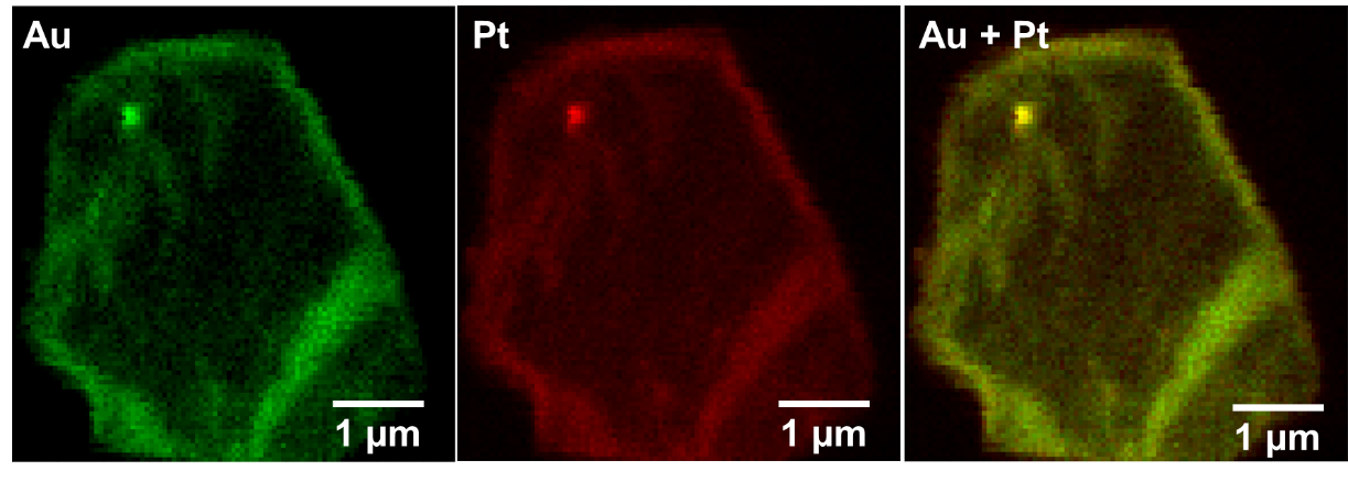
Figure 1. Tomographic slice generated (a) without and (b) with optical distortion correction.

 Segmentation: Supervoxels and Machine Learning

Computational segmentation of soft-tissue tomograms using traditional methods can be difficult. Humans, by contrast, are excellent at pattern recognition. This fact can be harnessed to make use of machine-learning software in which humans train algorithms to recognise patterns. Supervoxels – groups of similar, adjacent voxels – were used to speed up the process (Fig 2a). SuRVoS [4] was used for complete segmentation of grey and white matter and vasculature (Fig. 2b,c). Area measurements were taken along the length of the spinal cord (Fig. 2d). Such measurements could be useful for assessing changes in a variety of pathologies, such as spinal cord or brain injuries [5]. 

Uncaptioned visual

Figure 2. (a) Supervoxels. (b) Segmented grey and white matter. (c) Segmented vasculature. (d) Rostro-caudal measurements.

Correlative Imaging: Post-Tomography Histology

Histology can add molecular specificity to structures identified in 3D via tomography. After tomography, iodine was removed from the samples, which were then re-embedded in wax for sectioning and histology. This included the pan-neuronal marker NeuN, and generic tissue stains such as haematoxylin and eosin which stains cell nuclei (Fig. 3).

Uncaptioned visualFigure 3. Histological images can be aligned to tomograms, with remarkable consistency of features. (a) Haematoxylin and eosin staining of 7μm-thick transverse tissue section. (b) Equivalent ‘6.4μm’ tomographic slice.

Tomography of Unstained Central Nervous System Tissues

Good contrast – albeit via Fresnel fringes – can be achieved with unstained soft tissues via inline phase contrast alone. Spinal cords were sourced either from thick-toed geckos that had spent 30 days in space onboard unmanned spacecraft Bion-M1 (2013, Russia) – or a control group – as part of a blind study into the effects of spaceflight on animal tissues. Fixing and paraffin embedding conferred excellent stability to spinal cords and also whole mouse brains, and cracking could be avoided with suitable beam filtration. Various features could be clearly resolved (Fig. 4a,b,d). Cell bodies, grey matter and white matter were segmented from the spinal cords with SuRVoS (Fig. 4c), and these were used for rostro-caudal area measurements.

Uncaptioned visual

Figure 4. Unstained central nervous system tissues. (a) Transverse and (b) longitudinal sections of gecko spinal cord. (c) Segmentation of grey and white matter, and cell bodies. (d) Mouse brain tissue.

Acknowledgments

Tomographic imaging, reconstructions and analyses were conducted at I13-2 of Diamond Light Source, UK and its associated data beamline [6] (MT12538, MT14907 and MG23866). We thank A Maltsev and Е Shevtsova for preparing the mouse brain samples. The work was supported by King’s Bioscience Institute, Guy’s & St Thomas’ Charity Prize PhD Programme and MRC UK (G1002055).

Andrew Bodey and Merrick Strotton contributed equally to this project.

Keywords

soft tissue, central nervous system, spinal cord, synchrotron, tomography, CT, reconstruction, machine learning, segmentation

References

1. Strotton MC & Bodey AJ (joint first authors), Wanelik K, Darrow MC, Medina E, Hobbs C, Rau C, Bradbury EJ. Scientific Reports. 8 (2018).
2. Atwood RC, Bodey AJ, Price SW, Basham M, Drakopoulos M. Philosophical Transactions of the Royal Society A: Mathematical, Physical and Engineering Sciences. 373 (2015).
3. Vo NT, Atwood RC, Drakopoulos M. Optics Express. 23 (2015).
4. Luengo I, Darrow MC, Spink MC, Sun Y, Dai W, He CY, Chiu W, Pridmore T, Ashton AW, Duke EMH, Basham M, French AP. 198 (2017).
5. Strotton MC, Bodey AJ, Wanelik K, Hobbs C, Rau C, Bradbury EJ. Experimental Neurology. 336 (2020).
6. Bodey AJ, Rau C. Journal of Physics: Conference Series 2017. 849 (2017). IOP Publishing.


16:36 - 16:39

323 Revealing the 3D local atomic structure in an undercooled binary Al alloy melt

Mr Shi Huang1, Mr Shifeng Luo2, Dr Ling Qin1, Prof. Jiawei Mi1
1University of Hull, Hull, United Kingdom. 2Hefei University of technology, Hefei, China

Abstract Text


Understanding of material atomic structure transition from a disordered state to an ordered state is a fundamental issue in materials science. Using the synchrotron X-ray total scattering (SXTS) capability at the beamline I15 of the Diamond Light Source, UK, we studied systematically the temperature-dependent local atomic structures of a liquid Al and an Al-0.4wt% Sc alloy in a wide temperature range from the liquid to solid state. We also used the empirical potential structure refinement (EPSR) modelling method to reconstruct the atomic structure according to the pair distribution functions obtained from SXTS at different temperatures.  Here, we present the generic procedure developed to extract the scattering information of the liquid part from the solidifying semisolid melt, and the temperature-dependent atomic structure evolution. With the decrease of temperature, in the Al-0.4wt%Sc alloy, the Sc-centered (red atoms) Al polyhedral clusters evolved into cluster groups with higher Al atom (the light blue atoms) number connected via vertex, edges and faces. The research shed more light on how a randomly distributed atomic cluster gradually evolved into a cluster with more crystallinity during the solidification processes. 

Uncaptioned visual


Keywords

3D local atomic structure ;synchrotron X-ray total scattering; EPSR modelling 

References

 [1]   A. C. Redfield and A. Zangwill, Physical review letters 58, 2322 (1987).

[2]   D. Faken and H. Jónsson, Computational Materials Science 2, 279 (1994).

[3]   A. Hirata, L. Kang, T. Fujita, B. Klumov, K. Matsue, M. Kotani, A. Yavari, and M. Chen, Science 341, 376 (2013).