To save this page as a PDF, click this button and choose the PDF destination.

Super-Resolution Microscopy

09:45 - 11:45 Friday, 25th June, 2021

Tracks Meeting Session

Session Chair Paula Sampaio

The observation of sub-cellular structures at the smallest scale is essential in biomedical research to put more light in the understanding of the biological processes. Techniques as PALM, STORM, STED or SR-SIM permitted to overcome the diffraction-limited resolution of light microscopy down to tens of nanometres and some new cellular nanostructures emerged. This session will focus on the latest developments of super-resolution microscopy for imaging living cells, which will allow to visualize the organization, dynamic activity and interactions of sub-cellular structures and molecules in its natural context at a scale close to molecular level. Abstracts are encouraged in and around this area.


09:45 - 10:10

95 Live cell compatible super resolution fluorescence microscopy for 3D imaging

Ilaria Testa
SciLifeLab

Abstract Text

The volumetric architecture of organelles and molecules inside cells can only be elucidated with microscopes featuring sufficiently high spatial resolution in 3D. However, current super resolution microscopy methods suffer from severe limitations when applied to live cell imaging such as insufficient resolving power along the optical axis, long recording times and/or photobleaching. Our lab developed MoNaLISA and 3D pRESOLFT techniques capable of 3D isotropic 50 nm imaging in living cells. The parallelization of the illumination module enables rapid (1-2 Hz) acquisition of large field of view (~40-100x40-100 µm2), which allow full-length cellular imaging without compromising speed and spatial resolution. It is achieved with the creation of a new interference pattern featuring an array of 3D-confined and equally spaced intensity minima. This pattern is used to transiently switch photochromic fluorescent probes such as reversible switchable fluorescent protein in dark states allowing a targeted 3D confinement of the fluorescence emission. 

The 3D‑organization of several cellular structures can be visualized, even dynamically, which open new possibilities for 3D in situ biological studies of proteins and organelles in living cells. We are currently using our methods to study organelle such as exosomes, ER and mitochondria proteins trafficking as well as synaptic plasticity in firing neurons.  


10:15 - 10:27

7 Combining photoactivated localization microscopy and spatiotemporal image correlation spectroscopy to Characterize the spatial-temporal dynamics of Aquaporin-2

Eva Arnspang Christensen
University of Southern Denmark

Abstract Text

Aquaporin proteins are homo-tetrameric proteins which transport water and are crucial to the functioning of most organs in the body, especially the kidney. Here, we studied the nanoscale dynamics and organization of the water channel aquaporin-2 (AQP2) in kidney collecting duct cells (MDCK cells) using single particle tracking photoactivated localization microscopy (sptPALM). AQP2 functions in the kidney to help concentrate urine. In kidney cells, AQP2 normally resides in intra-cellular vesicles in the cell cytoplasm, but upon phosphorylation the protein is sent to the membrane, where it exerts its function in transporting water. To characterize both the diffusion coefficient and velocity vectors of AQP2 molecules on the plasma membrane of kidney cells, we acquired sptPALM datasets and then analyzed them with spatiotemporal imagine correlation spectroscopy (STICS). STICs uses a filtering mechanism to remove frequencies associated with immobile components, allowing measurements of protein dynamics even in the presence of a large fraction of immobile species. Using sptPALM and STICS, we acquired transport and spatial maps of AQP2 molecules in the kidney cell plasma membrane. Our results provide the first look at the broad, nanoscale organization and dynamics of AQP2 in kidney cells, opening the door for a deeper understanding of AQP2 function in urine concentration.


10:27 - 10:39

36 Understanding viral assembly with quantitative super-resolution microscopy: from single-molecule imaging to fast live-cell imaging

Romain Laine
MRC-LMCB, UCL. Francis Crick Institute

Abstract Text

Understanding viral assembly with quantitative super-resolution microscopy: from single-molecule imaging to fast live-cell imaging


Romain F. Laine 1,2, Anna Albecka-Moreau 3, Yuen Yuen 1, Caron Jacobs 1, Tommaso Galgani 4, Colin Crump 3, Clemens F. Kaminski 5, Bassam Hajj 4, Mark March 1, Ricardo Henriques 1,2,6


1 MRC-LMCB, UCL, London, UK

2 Francis Crick Institute, London, UK

3 Division of Virology, Department of Pathology, Cambridge University, Tennis Court Road, Cambridge, CB2 1QP, UK

4 Laboratoire Physico-Chimie, Institut Curie, CNRS UMR168, Paris, France

5 Department of Chemical Engineering and Biotechnology, Laser Analytics Group, Cambridge University, Pembroke Street, Cambridge, CB2 3RA, UK

6 Instituto Gulbenkian de Ciência, 2780-156 Oeiras, Portugal


Summary

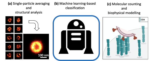
Due to their small sizes, the study of viral assembly has historically been carried out using electron microscopy. In the past decades, super-resolution technologies have allowed the imaging of viral replication with fluorescence microscopy while maintaining high molecular specificity and live-cell compatibility. Here, we show that nanoscale imaging can be combined with bespoke quantitative analysis to unravel detailed viral assembly observations at the molecular level. In particular, we show applications of single-particle averaging, machine learning and molecular counting as powerful tools for peering into the molecular life of viral replication. We finally show an outlook at how the development of fast live-cell super-resolution may also contribute to observations of dynamics nanoscale events during infection. 


Introduction

Viruses are obligate pathogens that commonly infect eukaryotic cells or bacteria. They typically have sub-micron sizes that make them difficult to image with standard light microscopy. The method of choice to study the structure of viral components has historically been electron microscopy. This approach, however, has the downside of having relatively poor molecular specificity and no live-cell compatibility. This, therefore, limits what can be observed during viral replication. 

In the past decades, the development of super-resolution microscopy (SRM) has uniquely allowed observations of viral components at the nanoscale 1,2. It has shown great potential for viral replication imaging in relevant biological contexts. Beyond achieving nanoscale resolution, SRM can be combined with quantitative approaches and provide a deep understanding of biological systems. Here, we show that combining SRM with single-particle averaging 3, machine learning 4 and molecular counting 5 can provide ultra-structural information about viral assembly, large scale understanding of viral population and insights into receptor reorganisation upon infection.


Materials and Methods

Here, we combine SRM methodologies, such as single-molecule localization microscopy (SMLM) and Structured Illumination Microscopy (SIM), to decipher nanoscale structural features involved in viral replication. Image and data analysis tools based on single-particle averaging, machine learning and molecular counting allow the extraction of quantitative understanding of the systems. 


Results

First, by combining SMLM with single-particle average and modelling, we show such data can extract the position of protein layers with near-Angstrom resolution within the tegument layer of Herpes Simplex Virus type 1 (HSV-1) 3. Second, we show that fast and high-throughput SIM imaging combined with machine-learning-based classification can describe the structural features of heterogeneous viral populations such as influenza and Newcastle Disease Virus (NDV) 4. This has a characterisation application in the field of biomedical industry. Last, we exploit the quantitative single-molecule imaging capabilities of SMLM to observe and quantify the nanoscale re-organisation of viral receptors at T cells’ surface upon HIV-1 binding 5


Uncaptioned visual

Figure 1: Quantitative analyses applied to virus replication study. (a) Single-particle averaging, (b) machine learning and (c) molecular counting.


Conclusion and outlook

Taken together, we show that SRM and quantitative analysis have great potential in understanding viral replication at the nanoscale. Additionally, we propose that refinement of live-cell compatible SRM methods like SRRF and in particular a novel implementation of it allowing 3D imaging may offer in the future an avenue for studying important stages of infection at the nanoscale within infected cells, therefore giving access to much-sought-after dynamics. 


References

1.    Müller, B. & Heilemann, M. Shedding new light on viruses: super-resolution microscopy for studying human immunodeficiency virus. Trends Microbiol. 21, 522–533 (2013).

2.    Touizer, E., Sieben, C., Henriques, R., Marsh, M. & Laine, R. F. Application of Super-Resolution and Advanced Quantitative Microscopy to the Spatio-Temporal Analysis of Influenza Virus Replication. Viruses 13, 233 (2021).

3.    Laine, R. F. et al. Structural analysis of herpes simplex virus by optical super-resolution imaging. Nat. Commun. 6, 5980 (2015).

4.    Laine, R. F. et al. Structured illumination microscopy combined with machine learning enables the high throughput analysis and classification of virus structure. eLife 7, (2018).

5.    Yuan, Y. et al. Single-Molecule Super-Resolution Imaging of T-Cell Plasma Membrane CD4 Redistribution upon HIV-1 Binding. Viruses 13, 142 (2021).


References


10:50 - 11:02

79 Multi-modal, label-free, polarisation-resolved super-resolution multiphoton microscopy

Peter Johnson1,2, Artemios Karvounis3, H. Johnson Singh4, Christopher J Brereton5, Konstantinos Bourdakos1,2, Kerry Lunn6, James JW Roberts6, Donna E Davies5,2, Otto L Muskens4, Mark G Jones5,2, Sumeet Mahajan1,2
1School of Chemistry, Faculty of Engineering and Physical Sciences, University of Southampton, Southampton, United Kingdom;. 2Institute for Life Sciences, University of Southampton, Southampton, United Kingdom. 3Optoelectronics Research Centre and Centre for Photonic Metamaterials, University of Southampton, UK. 4Physics and Astronomy, Faculty of Engineering and Physical Sciences, University of Southampton, Southampton, UK. 5NIHR Southampton Biomedical Research Centre, Clinical and Experimental Sciences, Faculty of Medicine, University of Southampton, Southampton, United Kingdom. 6Synairgen Research Ltd, Southampton, United Kingdom

Abstract Text

Summary: In this work the photonic nanojet (PNJ) phenomenon is exploited to achieve super-resolution imaging using endogenous contrast sources. Second harmonic generation (SHG) and two-photon excited autofluorescence (TPEAF) were used to image fibrillar collagen and elastin respectively. By manipulating the polarisation of the PNJ, polarisation resolved images were acquired and used to measure the structural disorder of the sample. It was observed that upon treatment with a cross linking inhibitor the degree of disorder within fibrillar collagen increased, furthermore the degree of disorder measured in elastin was similar to that of fibrillar collagen indicating an unexpectedly ordered structure.

Super-resolution (SR) optical microscopy has allowed the investigation of many biological structures below the diffraction limit, however, most of the established super-resolution techniques are hampered by the need for fluorescent labels. Multiphoton label-free techniques such as second harmonic generation (SHG) and two-photon excited autofluorescence (TPEAF) provide structurally and chemically selective contrast without the addition of exogenous labels, allowing observation of unperturbed biological systems. Attempts to achieve super-resolution imaging using these methods have seen limited success. 

High refractive index microspheres can be used to focus light into a jet with a waist narrower than the diffraction limit, so called, photonic nanojets (PNJs) (Figure 1). Unlike many of the common super-resolution techniques the PNJs do not rely on manipulating fluorescence emission to overcome the diffraction limit, as such they are amenable to multiphoton label-free imaging such as SHG and TPEAF. 

We use the photonic nanojet (PNJ) phenomenon to achieve super-resolution SHG (SR-SHG)1. A resolution of ~λ/6 with respect to the fundamental wavelength, that is, a ~2.3-fold improvement over conventional or diffraction-limited SHG under the same imaging conditions is achieved. Crucially we find that the polarisation properties of excitation are maintained in a PNJ. This is observed in experiment and simulations. This may have widespread implications to increase sensitivity by detection of polarisation-resolved measurements by observing anisotropy in signals. These new findings allowed us to visualise biological SHG-active structures such as collagen at an unprecedented and previously unresolvable spatial scale. Moreover, we demonstrate that the use of an array of self-assembled high-index spheres overcomes the issue of a limited field of view for such a method, allowing PNJ-assisted imaging to be used over a large area. Furthermore, PNJ imaging is independent of the contrast mechanism. This allowed us to apply the method to multimodal imaging with multiphoton techniques. We imaged elastin (via TPEAF) and collagen (via SHG) in lung cell spheroids (Figure 2). We observed unexpectedly high organisation of elastin molecules, similar to those is the highly organised fibrillar SHG. Thus, PNJ assisted multiphoton imaging was able to provide new biophysical insight and has implications for understanding the mechanisms of fibrotic disease progression and treatments. 

The ability to non-destructively image biological structures without labels at the nanoscale with a relatively simple optical method heralds the promise of a new tool to understand biological phenomena and drive drug discovery.


References

Johnson, P. et al. Super-resolved polarisation-enhanced second harmonic generation for direct imaging of nanoscale changes in collagen architecture. bioRxiv (2020). doi:10.1101/2020.02.07.934000


11:02 - 11:14

87 Mechanistic insights on the autoinhibition of the epidermal growth factor receptor (EGFR) and the formation of EGFR dimer and multimers by agonist ligand and tyrosine kinase inhibitors.

Sumanth Iyer, Sarah Needham, Laura Zanetti Domingues, Selene Roberts, David Clarke, Benjamin Davis, Daniel Rolfe, Marisa Martin-Fernandez
UKRI: Science & Technology Facilities Council

Abstract Text

Summary

The epidermal growth factor receptor (EGFR) forms autoinhibited and ligand-activated dimers and multimers to elicit its many crucial cellular functions. Neither the mechanisms of formation nor the function of multimer complexes is yet well understood. We have developed and automated a super-resolution method that can provide population-based quantitative information of the structure of the multimers at ~4 nm resolution. Recently we obtained new insights on the mechanisms of EGFR multimer formation, and how the transition between autoinhibited and activated oligomers is catalysed by the presence on the cell surface of a different autoinhibited dimer structure. We also found how this mechanism is implicated in the resistance to some anti-cancer drugs.


Introduction

A crucial step in the evolution of multi-cellular organisms was the emergence of receptor tyrosine kinases (RTKs). Trafficked to the plasma membrane, their role is to transduce signals in response to cognate growth factor ligands. EGFR was the first RTK to be cloned and this receptor is of paramount importance to cell function and human health as mutations in EGFR and gene amplification are observed in many human cancers. Indeed, cancer is the second cause of death worldwide, accounting for an estimated 9.6 million deaths in 2018. Despite ~4 decades of EGFR research, the information on EGFR structure-function relationships derived from cell-free methods still does not explain the mechanisms underpinning either its normal function, or its dysregulation in cancer. Of particular interest is the distinct roles of dimers and multimers in activation and cancer treatment autoinhibition. Determining the structure-function relationships of EGFR in cells is of paramount importance to find better cancer treatments.


Methods/Materials

As part of efforts to determine the structure of the EGFR in the physiological cell context, we developed a single molecule localisation super-resolution method, coined fluorophore localisation imaging with photobleaching (FLImP). By exploiting molecular fluorescence-photobleaching steps to localise molecules co-located within diffraction-limited spots, this method measures the lateral separation (r) between such molecules with ~4 nm resolution. Using FLImP in concert with fluorescence resonance energy transfer (FRET), to measure EGFR residue-to-plasma membrane separation (z dimension), and long-time-scale molecular dynamic simulations, we previously determined the atomic resolution structures of ligand-bound multimers, and those of ligand-free EGFR dimers and oligomers coexisting at the plasma membrane of cells (Needham et al., Nat. Commun. 2016; Zanetti-Domingues et al., Nat. Commun. 2018). These results shed new light on the mechanisms of EGFR autoinhibition and phosphorylation in both normal and dysregulated signalling. We have since automated data collection and analysis processes, and added a second spatial dimension to FLImP (from lateral r to independent measurement of x,y dimensions).


Results and Discussion

Using a palette of strategic mutations, we have found that ligand-free EGFR oligomers uses a fail-safe, autoinhibitory mechanism that relies on a triple-safe mechanism combining extracellular, transmembrane, and intracellular contacts, via which the receptor avoids serendipitous activation. We have also found that by achieving such stability, ligand-free oligomers pay a price, which is that on binding agonist ligand, autoinhibited oligomers can form ligand-bound dimers, but not the higher energy multimer structures essential for its physiological function. To achieve the latter, EGFR relies on the presence of catalytic amounts of a different inactive dimer structure, previously proposed by crystallography to be the autoinhibited dimer moiety. This structure conveyed the system with enough free energy to form ligand-bound EGFR multimers. We also found that the failure of anti-cancer drugs in the form of tyrosine kinase inhibitors is related to the mechanism of EGFR oligomerisation.


Conclusion

The super-resolution data at >4 nm resolution to be presented will reveal new insights into the much sought after mechanisms underpinning the formation of autoinhibited, activated and anti-cancer drug-treated EGFR multimers. By these results we are currently beginning to unravel the structure-function relationship encoding the behaviour of cancer mutants responsible for the development of therapeutic resistance, thus revealing potential new candidates for therapeutic intervention.


References

Needham et al., Nat. Commun. 2016

Zanetti-Domingues et al., Nat. Commun. 2018


11:14 - 11:39

101 All-optical imaging of molecules in their nanoscale cellular context

Joerg Bewersdorf
Yale School of Medicine, Yale University

Abstract Text

Super-resolution microscopy has become a powerful tool to study the nanoscale spatial distribution of proteins of interest in cells over the last years. Imaging any of these distributions in the context of other proteins or the general cellular context is, however, still challenging. I will present recent developments of our lab which tackle this challenge: A new fluorogenic DNA-PAINT probe enables fast, 3D whole-cell imaging without the need for optical sectioning, adding a versatile tool to the toolbox of single-molecule super-resolution probes [1]. Labeling proteins and other cellular components in bulk in our recent pan-Expansion Microscopy method provides ultrastructural context to the nanoscale organization of proteins, replacing complex correlative light/electron microscopy by an all-optical imaging approach [2]. 

Financial Interest Disclosure: J.B. has financial interest in Bruker Corp. and Hamamatsu Photonics and is co-founder of a startup company related to Expansion Microscopy.

[1] Chung, K.K.H. et al. “Fluorogenic probe for fast 3D whole-cell DNA-PAINT”. bioRxiv (2020). https://www.biorxiv.org/content/10.1101/2020.04.29.066886v1 

[2] M’Saad, O., Bewersdorf, J. Light microscopy of proteins in their ultrastructural context. Nat Commun 11, 3850 (2020). https://doi.org/10.1038/s41467-020-17523-8