Abstract Text
Coherent Raman Scattering microscopy (CRS) is a powerful new approach for label-free, chemically specific imaging, based on the characteristic intrinsic vibrational contrast of the sample molecules.
CRS provides high-resolution (sub-cellular level) and dynamic (up to video-rate) information on the biochemical composition and metabolic processes in cells, tissues, and intact model organisms, and it enables imaging of small molecules without perturbing their function. This information is highly synergistic with the molecular contrast provided by fluorescence microscopy. Unsurprisingly, CRS is finding a growing number of applications in fields like neurodegenerative disease, cancer, 3D biology, stem cell and developmental biology, and pharmacology.
Here, we introduce the all-new STELLARIS 8 CRS – Leica’s hands-free Coherent Raman Scattering microscopy platform. It offers the two popular CRS modalities – Stimulated Raman Scattering (SRS) and CARS – and allows for the simultaneous acquisition of two-photon fluorescence and second-harmonic generation signals. Importantly, the seamless integration of CRS with the STELLARIS visible confocal fluorescence microscopy platform results in a true multi-modal optical discovery platform that is capable of capturing a unique combination of biochemical, biophysical and molecular contrasts.
With its intuitive user interface, Image Compass, it is application-oriented and easy to use, enabling spectroscopic CRS imaging, 3D-and 4D-imaging modes, label-free high-content screening applications, and large specimen tile-scans.
Join our workshop to learn how the vast potential of STELLARIS 8 CRS can be a game-changer in your research!
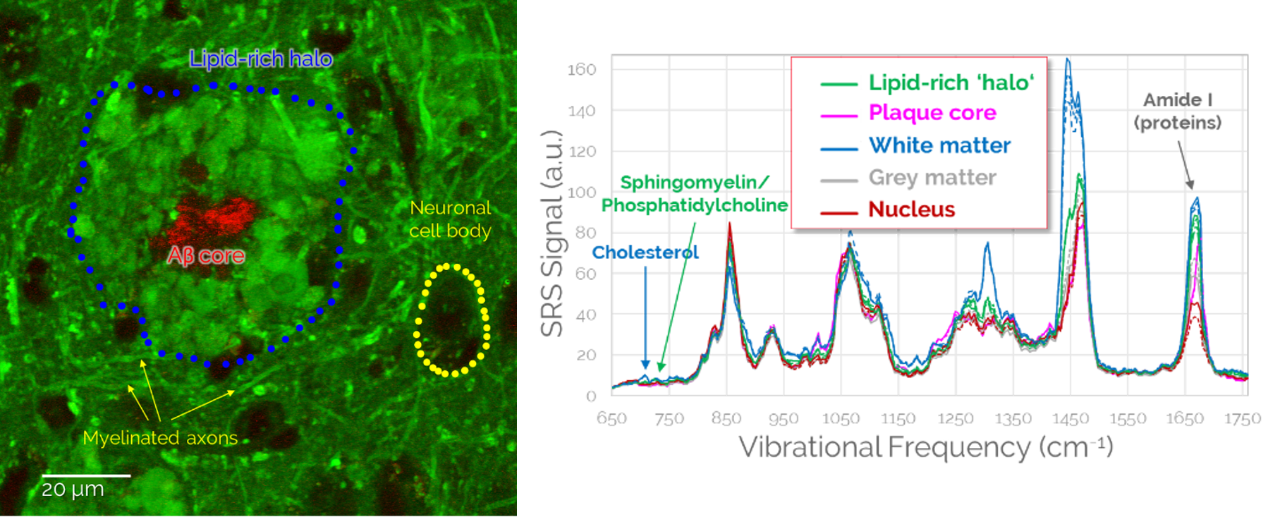
Application example of CRS in neurodegenerative disease.(1) Left: Label-free imaging of Alzheimer’s pathology in unstained mouse brain tissue. Green: Lipids (SRS, 2850 cm-1) Red: Amyloid-β (SRS intensity ratio 1675/1665 cm-1). Right: SRS spectroscopic imaging reveals the biochemical composition of healthy and diseased brain structures. Sample courtesy of Dr. Martin Fuhrmann, German Center for Neurodegenerative Diseases, Bonn.
References
(1) Volker Schweikhard, Andrea Baral, Vishnu Krishnamachari, William C. Hay, Martin Fuhrmann, "Label-free characterization of Amyloid-β-plaques and associated lipids in brain tissues using stimulated Raman scattering microscopy", bioRxiv (2019), doi: https://doi.org/10.1101/789248