Round Table Program & Brunch

11:30am - 1:30pm Thursday, 4th November, 2021


146 Hula Hooping as a Recreational Activity to Facilitate Renal Calculi Passage: Technique Report

Emily Ton BS, Madison Lorenz BA, Sina Sobhani BS, Cu Phan MD
Urology Care Center, Newport Beach, CA, USA

Abstract

Introduction

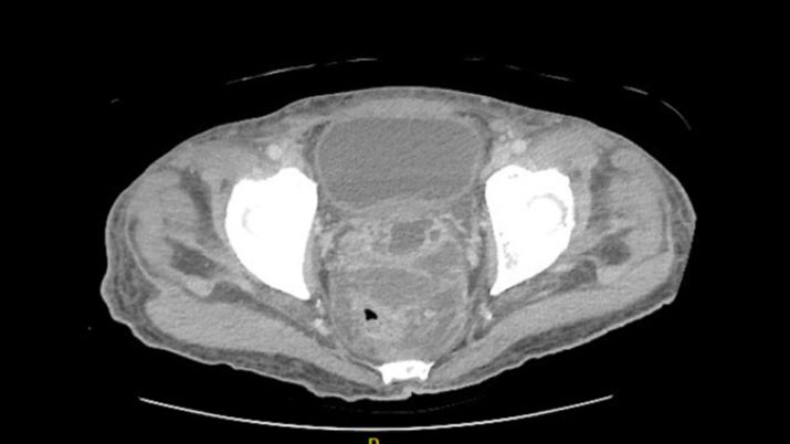
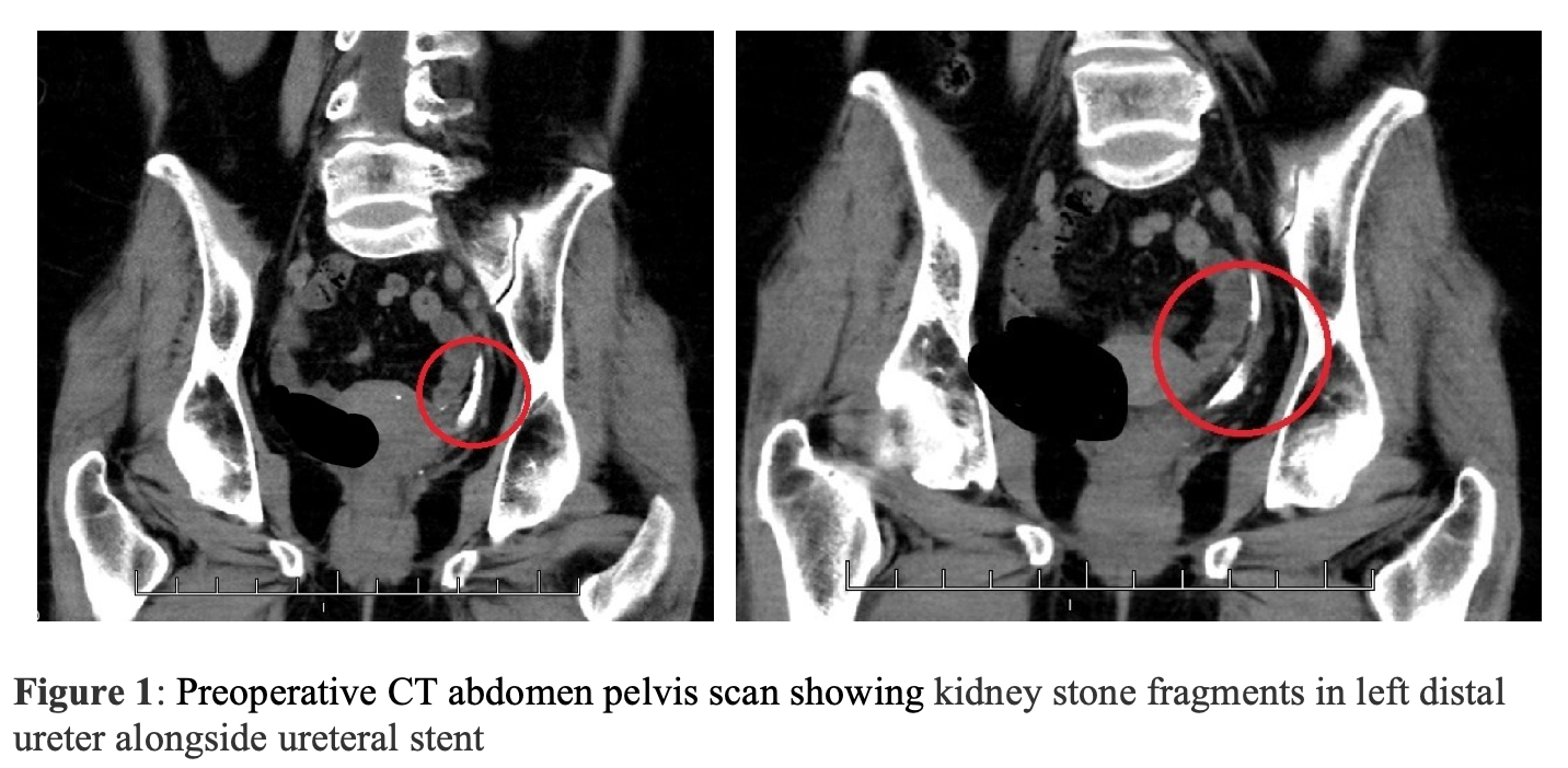
A 69-year-old woman presented to our clinic with bilateral kidney stones. A preoperative computed tomography (CT) scan showed multiple stone fragments in the left distal ureter alongside the stent (Figure 1). The largest stone on the left side measured up to 2 cm. The patient underwent left ureteroscopy with Holmium laser lithotripsy and placement of ureteral stent on April 28, 2021. She did not pass any stone fragments up to 2 weeks after her procedure. With consideration of the COVID-19 pandemic and the patient’s limited physical ability, the patient inspired our novel implementation of hula hooping for facilitating her passage of stone fragments. 


Objective:

To evaluate hula hooping for facilitation of passage of kidney stone fragments. 


Materials and Methods:

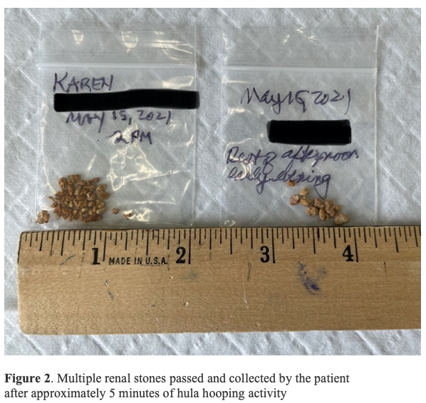
The patient hula hooped for approximately only 5 minutes on May 15, 2021 and passed an impressive amount of stone fragments.


Results:

After hula hooping, the patient passed a multitude of stone fragments and continued to pass fragments afterward (Figure 2). 


Conclusion

Hula hooping is a fun activity that can be added to the urologist armamentarium. Although we provide anecdotal evidence, it presents hula hooping as a viable technique for facilitating passage of kidney stone fragments. Alternatively, mechanistically similar activities, such as Hawaiian belly dancing and twist dancing, may also facilitate passage. As urologists, it is important to be open-minded about fun everyday activities that can also have clinical benefits for patients.


Uncaptioned visual


Uncaptioned visual


If funding provided, type in source company / entity name(s):

None

230 The No Touch Technique for Primary Artificial Urinary Sphincter Placement: Outcomes and Historical Comparison

Matthew J Ziegelmann MN, Brian J Linder MN, Daniel S Elliott MN
Mayo Clinic, Rochester, MN, USA

Abstract

Objectives: To describe our modified “minimal-touch” technique for primary artificial urinary sphincter (AUS) placement and evaluate early device outcomes compared with a historical cohort. 

Materials and Methods: We identified patients who underwent primary AUS placement at our institution from 1983 to 2020. A “minimal-touch” technique was implemented in 2014 wherein an Ioban® dressing is placed over the exposed abdominal and perineal skin to the AUS components from touching the patient’s skin, along with copious antibiotic irrigation and glove changes. Statistical analysis was performed to identify the rate of postoperative device infection in patients who underwent minimal-touch versus those who underwent our traditional technique.

Results: 526/2601 total procedures (20%) were performed using our “minimal-touch” approach, including 271/1554 patients (17%) who underwent primary AUS placement over the study period. 2.3% of patients experienced device infection after AUS procedures. In the “minimal-touch” era, 3/526 patients (0.7%) experienced device infection, including 1/271 (0.4%) of those with primary AUS placement. In comparison, 46/2075 patients (2.7%) experienced device infection using the historical approach, with 29/1283 (2.3%) of primary AUS placements result in removal for infection. Notably, 90% of device infections occurred within the first 6 months after primary placement. The difference in cumulative incidence of device infections at 12-months did not meet our threshold for statistical significance for either the total cohort of all AUS procedures (primary and revision) or the sub-group of only those patients undergoing primary AUS placement (Gray K-sample test; p=.13 and 0.21, respectively).

Conclusion: The “minimal-touch” approach for AUS placement represents an easy-to-implement modification with potential implications on device outcomes.   While early results appear promising, longer-term follow with greater statistical power is needed to determine whether this approach will lower the infection risk.     

If funding provided, type in source company / entity name(s):

none

149 Fieros to Fournier’s

Nikola Teslovich MD
Stanford Hospital, Stanford, CA, USA

Abstract

FIEROS TO FOURNIER’S

Nikola Teslovich MD

Stanford, CA

Objectives: Studies have shown that 100% of Urology residents with my name have an enduring desire to share with the world the importance of skinned knees, bruised knuckles, and dirty fingernails in childhood, and the beautiful way that working with your hands can lead to a career in Surgery.

Materials and Methods: For this study, a cohort of one Urology resident was allowed to postulate wildly in a fairly unscripted form. Data was collected using Microsoft Word (Microsoft, Seattle WA) with data analysis performed with assistance of Longboard Island Lager (Kona Brewing Company, Hawaii).

Results: Analysis showed that within our cohort, every member enjoyed writing this short essay for your [hopeful] enjoyment.

Conclusions: Let your kids get dirty, fix things, build things, and do things. Your amateur tinkerer can become a professional tinkler like me.

Source of Funding: hah, no.


84 Fishing in the OR: An Unexpected Catch

Gabriel E. Martin M.D., Cayde Ritchie M.D., Stephanie Jensen M.D., Joshua D. Chamberlin M.D.
Loma Linda University, Loma Linda, CA, USA

Abstract

Objectives: In clinical practice, urologists may encounter urethral foreign bodies. Motivation for self-insertion includes erotic impulses, psychiatric disturbances, and sexual curiosity. We highlight the case of a 15-year-old male who presented with a fishhook in his urethra. We present an atraumatic endoscopic method for urethral fish hook removal. 


Materials and Methods: This is a case report of a 15-year-old male who was hospitalized after a suicide attempt via KCl tablet ingestion. He had a history of depression, ADHD, and autism. PICU evaluation included serial KUBs to evaluate KCl tablet passage, which incidentally demonstrated a fishhook in his prostatic urethra. A foley catheter had already been placed. With further inquiry, the patient attested to placing a fishhook in his urethra attached to a 1 cm length of fishing line. The fishhook was inserted with the bend of the hook first, the eye and point of the hook located distally. 

The patient was taken to the OR and placed in dorsal lithotomy position. The foley catheter was removed atraumatically. A 14 French adolescent cystoscope was inserted into the patient's urethra. The fishhook was wedged in the prostatic urethra, preventing direct retrograde removal without hooking the prostatic tissue. The bend of the hook was grasped with the 5 Fr grasper and advanced atraumatically into the bladder. The hook was flipped 180 degrees, angling the point of the hook towards the center of the lumen and away from the scope, limiting urethral trauma during removal. Under direct cystoscopic vision, the fishhook was removed atraumatically. There was a small string of fishing line attached to the eye of the fish hook. The urethra was again inspected and noted to have no injury. 


Results: The patient tolerated the endoscopic removal of the fishhook after repositioning of the hook within the bladder. He was able to void post-operatively without dysuria or hematuria and was instructed to avoid placing objects in his urethra. 


Conclusions: To our knowledge, this is the first case report describing an atraumatic fishhook removal from a pediatric patient using a cystoscope and grasper. 

If funding provided, type in source company / entity name(s):

NONE

152 A Unique Case of Bladder Injury Following a Urethral Stimulation Misadventure

Kayla Schilling BS1, Ryan Werntz MD2, William Flanagan MD2
1University of South Carolina School of Medicine Greenville, Greenville, SC, USA. 2Prisma Health, Greenville, SC, USA

Abstract

Introduction: Introduction of foreign bodies into the urethra is a rare practice that is often done for autoerotic stimulation in patients with psychiatric illness, maintenance of erection, relief of urinary symptoms, or by intoxicated patients. Although current literature shows the occurrence of intra-urethral foreign body insertion is more common in males than females, the short and straight tract of the female urethra makes complications more likely since urethral foreign bodies may migrate to, and become retained in, the bladder. A wide variety of items have been reported in these cases including wiring, nail clippers, writing instruments, batteries, utensils, and more. 


Methods: The patient’s records were utilized to describe this unique case and considerations for treatment. 


Case Report: This report documents an intoxicated 35-year-old female who presented with dysuria, hematuria, and intermittent difficulty urinating after a magnetic cell phone stylus was inserted into her urethra during sexual activity and migrated into her bladder. A unique consideration was the force of insertion by the patient’s boyfriend, which caused the stylus to become lodged into the patient’s bladder wall. Careful evaluation for bladder perforation and urinary extravasation, or concomitant vascular or bowel injury was necessary prior to treatment planning. Diagnostic imaging, treatment, and outcome are discussed.


Discussion: This report brings attention to the increasing popularity of urethral stimulation and foreign body insertion and the methods that have been successfully used for diagnosis and treatment. The demographics and data on the populations of patients that typically present with this complaint should influence considerations for treatment, prophylactic antibiotics, psychiatric evaluation, patient counseling, and the necessity for long-term urologic follow up. 


86 Torsion of an Epididymal Cyst in a Pediatric Patient

Paul Jones MD, Casey Seideman MD
OHSU, Portland, Oregon, USA

Abstract

Introduction 

Testicular torsion is a common emergency in pediatric urology. It is important to review history, physical exam, and testicular ultrasound when considering treatment. Even in the setting of a normal ultrasound, clinical exam can indicate surgical exploration. 


Methods 

In this case report we review torsion of an epidydimal cyst. The patient is a 10-year-old male who presented to the emergency department with 4 hours of right scrotal pain and emesis. TWIST score was 3 for emesis and testicular swelling. Exam showed focal pain on the lateral testis. Scrotal ultrasound demonstrated flow to the testicle. Based on the clinical exam, the decision was made to proceed with surgical exploration. 


Results 

Scrotal exploration of the scrotum revealed an epidydimal cyst with torsion of its stalk. The cyst was discolored with greater than 720 degrees of rotation. The cyst was untwisted, excised at its stalk, and oversewn with 4-0 Polysorb suture. We did not identify a bell clapper deformity on either side. Testicular fixation sutures were placed bilaterally. The patient did well postoperatively and discharged the day of surgery.  


Conclusion 

Torsion of an epidydimal cyst is uncommon. If there is high clinical suspicion of ischemia then surgical exploration should be performed, even if scrotal doppler ultrasound demonstrates blood flow.  


121 Once in Blue Moon in Urology Prospective , Cases Rivew

KAMRAN HASSAN BHATTI MS
CITY HOSPITAL, PAKPATTAN, PAKPATTAN, Pakistan

Abstract

CASE-1

Urinary retention in women is a rare occurrence, with an overall incidence of 0.07 per 1000 inhabitants per year. Herein we describe our experience with a female patient who presented to the Emergency Department [ED] with acute urinary retention.

Although uterine leiomyoma is a common finding in the general population, it is an extremely rare cause of acute urinary retention in women with just a handful of reported cases in the literature

  • 40 years old, Married - Nullipara-
  • Retention of urine 1 day followed by catheterization about 500 ml Operation -Laparotomy with partial removal of adenomyoma and right salpingoophorectomy
  • Histopathology; Findings consistent with leiomyoma measuring 1.5 cm with adenomyosis.

CASE; 2

  • Cauda equina syndrome (CES) is an uncommon but potentially devastating condition caused by compression of the cauda equina in the spinal canal. CES is a rare condition. The incidence quoted in the literature varies from 1 in 33 000 to 7 in 100 000 in a recent North American study. 

 . It is also one of the emergency surgical indications. 27 years/ married

  • No   neurological disorder, no drug abuse
    no trauma or RTA ,no neurological deficit or symptom suggestive of multiple sclerosis, normal perianal sensation
     physical examination.
  • Higher Mental Function, cranial nerves, motor system, tone, power, reflexes, sensation, coordination, gait
     all were normal.
  • MRI, L1-L2 large posto-central disc with significant cord compression.
  • Operation ;Right L1 hemi Laminectomy and L1/L2 Micro diskectomy
  • Follow up no urinary symptoms.

 CASE -3


A 34 years old male patient who presented with acute urinary retention 3 days after developing blister like rash typically of chickenpox, there was no similar preceding history, trauma, use of anti-cholinergic medication, or other urinary or neurological signs or symptoms. Urinary bladder was fully distended and palpable at the time of presentation and was catheterized and passed 1000ml of clear urine.

  • He experienced vesicular rash 2 days prior to admission which started over the face and disseminated to the trunk and upper extremities, sparing perennial and perianal region. At that time, he was started on Acyclovir treatment.
  • Chickenpox can be more serious in adults than in children. 

219 Dusting or Basketing it’s  on urologist mood

Kamran Hassan Bhatti Ms urology
Hmc, Alkhor, Alkhor, Qatar

Abstract

Introduction 

Renal stones are broken into tiny fragments using a small laser called a Holmium laser. While this treatment is a well- established option for treatment of these stones, there are several different techniques used to help eliminate them from the kidney. Some urologists treat the stone by a method called "active" extraction whereby the ureteroscope is passed back and forth into the kidney to remove all visible stone fragments. Others use a method called "dusting" whereby the stones are broken into tiny fragments or "dust" with the intention that achieving such a small stone size will allow the stones to pass spontaneously. There has not been a systematic and rigorous comparison of these techniques in terms of treatment outcomes. By collecting information on the success of treatment, the investigators hope to provide benchmark data for future studies of kidney stone treatment and improve the care of all patients who need surgery for their kidney stones.

Objects.To compare dusting or basketing in flexible ureteroscopy .

Results .
200 patients data were evaluted (N1⁄106 Basketing, N1⁄94 Dusting). Overall 30.7% were female and 69.3% were male. The stones were slightly larger in the dusting group and significantly more laser energy was used in the dusting group. There were fragments present in 13.7% in the basketing group compared to 40.8% of the dusting group. This equates to a stone free rate of 91.8% in the bas- keting group and 83.8 % in the dusting group. There was no difference in readmission to ER or hospital between the groups (14.5%- dusting vs 14%-basketing) or in reintervention rates (2 patients in dusting, 3 in basketing). There were no differences between the groups in post-operative labs work

Conclusion 

In patients undergoing ureteroscopy for renal stones between 5-20 mm active extraction of all fragments with a basket produces a higher stone free rate (91.8%) than dusting the stone (83.8%). However, there was no difference in readmissions, reinter- vention rates, or patients becoming symptomatic from their residual fragments. Long-term follow-up of these patients will also determine impact of residual fragments in this group of patient

Funding . Nil 


223 The Teacher and the Master: George Goodfellow, Hugh Hampton Young and the Perineal Prostatectomy

Unwanaobong Nseyo MD, MHS
USC Institute of Urology, Keck School of Medicine of USC, Los Angeles, CA, USA

Abstract

Upon a cursory review of the annals of urologic history one name dominates the field -- Hugh Hampton Young, widely celebrated as the "Father of Modern Urology" in the United States. Also credited to his name and the likely foundation of his urologic celebrity is performing the "first" perineal prostatectomy. Hugh Hampton Young, however, recognized that had he not modified George Goodfellow’s operation “no one would have ever heard of [him] as a prostatectomist." So how was it that Young and not Goodfellow, who had amassed his own fame as the surgeon to gunslingers at the O.K. Corral, became the famous prostatectomist? The impact of power, fame and institution on the history of the perineal prostatectomy will be explored through the relationship between these two men, the teacher, George Goodfellow, and the master, Hugh Hampton Young. 


The data for this research consisted of both primary and secondary sources, including articles written by George Goodfellow and Hugh Hampton Young, archival material, and secondary sources obtained from the Didusch Museum. 


On October 13, 1891 likely at St. Mary's Hospital in Tucson, Arizona where he was serving as the Chief Surgeon for the Southern Pacific Railroad, George E. Goodfellow performed “a pure perineal prostatectomy, the first so far...deliberately devised and carried out.” However, his formal publication of his surgical technique was delayed by nearly 13 years lending controversy to his claim. Unbeknownst to many, in the interval prior to publication, Goodfellow participated in a visiting professorship of his own, demonstrating his surgical technique for the perineal prostatectomy with Dr. Hugh Hampton Young, at the time a Professor of Urology at Johns Hopkins, while in Baltimore on his return from the Spanish-American War. 


The perineal prostatectomy was initially developed as a treatment for benign prostatic enlargement and was further refined to address prostate cancer. The story of Hugh Hampton Young, George Goodfellow and the development of the perineal prostatectomy highlighted the way in which institution and celebrity shape the historical narratives of surgical techniques. May their story prompt us to continue to look for all of the George Goodfellows behind our Hugh Hampton Youngs.


231 A Novel Full Core 18 Gauge Prostate Biopsy Needle Collects More Tissue Volume by Weight: Results from Initial Comparative Study.

Bela S Denes MD1, Jeffrey Proctor MD2, Eric Gwynn MD3, Dan Wiener MD4
1Lantheus Medical Imaging, N. Billercia, MA, USA. 2Georgia Urology, Marietta, GA, USA. 3New River Urology, Blufton, SC, USA. 4Cartersville Medical Center, Cartersville, GA, USA

Abstract

Prostate biopsy is the definitive test to diagnose prostate cancer.  Most core-needle biopsy devices employ a core collector design that captures tissue in a notch located just proximal to the tip of the collection needle. A recent study demonstrates that deflection of the needle tip as it encounters tissue of varying densities, may impede accurate sampling of cancerous areas, and reduce the volume of tissue captured within the notch up to 50%.

A novel needle design in which the collection needle is centered within the outer cannula and minimizes potential needle deflection has been developed. Physical dimensions of this 18ga needle including the tissue core collection area, are similar to current 18ga standard of care (SoC) needles. We compared tissue cores sampled with an SoC needle and with this novel needle in a series of prostate biopsies.

Methods

Patients undergoing either trans-rectal (TR) or trans-perineal (TP) prostate biopsy participated in the study. An extended pattern 12 core plus 4 ultrasound-guided prostate biopsy method was used. An SoC biopsy needle was used to sample the standard twelve areas. Before the procedure, the physician chose 4 distinct areas from which a second tissue core would be obtained using the test needle. Each tissue core from the areas of comparison was weighed immediately after sampling in a similar fashion.


Uncaptioned visual


Uncaptioned visual


Results

The following are the results from 104 pairs of tissue cores from 16 TR and 10 TP procedures: 

Comparison of mean weight

Trans-Rectal 


Trans-Perineal


SoC Needle

Test


SoC Needle

Test

Mean weight (mg)

5.54

6.21


Mean weight (mg)

4.73

5.71

Variance

2.3894

2.1752


Variance

3.0536

1.1739

% difference

12%


% difference

21%

Observations

64


Observations

36

t Stat

-3.0130


t Stat

-3.2872

P(T<=t) two-tail

0.004


P(T<=t) two-tail

0.002


Conclusion:

Uncaptioned visual
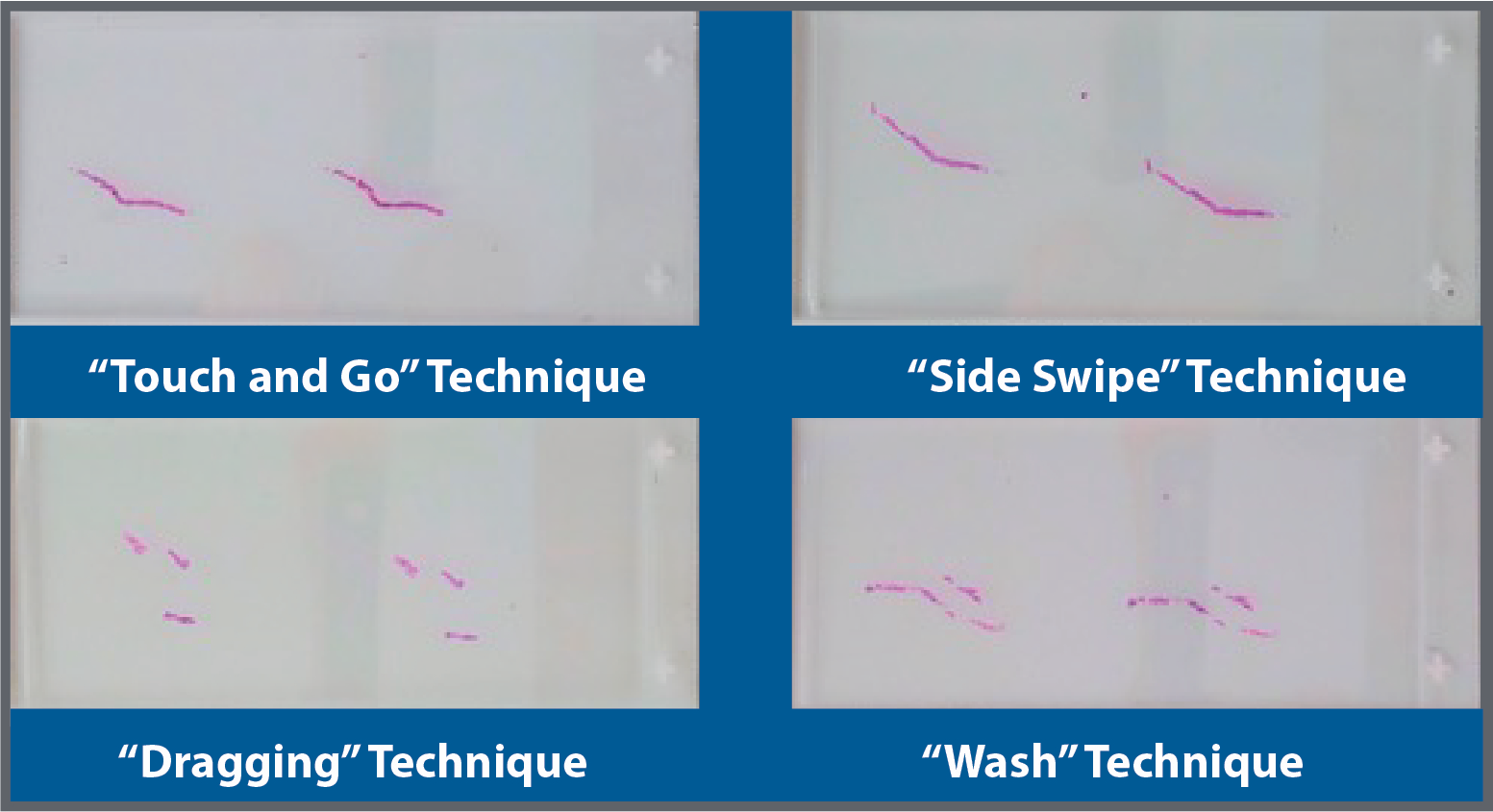
In this head-to-head comparator study, the novel test needle collected significantly more tissue volume by weight than the SoC needle, 12% in TR and 21% in TP.  Additional studies are continuing to assess the impact and utility of this novel biopsy needle design on quantitative and qualitative aspects of histologic processing and diagnosis.


If funding provided, type in source company / entity name(s):

URO-1, Inc.

232 A Novel “Touch & Go” Method for Retrieving Tissue Specimens from Prostate Biopsy Needlesthat Prevents Core Fragmentation and Preserves Core Integrity.

Bela Denes MD1, Jeffrey Proctor MD2, Dan Wiener MD3
1Lantheus Imaging, North Billerica, MA, USA. 2Georgia Urology, Marietta, GA, USA. 3Cartersville Medical Center, Cartersville, GA, USA

Abstract

Introduction and Background

Tissue fragmentation while retrieving specimens from prostate biopsy needles can adversely affect histologic analysis. Degree of fragmentation is a function of the retrieval method and the skill of the assistant. Retrieval by swiping the needle laterally against a foam pad is reported to result in less core fragmentation than dragging the needle proximally on the pad. Vigorous washing of needle tip dislodges the specimen without fragmentation but causes the tissue to warp and curl up.  We describe a new method for specimen retrieval utilizing a biocompatible material currently in clinical use. Touching the needle against it lifts the specimen from the needle due to surface tension without shearing movement or direct pressure that can fragment the sample. We report results of comparing the “Touch & Go” method with the three conventional methods to assess differences in specimen quality and integrity for histologic analysis.


Methods

Bovine prostate tissue cores were collected using an 18ga prostate biopsy needle. The specimens were retrieved by swiping, dragging, washing or “Touch & Go” method. Histologic analysis was performed following standard protocol to assess tissue artifact, fragmentation, and sample integrity.


Results

Histologic analysis demonstrated consistently less fragmentation and improved integrity of the cores with “Touch & Go” compared with the other methods with the exception of the "side swipe" method which was deemed to be equal in performance. 


The following are representative slides of specimens retrieved:

Uncaptioned visual

No deleterious effects on tissue collected with the “Touch & Go” method were detected in its use in other assays.


Conclusion

The novel “Touch & Go” method of tissue specimen retrieval has the potential to simplify tissue collection while preserving tissue integrity, reducing tissue artifact and improving diagnostic performance. Further studies have been initiated to determine if “Touch & Go” consistently preserves core integrity over the “side swipe” and other methods.


If funding provided, type in source company / entity name(s):

URO-1, Inc.

216 What is the color of kidney function? A brief history of race and estimating the glomerular filtration rate.

Fernandino L Vilson MD, John T Leppert MD MS
Stanford University School of Medicine, Stanford, CA, USA

Abstract

Abstract: 

To most, the kidney is reddish brown. Its function knows no race. However, data from landmark studies from the past 30 years suggest there are key differences in kidney function which estimates Black patients as having 16 – 21% higher kidney function compared to white or “non-Black” patients. These differences were captured in the MDRD and CKD-EPI equations in 1999 and 2009 which have since gained widespread use and acceptance in the medical community. Over the years, many have challenged the inclusion of the race correction in these equations, but recent racial tensions and pushes from medical students have brought the equations under heavy scrutiny with many institutions recognizing the inherent racism and selecting to remove the race coefficient from clinical practice altogether. But how did we get to this point? What were the findings that led researchers to factor in race as a coefficient?  In this paper, we review the key studies from 1976 – 2009 in order to answer the question, “what is the color of kidney function?” 


233 Prostate Abscess: A Rare Causative Organism

Kai Wen Cheng MD, Muhannad Alsyouf MD, Brian Hu MD
Loma Linda University, Loma Linda, CA, USA

Abstract

We present a case of 60 year old man who presented to the emergency room with 2 weeks of pelvic pain, urinary frequency and urgency, and weight loss. He resides in Southern California with a history of poorly controlled diabetes mellitus. Examination revealed a cachectic male with suprapubic tenderness with laboratory findings of leukocytosis (16.4 bl/L), hyperglycemia (463 mg/dl), and sterile pyuria. Treatment for urinary tract infection was initiated; however, symptoms persisted which prompted a CT scan.  This showed a multi-loculated prostatic abscess (see image 1). There were also miliary pulmonary nodules and splenic lesions. CT-guided drainage of the prostatic and splenic abscesses was performed, and fluid cultures grew white mold. Results confirmed Coccidioides immitis infection. Liposomal amphotericin B and voriconazole was initiated with transition to fluconazole. The patient’s symptoms improved and drains were removed.  


Conclusions 

Coccidioidomycosis is endemic to Southwestern United States, Northern Mexico, and parts of Central and South America. Prostatic involvement is rare and occurs primarily in immunocompromised patients, indicating disseminated disease. Prompt diagnosis requires recognition of common irritative and obstructive lower urinary tract symptoms of dysuria, frequency, urgency, incomplete voiding, urinary retention, and suprapubic pain in patients who have resided in endemic areas, especially those who are immunocompromised. Diagnosis of prostatic involvement is confirmed with culture or biopsy. Treatment requires aggressive antifungal therapy, drainage of sources, and optimization of underlying immunocompromised state. 


Uncaptioned visual

Image 1: Computed tomography demonstrating 11 x 8 cm multi-loculated prostatic abscess with associated regional cystitis. 


If funding provided, type in source company / entity name(s):

None