BACKGROUND: Recent practice patterns suggest continent cutaneous urinary diversion is becoming increasingly less commonly performed for patients with bladder cancer. While the reason for this is unclear, lack of surgeon experience and comfort with the technique are common barriers.
OBJECTIVE: To provide a step by step description of the Indiana pouch continent cutaneous urinary diversion technique and to report a 16-year experience of functional outcomes and complications in patients undergoing robotic radical cystectomy with Indiana pouch continent cutaneous urinary diversion.
METHODS: A prospectively maintained bladder cancer database was queried for patients undergoing radical cystectomy with Indiana pouch urinary diversion at a tertiary referral center from 2004 to 2020. All cystectomies were performed using the DaVinci (Intuitive Surgical, Sunnyvale, CA) robot with extended bilateral pelvic lymphadenectomy. Urinary diversion was performed by a single surgeon in an extracorporeal fashion. Thirty and 90-day complications were recorded by Clavien grade as well as urinary continence.
Objectives: Catheterizable channels are used as a surgical treatment for pediatric patients with neurogenic bladder or congenital malformation of the urinary tract. There are several approaches for creation of catheterizable channels in pediatric urology using the Mitrofanoff principle. The appendix is most commonly utilized, however, the Monti ileovesicostomy can be performed when the appendix is unavailable for use. In this video we cover the important points in construction of the various channels.
Materials and Methods: The key steps of variations in approach for appendicovesicostomy and ileovesicostomy were recorded in pediatric patients. Procedure types include appendicovesicostomy with extravesical and split appendix approaches as well as ileovesicostomy with traditional Monti and spiral Monti approaches.
Results: The presented surgical techniques allow for durable results in pediatric patients and the variations in approach address the anatomical needs of each patient.
Conclusions: Creation of catheterizable channels via the presented techniques allow for intermittent catheterization via continent catheterizable stoma for pediatric patients.
Source of Funding: None
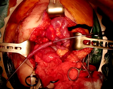
Figure 1 - Preparation of appendicovesicostomy channel.
Objectives
Radical nephroureterectomy (RNU) with bladder cuff excision is the gold standard treatment for upper tract urothelial carcinoma (UTUC). The distal ureter and bladder cuff techniques have not yet been standardized. The aim of this video is to present the Keyhole technique for the management of bladder cuff during robotic RNU.
Materials and Methods
The illustrative case in the video is that of a 79-year-old male with right UTUC, who underwent robotic RNU and bladder cuff excision with Keyhole technique. We also reviewed the records of 19 patients who underwent robotic RNU by a single surgeon to evaluate the feasibility and outcomes of this technique. Patients with a history of bladder cancer (n=5) were excluded.
Results
A single-dock single position robotic RNU using Xi robot platform was performed for this patient. Operative time and estimated blood loss were 350 minutes and 50 mL, respectively. Surgery was uneventful with no need for perioperative blood transfusion. The patient was discharged on postoperative day 5. Pathology revealed low-grade, with focal high-grade, papillary urothelial carcinoma (pTa N0) with negative margins. No complication was reported during the 90-day follow-up. The patient was disease-free at 1-year follow-up. A total of 14 patients with a median age of 75 years were included in the case series. No intraoperative complications were reported, and all margins were negative. Genitourinary-related 90-day complications were reported in 1 patient (urine leak managed by Foley catheter). In a median follow-up of 20 months, 3 patients developed bladder recurrence.
Conclusions
Keyhole technique for the management of distal ureter and bladder cuff during robotic radical nephroureterectomy is feasible with minimal 90-day complications and low bladder recurrence.
Source of Funding: None
OBJECTIVE
Midurethral slings are the gold standard for stress urinary incontinence. Complications after a retropubic midurethral sling are rare, and mesh erosion into the bladder or urethra occur in less than 1% of cases. Our case presents a robotic technique used to excise eroded midurethral sling mesh into the bladder and extract the associated bladder calculus.METHODS
A 65-year old female presented with complaints of gross hematuria, dysuria, incomplete bladder emptying and history of recurrent UTIs for the last 2 years. She had a history of a total vaginal hysterectomy, uterosacral ligament fixation, retropubic midurethral sling and cystoscopy performed approximately 2 years ago. On examination, she was found to have a 3mm area of vaginal mesh erosion. CT scan showed that she had a large bladder calculus and an office cystoscopy was performed preoperatively. During cystoscopy, an eroded segment of mesh was encountered at the left lateral bladder wall with associated calculus. Decision was made to proceed as a combined case with FPMRS Urology to excise the bladder mesh and stone via a robotic approach.
RESULTS
Robotic dissection of the space of Retzius exposed the left lateral dome of the bladder. With cystoscopic guidance, the eroded left arm of the retropubic midurethral sling was identified and excised. Associated bladder stone was extracted through a left lateral bladder dome cystotomy. The cystotomy and surrounding peritoneum were closed and the bladder calculus was removed through an extended umbilical trocar site. The patient was discharged home with foley catheter in place for 13 days and returned for removal after cystogram performed, which was negative for leak.
CONCLUSIONS
In conclusion, we found that a complex complication of midurethral sling can be managed safely via a robotic approach. This method allowed for faster recovery time, better visualization and more mesh removal. We found that concurrent cystoscopy throughout the robotic portion of procedure can help identify precise location for cystotomy creation and for faster identification of the eroded mesh. We found that using a robotic technique to remove mesh erosions even with associated large calculus can be performed and lead to successful outcomes for patients.
Tips and Tricks for Robotic Retroperitoneal Lymph Node Dissection in Testicular Cancer
David G Ortega*, Alireza Ghoreifi, Michael Chevinsky, Laura C Perez, Akbar Ashrafi, Kailyn Koh, Wesley Yip, Luis G Medina, Inderbir Gill, Hooman Djaladat, Los Angeles , CA
INTRODUCTION AND OBJECTIVE: Retroperitoneal lymph node dissection (RPLND) is a mainstay of surgical management for advanced testicular cancer, both in the first line and post-chemotherapy setting. Robotic RPLND has been increasingly utilized in recent years as an alternative to the open approach. The aim of this video is to demonstrate some of the tips and tricks for a successful robotic RPLND.
METHODS: Two patients are illustrated in this video. First case is a 24-year-old male with a history of right testicular mixed germ cell tumor (99% seminoma, 1% embryonal carcinoma), who underwent robotic bilateral nerve-sparing RPLND for a 1.8 cm lesion in the retroperitoneum (IIA) and elevated ßHCG to 4.4 mIU/mL. The second is a 37-year-old male with a history of left testicular cancer 15 years prior (unknown pathology) who also received chemotherapy (Bleomycin/Etoposide/Cisplatin x 3) following orchiectomy for advanced stage. He underwent robotic post-chemotherapy RPLND due to a 9.6 cm retroperitoneal mass with negative tumor markers.
RESULTS: Initially, we outline the appropriate patient positioning, port placement, and pneumoperitoneum for a feasible and oncologically sound surgery. Our first tip describes securing the posterior peritoneum to the anterior abdominal wall. Second tip delineates the nerve-sparing of the postganglionic sympathetic nerves. Then we present the minimal tumor touch as well as the robotic split and roll techniques for thorough lymph node dissection. The last tip includes maneuvers that help with the dissection around the great vessels as well as cysterna chyli in both primary and post-chemotherapy settings. In both aforementioned cases, no intra/perioperative complications were reported, and they were discharged the day after surgery.
CONCLUSIONS: We present some of the tips and tricks for an efficient robotic RPLND that are helpful in achieving optimal oncological and functional outcomes, while minimizing the perioperative complications
Objectives: We present the case of fluoroless ureteroscopy and laser lithotripsy in a patient with Body Mass Index (BMI) of 88 kg/m2 with 13mm x 15mm right mid-ureteric stone and moderate hydroureter.
Materials and Methods: The patient was placed under general anesthesia and in supine lithotomy position. Panniculus retracting tape and pads were used to retract the lower abdominal pannus and thigh adiposity to expose the urethral meatus. Using a semirigid ureteroscope, the right ureteric orifice was cannulated, and the stone was encountered in the mid-ureter. A 0.035 hydrophilic glide wire was passed by the stone under direct vision. Tactile feedback helped to confirm wire placement within the renal collecting system. The end of the wire was marked on the sterile drape as a reference for the distance to the kidney. The scope was removed, an open ended 6 F catheter was passed and 0.038 PTFE standard wire placed until the end of wire matched the marking on the drape. The ureteroscope was reinserted and stone dusting with 150-micron thulium laser fiber at .4 J and 40 Hz with a thulium Soltive premium 60W machine was performed. One stone fragment was removed with a Ncircle 2.2 F basket and sent for analysis. Flexible ureteroscopy was performed confirming no residual ureter or renal stones and tip of safety wire in the upper pole. Fluoroless stent insertion was performed with use of flexible ureteroscope to measure the length of the ureter (21 cm). The 6 F double pigtail ureteral stent was inserted over the safety wire up to the 21 cm mark at the ureteric orifice and wire removed to deploy the stent. Semirigid ureteroscopy along the stent confirmed proximal curl in the renal pelvis and the distal curl in the bladder.
Results: Due to the patient extreme obesity (BMI 88 kg/m2) initial fluoroscopic image was of poor quality. We relied on tactile feedback and fluoroless stent insertion technique to perform successful fluoroless ureteroscopy and laser lithotripsy in this complex patient.
Conclusions: This is the largest reported BMI in a patient treated with completely fluoroless ureteroscopy, thulium laser lithotripsy and stent insertion.
Introduction and Objective:
Papillary renal cell carcinoma (PRCC) is the second most common type of renal carcinoma following clear cell renal cell carcinoma. PRCC is a heterogeneous tumor, which specifically targets the tubular epithelial cells of the kidney. Most PRCC tumors are found incidentally; radiographic imaging, including ultrasonography and computed tomography, has not only increased the renal cell carcinoma detection frequency, but also aided early detection of asymptomatic or incidental localized RCCs. Surgery, the standard curative treatment for localized RCCs involves the excision of the entire tumor with an adequate surgical margin. Multi-focal bilateral renal cell carcinoma is a unique surgical challenge, that necessitates accurate tailoring of available surgical choices. We demonstrate a robotic approach to a multi focal renal mass with acceptable warm ischemia.
Methods/ Case Presentation:
Here we report a case of multifocal (total 11) papillary renal cell carcinoma excision with the aid of robot assisted. Initially, the patient reported 2-month history of a small mass in his right upper quadrant abdomen with no associated pain or change in bowel habit. CT scan with contrast showed multiple bilateral renal cystic masses with significant size of 10x9x10.6cm on the right kidney and 5.4 x 5.6 x 5.7 cm on the upper pole of the left kidney with internal calcification, and septa. Additional smaller 3 to 4 cysts were captured in the CT scan. No other discrete intra-abdominal mass, fluid or adenopathy were seen. Treatment included radical vs. partial nephrectomy utilizing Da-Vinci surgical system.
Results:
Stage procedure started with robotic left partial nephrectomy having 11 tumors identified intra-op. Intra-operative sonogram was performed to identify the margins of the tumor. All tumors were successfully enucleated. Subsequently, right robotic total nephrectomy performed. Post-surgery CT chest, abdomen, and kidney were unremarkable for recurrence. Left renal functions were preserved.
Conclusions:
In this case report, we demonstrated; in terms of perioperative and postoperative outcomes, the favorable approach of a robotic-assisted excision in a patient with multi-focal papillary renal cell carcinoma and showing pronounced long-term clinical response.
Background: The aim of our study is to present the technique and results of a robotic partial nephrectomy for a complex hilar tumor.
Methods: A 45-year-old healthy woman presented with an incidentally discovered 6 cm left heterogeneously enhancing lower pole renal mass with central and cephalad projection to closely encroach on a complex renal hilum. Renal function was normal and staging was negative for metastatic disease. She underwent left robotic partial nephrectomy with intraoperative ultrasound guidance to identify tumor borders in relation to hilar structures. An enucleo-resection technique was required to obtain negative margins and preserve hilar vessels.
Results: Operating time was 3 hours and 31 minutes. Warm ischemia time was 31 minutes. Estimated blood loss was 200 milliliters. Final surgical pathology was chromophobe renal cell carcinoma, pT1bNx, 6.0 x 5.0 x 4.5 cm with negative margins. Blake drain and foley catheter were removed prior to discharge on post-operative day #1. At one-month post-op follow up, she had normal renal function and no events or complications from the procedure.
Conclusion: Robotic partial nephrectomy for complex hilar renal tumors can achieve negative margins with minimal post-operative morbidity and maintenance of normal renal function. Meticulous exposure of renal hilum and use of intraoperative ultrasound was key to defining hilar structures in relation to tumor borders.