Objectives:
We wanted to understand the impact of late injections of GnRH agonist(GnRHa) treatments for prostate cancer(PC) patients that might occur during a pandemic due to patient and provider concerns about coming to clinic and laboratory appointments. We hypothesized that late injections would result in loss of optimal testosterone(T) suppression and that this would occur less frequently over one year’s time with a longer-acting formulation than with shorter-acting formulations.
Methods:
Analysis(1/1/07-6/30/16) of US oncology/urology EMR of PC patients receiving GnRHa injections was conducted to evaluate frequency of late injections and T >50/20ng/dL. Late dosing was defined as occurring after days 33,98,129,195 for the 1,3,4,6-month formulations respectively. The mean number of late doses/year for 1,3,4,6-month formulations was calculated by multiplying the proportion of late doses by the number of doses/year for each formulation. T tests prior to a subsequent injection and after the previous injection served as the individual data points to analyze the impact of delayed dosing.
Results:
Over ten years, 22,860 PC patients received 85,030 GnRHa injections: 9,420 1-month, 30,256 3-month, 28,131 4-month, and 17,223 6-month formulations. 11,284 T tests were evaluated. 84% of doses were administered late. Mean late doses/year for 1,3,4,6-month formulations were 5.4,0.8,0.8,0.6 respectively. 27% of T tests were >50ng/dL with late dosing vs. only 4% with early/on-time doses. 43% of T tests were >20ng/dL for late injections vs. only 21% for early/on-time injections.
Conclusions:
6-month GnRH formulations reduce the number of treatment visits required to maintain an optimal therapeutic T level. 6-month androgen deprivation therapy formulations had the least number of late doses/year compared to the 1,3,4-month formulations. Late injections were often correlated with ineffective T suppression and an increased proportion of patients with T >20ng/dL compared to early/on-time dosing. As 6-month formulations of GnRH agonists require fewer clinic and laboratory visits to maintain optimal T suppression and are associated with fewer late doses, clinicians should consider using 6-month formulations during a pandemic or other circumstances where limited visits are desired by the patient.
Objectives
The ExoDx Prostate(IntelliScore) is a urine test that detects exosomes of prostate origin and uses first catch urine. Exosomes are shed from many cells along the urinary tract, but first catch urine is believed to contain the most exosomes of prostate origin. We investigated the importance of first catch urine and the effect of time since the last void on prostate exosome signal.
Methods
We collected urine from healthy volunteers using a prototype collection cup (EZPZ), to sequester the first 20 mL of a urine donation. We collected samples from the same individuals over time, and assessed exosome signal from prostate- vs non-prostate origin, and isolated exoRNA from all fractions and determined the level of prostate- and bladder specific transcripts using RT-qPCR.
Results
From 5 healthy individuals, the first of 5 sequential 10 mL fractions from a 50 mL donation consistently had the highest prostate mRNA marker signal (SPDEF and PCA3). Averaging over all donors and two markers, fractions 1 through 5 contained 50%, 15%, 13%, 10% and 10% of the total signal, respectively. The corresponding distribution for the UPK2 marker was constant with 20%, 19%, 21%, 19% and 21%. Similarly, from 6 normal individuals and 8 confirmed prostate cancer patients donating 30-80 mL urine using the EZPZ cup, 15 mL of urine in the lower first-catch chamber contained 2.8x (1.8-5.9) higher signal than the upper chamber for normal individuals and 12.6x (1.5-59.3) for prostate cancer patients. From a single healthy subject, 4 consecutive donations at different times, resulted in continuously increasing exosomal signal in first catch samples. After 8.5 hours since the last void, the prostate signal increased to 300% relative to the 2-hour donation (SPDEF, FOL1 and PCA3) whereas the bladder-specific signal (UPK2 and UPK1B) had increased to 200%.
Conclusion
The majority of prostate exosomal RNA signal is in the first 10 mL of a urine donation. When collecting samples for urine exosome-based prostate assessment tests like ExoDx Prostate(IntelliScore) “EPI” it is important to collect the first catch. The prostate exosomal signal in the first catch increases with time since the last void.
Objectives
Active surveillance (AS) is an option for patients with low-risk prostate cancer (GG1, PSA<10 ng/mL), however sampling error, genetic variability, and multifocality complicate AS decision-making. ExoDx Prostate is a noninvasive urine assay for assessment of risk of high-grade prostate cancer (HGPCa) pre-diagnosis. We explored the potential value of EPI to indicate pathology upgrade risk on radical prostatectomy (RP), as an aid in identifying patients who may be less suitable for AS.
Methods
Patients had no history of PCa, >50yrs, PSA 2-10 ng/mL, and were scheduled for prostate biopsy (Bx). First-catch, pre-Bx urine was collected and analyzed with EPI. We focused on patients with Gleason Grade Group 1 (GG1) Bx pathology who underwent RP. EPI scores from pre-Bx urine in the GG1-group were compared to a multiparametric linear regression model using clinical covariates PSA, age, ethnicity and family history for correlation with upgrading post RP.
Results
Samples were collected from N=1563 patients from 2014 through 2020. A subset of patients (N = 295) proceeded to RP and a further subset (N=106, 36%) had GG1 on Bx. Between the Bx-GG1 (N=106) and the Bx>GG1 (N=189) groups, we found no significant differences in age (60 [57-65] vs 64 [59 - 68] years; p=0.61), PSA value (median PSA 5.32 [4.3 - 6.47] vs 5.48 [4.3 - 7.0] ng/mL; p=0.66), African American ethnicity (2.8% vs 7.4%; p=0.89) or family history of PCa (32% vs 25%; p=0.33). In the Bx-GG1 group, 45% (48) were confirmed GG1 on RP, whereas 55% (58) were upgraded – 41% (43) GG2, 11% (12) GG3, 1% (1) GG4, and 2% (2) GG5. The multiparametric model showed no significant differences between the groups (p>0.1). EPI scores for patients who remained GG1 vs those upgraded to GG2 showed no significant difference (p=0.45), whereas EPI scores were significantly higher (p<0.01) for patients upgraded to ≥GG3 on RP.
Conclusions
The current study provides the first evidence that the EPI test, validated for pre-Bx HGPCa risk stratification, may have prognostic value for assessing upgrading risk post GG1 diagnosis. A liquid biopsy assay might more appropriately address tumor heterogeneity compared to tissue-based molecular assays.
Introduction and Objective: This observational study evaluated the association between use of active surveillance (AS) and relevant covariates in patients with NCCN favorable intermediate-risk (FIR) prostate cancer (PCa) who received the 17-gene Genomic Prostate Score® (GPSTM) molecular assay.
Methods: Contemporary data were collected from both academic and large community urology group practices across the United States. Eligible patients had localized PCa classified as FIR per NCCN guidelines and received a GPS report between May 2017 and April 2019. Higher GPS results (scale: 0-100) are associated with higher risk of adverse outcomes. The proportions of patients selecting active surveillance was calculated with 95% confidence intervals (CI). Uni- and multivariable logistic regression analyses were performed to determine the association between AS selection and relevant covariates.
Results: 324 eligible patients had a median of 12 biopsy cores; 79% had grade group 2 tumors, 19% had PSA 10-20 ng/mL, and 2% were clinical stage T2b. GPS results for 76 patients were <20, 195 were 20-40, and 53 were >40. Overall, 31% (95% CI 26%, 36%) selected AS, with percentages decreasing as GPS results increased. In univariable models, Gleason Score, percent positive cores, PSA, and GPS result were significantly associated with AS selection. In a multivariable model including these variables, percent positive cores and the GPS result remained significantly associated with AS selection (Table 1).
Conclusions: Percent positive cores and the GPS result appear associated with AS use after controlling for relevant clinical variables in NCCN FIR PCa patients.
Objectives
Patients (pts) with nonmetastatic castration-resistant prostate cancer (nmCRPC) need therapy that prolongs survival with little added toxicity, thus preserving quality of life. The second-generation androgen receptor inhibitors (ARIs) including DARO, apalutamide, and enzalutamide offer durable survival in nmCRPC but differences exist in AE profiles (eg, fatigue, falls, fractures, rash, mental impairment, and hypertension) that can limit daily activities. These AEs may require dose modifications and limit pts’ willingness to continue treatment, with an adverse impact on efficacy. DARO is a structurally distinct ARI that significantly extended metastasis-free survival and overall survival (OS) vs placebo (PBO) in ARAMIS (NCT02200614), with minimal AE risk. We report tolerability from extended follow-up and treatment response analyses from ARAMIS.
Materials and Methods
Pts with nmCRPC (N=1509) were randomized 2:1 to DARO or PBO with androgen deprivation therapy. The ARAMIS trial was unblinded at the primary analysis, after which all pts could receive open-label (OL) DARO. Tolerability was assessed every 16 weeks. Pharmacodynamic modeling investigated the association between treatment response (maximum prostate-specific antigen [PSA] decline from baseline) and OS at 2 years using a Cox proportional hazards model.
Results
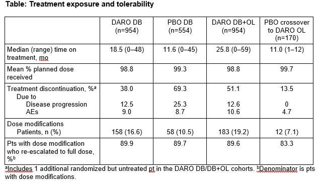
As shown in the Table, DARO remained well tolerated over the double-blind (DB) and OL periods: 98.8% of pts on DARO received the full planned dose and almost all pts with dose modifications were able to resume and re-establish the planned dose (DARO 89.6% vs PBO 89.7%). Discontinuation of DARO due to AEs increased slightly from the DB period (9.0%) to the DB+OL period (10.5%). Pharmacodynamic modeling showed that longer OS was positively associated with maximum PSA decline in DARO-treated pts.
Conclusion
DARO remained well tolerated with extended treatment at the recommended dose of 600mg twice daily. Almost all pts with nmCRPC were able to receive the full planned dose, increasing the likelihood of clinical benefit from effective disease control (PSA decline) and prolonged survival. Tolerability of different ARIs in the real world should be assessed.
Objectives: To present our experience in implementing a same-day discharge (SDD) protocol using enhanced recovery after surgery (ERAS) principles in patients who underwent robot-assisted radical prostatectomy (RARP).
Methods: This is a retrospective review that included all patients who underwent RARP at our institution between 06/01/2019 and 07/01/2020. Patients were stratified into two cohorts based on when COVID-19 restrictions were implemented at our institution : Pre COVID-19 era (06/01/2019-02/29/2020) and COVID-19 era (03/01/2020-07/01/2020). During the COVID-19 era, we implemented a protocol that supported SDD by minimizing opioid use, encouraging ambulation immediately after surgery, and early oral intake. Success of SDD was assessed and perioperative complications were compared between the cohorts.
Results: In 283 prostatectomies performed at our institution during this review, 83 (29.3%) were performed during the COVID-19 era. The pre COVID-19 era included more patients who had cT1 disease (56.5% vs 20.5%) and fewer patients with >cT2 (10.5% vs 30.1%, p<0.001). Same-day discharge was successful in 29 (34.9%) of patients in the COVID-19 era vs 13 (6.5%) in the pre COVID-19 era ( p<0.001). In the most recent 3 months since this paper was written, SDD rates have increased to 67.4%, with 100% success over the most recent month. Overall complication rates, including hospital readmission, emergency department visits, and telephone calls after surgery did not differ between cohorts (p>0.05).
Conclusions: We present practice changes that permitted the surgical management of prostate cancer patients to continue in an era where hospital bed capacity is limited. We utilized ERAS protocols to rapidly and safely increase the SDD rate after surgery without increasing the burden of ER visits, readmissions, and phone calls. Implications of such changes can be significant on the institutional and healthcare system level without compromising quality of care.
Introduction: Most patients considering RARP have a low risk of prostate cancer progression with short-term treatment delay and can expect a limited hospital stay. Multiple professional organizations recommended delaying most radical prostatectomies early during the COVID-19 pandemic to prioritize urgent cancer surgeries and preserve access to care.
Methods: Comprehensive clinicopathologic information was prospectively collected for patients undergoing RARP at a non-acute care comprehensive cancer center. Consecutive patients with newly diagnosed prostate cancer from a 12-month period beginning March 2020 (i.e., COVID) were compared to a retrospective cohort from March 2019 (i.e., pre-COVID). Same day discharge was encouraged in the COVID cohort, but a standardized program was not implemented.
Local epidemiologic data from the county public health department is reported to contextualize our center’s pandemic experience.
Results:
During the study period, COVID-19 bed occupancy at our facility ranged from 0 to 24 (11%). By comparison, among the county’s 70 9-1-1 receiving hospitals, COVID non-ICU bed occupancy ranged from 5% to 54%.
Of patients with D’Amico intermediate or high-risk disease, 136 (41%) of 330 were treated during COVID. Clinical grade group of 3 or greater was identified in 50% and 43% (p=0.02) of those in pre- and COVID cohorts, respectively. Age, ethnicity, and ASA score were similar, as was baseline PSA (7.0 ng/ml vs 8.0, p=0.5).
Median time from positive biopsy to surgery was 2.8 months (IQR 2.1-4.2) pre-COVID vs 3.4 months (IQR 2.0-8.0) COVID (p=0.1).
Histological upgrade on surgical pathology occurred in 18% vs 21% of patients pre- vs COVID (p=0.5). Pathologic upstaging was also infrequent, 22% vs 13% (p=0.1).
The percentage of patients discharged same day was 10% vs 15% pre- vs COVID, p=0.2. Distance from residence to facility was not correlated with same day discharge.
Similar results were observed when the COVID cohort was stratified into early (i.e., Mar, Apr 2020) vs. late (i.e., after Apr 2020) pandemic.
Conclusion: As a non-acute comprehensive cancer center, COVID bed utilization was lower than local rates. A small but non-significant treatment delay was observed without different rates of adverse pathologic outcomes among patients treated during COVID.
Introduction and Objective: Over the past two decades, the urologist’s armamentarium has rapidly grown to include newer minimally invasive treatment options for benign prostatic hyperplasia. One such technique is laser enucleation, which made its debut on the AUA, and EAU, guidelines. However, a closely related procedure, transurethral bipolar enucleation, TUBE, is briefly mentioned in the EAU guidelines but remains to be seen in the AUA guidelines. Despite this, several randomized controlled trials have shown decreased operative time, hospital stay, and blood loss in patients who underwent TUBE compared to simple prostatectomy. In conjunction, there have been no significant differences in functional outcomes. Although a plethora of research exists establishing enucleation equivalence, to our knowledge there is no data to date for patients who have undergone radical prostatectomy (RP) after TUBE. Herein we detail our retrospective, single institution two surgeon outcomes.
Methods: A total of 10 patients who underwent TUBE with subsequent RP between 2016 and 2020 were identified. Functional as well as oncological follow-up was retrospectively assessed. Patients with complete follow-up were matched by age and clinical stage with patients that did not have prior enucleation.
Results: All patients had final pathology of pT2, Gleason Grade Group 2 or higher. Follow up time was 2.4 and 3.1 years, respectively. There were no significant differences in comorbidities and no statistically significant difference in oncologic outcomes. The TUBE group demonstrated an absolute increase in the number of patients with post-operative stress urinary incontinence when compared to the matched RP only group, however this absolute difference was not significant. Among all comorbid conditions, obesity had the greatest association with post prostatectomy SUI, and this association was independent of TUBE.
Conclusions: Our study is the first publication to date that has shown RP after previous TUBE to be surgically feasible, with no negative impact on oncologic outcomes. Similarly, no statistical decrease in SUI or erectile function outcomes was observed in patients with prior TUBE. As a result, all patients with clinically significant, localized, prostate cancer after TUBE should be offered RP by high volume centers.
No source of funding.
Introduction: Focal cryotherapy is an attractive option to treat clinically significant localized prostate cancer while limiting side effects. We aim to define the oncologic and functional outcomes of focal cryotherapy.
Methods: We performed a retrospective analysis of patients undergoing focal cryotherapy at University of California, San Francisco. Inclusion criteria are defined as clinically localized, biopsy-proven Gleason Grade (GG) ≥2, stage ≥cT2a prostate cancer with >1 year of follow up. Patients with low volume GG1 cancer in the contralateral lobe were included. The cryotherapy treatment targets specific lesions only as opposed to hemi-lobe ablation. Follow up includes a PSA every 3 months followed by an MRI and biopsy at 1 year. The primary outcome is failure free survival, defined as no progression to definitive or systemic therapy or metastasis/death. Secondary outcomes include changes in GG, MRI findings and international prostate symptom (IPSS) / sexual health inventory for men score (SHIM). Kaplan Meier survival analysis was used to calculate failure free survival.
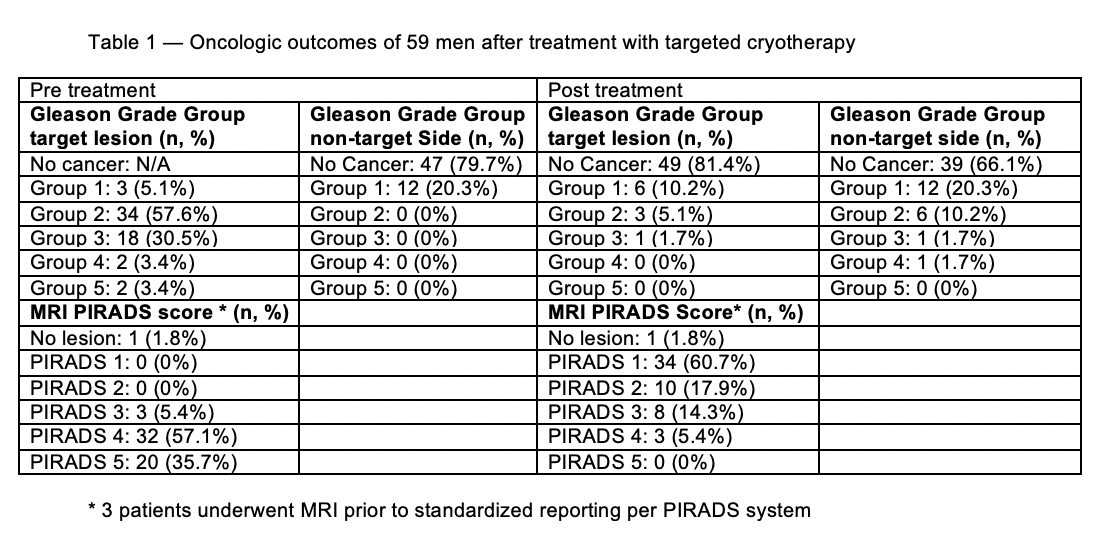
Results: 59 men underwent focal cryotherapy and all men completed post cryotherapy biopsies with a median follow up of 2.48 years (IQR 1.49-3.11) The median age at diagnosis was 67.0 years (range 48-81). 51 patients (86.4%) were cT2a-c and the remaining 8 were cT3a. Median (SD) PSA pretreatment was 6.7 ng/mL (3.2). 48 patients (81.4%) met the primary outcome and remained free of progression at the end of the follow up. Eight patients underwent definitive radiation or prostatectomy, and 3 patients were found to have metastases. Table 1 shows pre and post cryotherapy MRI and GG.
Conclusions: Over 80% of patients remain on active surveillance after focal cryotherapy during a median follow up of 2.5 years. Focal cryotherapy for localized prostate cancer shows encouraging oncologic and functional outcomes. Men must be counseled that while cryotherapy is a treatment option for prostate cancer which is largely focal in nature, they should be followed closely for recurrence.
OBJECTIVE: Multiparametric MRI (mpMRI) has not been quantitatively studied in prostate cancer with neoadjuvant androgen deprivation therapy (NADT). We selected men with extracapsular extension (ECE) based on mpMRI who were administered NADT prior to robotic-assisted radical prostatectomy (RARP). We examined changes in tumor characteristics on MRI over time.
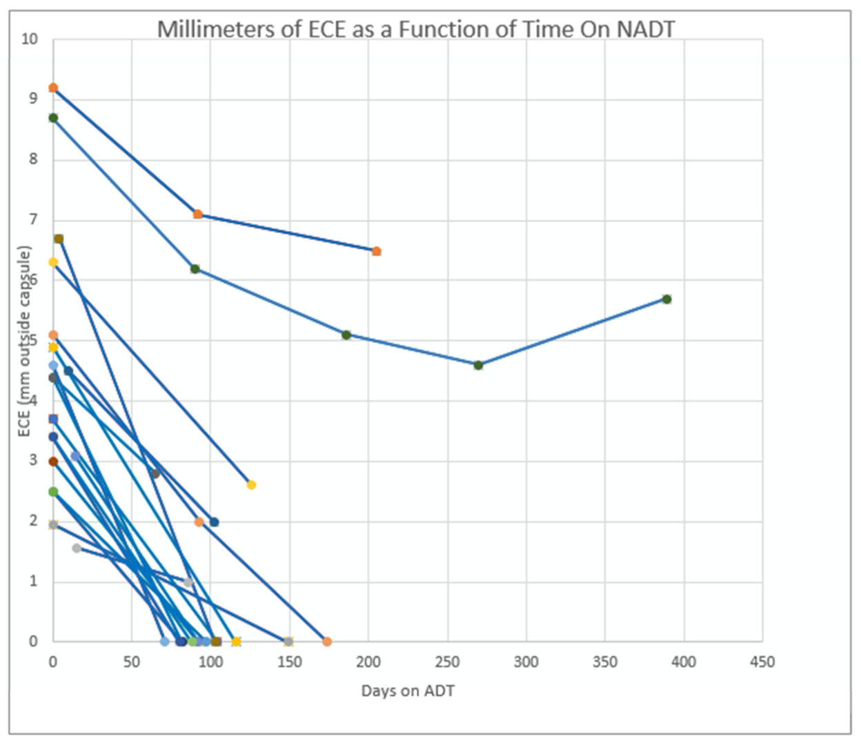
METHODS: 19 patients with radiographic T3a prostate cancer underwent NADT and serial MRI prior to RARP. The decision to proceed with surgery was based on sufficient reduction of ECE on MRI to achieve a negative margin. A total of 45 MRIs were reviewed by a single reader using computer-aided detection software (DynaCAD) to obtain precise spatial measurements of various tumor characteristics [e.g. tumor volume (TV on T2-weighted imaging cm3), maximum axial tumor dimension (mm) on diffusion weighted imaging (DWI), extracapsular TV (cm3), and extent of ECE (mm)]. Spider plots were constructed and cytoreductive velocity (CRV, mm tumor shrinkage/mo) was calculated from the linear regression slope of each patient’s tumor response curve.
RESULTS: The degree of ECE on initial MRI averaged 4.58mm (range 1.6mm – 9.2mm). 13/19 (68.4%) patients achieved radiographic resolution of ECE after an average of 3.86mo on ADT (SD=0.89). The average CRV among all patients was 0.98 mm/mo (SD=0.51mm/mo). On pathologic evaluation, 10 patients were ypT2 (52.6%), 4 were ypT3a (21.1%), and 5 were ypT3b (26.3%); 2 patients were node positive. Negative margins were obtained in 18/19 patients (94.7%). Over a median follow-up of 30.1 months, 11 patients (57.9%) remained biochemically free of disease in the setting of normalized testosterone, whereas 8 failed (42.1%). When comparing the two groups, patients with BCR had a larger initial tumor volume (9.46cc vs. 3.33cc, p=0.03), larger extracapsular tumor volume (0.62cc vs. 0.32cc, p=0.04), and a higher proportion of seminal vesicle invasion (4 vs. 1).
CONCLUSIONS: In our cohort, the MRI-based NADT induced cytoreductive response of ECE occurred at a velocity of 1mm/mo over 3-4 months.
Objective: To evaluate the rate at which clinically significant prostate cancer would go undiagnosed by performing an MRI-targeted biopsy alone.
Materials and Methods: Retrospective review of all prostate MRIs performed at Madigan Army Medical Center between March 1, 2018 and September 30, 2020. Prostate MRIs were excluded if they were performed for the purpose of radiation planning or cancer staging in individuals who had previously undergone prostate cancer treatment. After identification of qualifying imaging studies the corresponding medical records were reviewed for demographic and medical data. Individual prostate MRIs were considered positive if a radiologist identified a PI-RADS 3, 4, or 5 lesion. Individuals with a positive MRI were separated into two groups: those who underwent a systematic 12-core biopsy alone and those who had additional cores by MRI-targeted biopsy. Rates of clinically significant prostate cancer defined as Gleason Grade 7 or higher diagnosed on a systematic biopsy in the setting of a negative MRI-targeted biopsy were ultimately identified.
Analysis was performed with R Core Team (2020).
Results: Of the 337 prostate MRIs performed in the defined time frame, 291 were included. 145 of those individuals had a positive MRI prompting a prostate biopsy which included both a systematic and MRI-targeted sampling of the suspicious PI-RADS lesion. A total of 106 individuals had pathology return with a negative MRI-targeted index lesion. Of those individuals with a negative targeted biopsy 31 had identified prostate cancer outside of the PI-RADS lesion through systematic biopsy, 7 (22.6%) revealing clinically significant prostate cancer.
Conclusion: Recent updates to the AUA guidelines support the use of MRI-targeted prostate biopsies, either cognitive or software-based fusion, to aid prostate biopsy performance by aiming to increase detection rates of clinically significant prostate cancer. As the knowledge within this field is rapidly growing, this data supports the continued use of standard systematic biopsies even in the setting of an identified PI-RADS lesion due to the high rate of missing the diagnosis of clinically significant prostate cancer.
Objective: Prostate cancer is one of the most common cancers, affecting up to 11% of men over their lifetime. The diagnosis can be challenging, as most with early prostate cancer are asymptomatic and diagnosed based on PSA screening. The standard technique for prostate biopsy (bx) is with transrectal ultrasonography (US) and either a transrectal or transperineal needle approach with 12-core sampling. In this study, we examined our initial experiences with high-frequency micro-ultrasound machine for the detection of prostate cancer after mpMRI and the value of obtaining off-target biopsy cores.
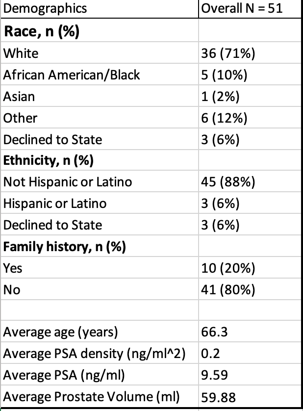
Methods: We retrospectively examined medical records of 51 patients at UC Davis Medical Center who underwent a prostate mpMRI followed by transrectal bx using the high-frequency US system. On and off target cores were taken with MRI lesions targeted using the integrated fusion assist targeting system. The primary outcome was the determination of the percentage of highest-grade lesions that were on-target, stratified by PIRADS score.
Results: Out of 51 patients, 15 had either a negative MRI (PIRADS 1 or 2) or no MRI. Out of the 5 patients who had a PIRADS 3 lesion on MRI, the highest-grade lesion was on-target 60% of the time. The other 40% of lesions were benign. Out of the 17 patients who had a PIRADS 4 lesion, the highest-grade lesion was on-target 76% of the time. The other 24% of lesions were benign. Out of the 14 patients who had a PIRADS 5 lesion, the highest-grade lesion was on target 79% of the time. The other 21% of lesions were off-target or benign.
Conclusions: Our study demonstrated that, in patients with PIRADS 4 or 5 lesions, the highest-grade biopsy lesion was on-target at least 76% of the time. Obtaining only on-target biopsy lesions would allow for fewer biopsy cores, which could decrease the risk of pain, bleeding, and even infection. Our findings are consistent with other series describing technology for MRI lesion targeting.
INTRODUCTION AND OBJECTIVES:
MRI-ultrasound fusion biopsy (Fus-Bx) is increasingly being utilized for prostate cancer detection, however, not all patients have access to the technology. MRI cognitive fusion biopsy (Cog-Bx) enables targeting of MRI visible lesions without the cost of Fus-Bx systems. We compare detection of prostate cancer between Fus-Bx and Cog-Bx modalities from a prospectively collected database of patients who underwent both fusion modalities.
METHODS:
From August 2017 - January 2021, patients with at least one PIRADS 3 or greater lesion on multiparametric 3.0 Tesla prostate MRI underwent both Fus-Bx and Cog-Bx as part of a prospective within patient protocol. Cog-Bx was performed first for each region of interest (ROI), followed by Fus-Bx using the UroNav™ system. 2 targeted cores were obtained from each ROI for each modality. Following targeted biopsies, standard 12 core systematic biopsy was performed. The primary endpoint was clinically significant cancer detection (≥Gleason grade group 2) and the secondary endpoint was any cancer detection. Agreement between the two modalities was assessed using McNemar's test and Cohen's kappa coefficient for each ROI.
RESULTS:
91 patients with 98 lesions were included. The mean age was 69 years (range 47-87). Median PSA was 7.31 ng/dl (range 0.9-33.6). Mean gland volume was 51.7cc (range 20-198). Mean lesion size was 1.6cm (range 0.5-4.5) in largest dimension, and mean volume was 1.6 cc (range 0-24). Bx (McNemar’s test, p = 0.285, p = 0.405, respectively). The detection rate for any cancer for Fus-Bx and Cog-Bx was 46.9%, and 39.8% for GG2 cancer, respectively. Concordance between Fus-Bx and Cog-Bx was 85.7% (84/98) for clinically significant cancer.There was no significant difference for detection of either cancer type between Fus-Bx and Cog-any cancer detection and 86.7% (85/98) for clinically significant cancer. There was strong agreement between modalities for both any cancer and clinically significant cancer (kappa statistic of 71.5% (95% CI 58%-85%) and 72.7% (95%CI 59%-86%), respectively).
CONCLUSIONS:
There is high concordance for prostate cancer detection between Fus-Bx and Cog-Bx, and detection rates are not significantly different. Cog-Bx is a viable alternative to target suspicious lesions seen on MRI where Fus-Bx is not available.
Objective
To evaluate the association between intravesicular prostate gland protrusion and the detection rate of clinically significant prostate cancer (csPCa) on MRI TRUS fusion targeted biopsy (TB).
Materials and Methods
538 consecutive men who underwent MRI TRUS fusion TB and concomitant systematic biopsy at a single academic center between 2014 and 2018 were evaluated. Intra-prostatic protrusion (IPP) on MRI was independently measured by four blinded reviewers. IPP was measured on T2-weighted mid-sagittal MRI in millimeters (mm). Patients were grouped by IPP: zero, small (>0-<5mm), medium (5-10 mm), and large (>10mm). csPCa was defined as GS ≥ 3+4=7. The primary outcome was per-lesion detection of csPCa on TB. We used Chi-square test to compare detection rates between protrusion groups, and multivariable logistic regression to assess the association between IPP and csPCa detection on TB, controlling for age, PSA, PIRADS, prostate volume, targeted cores sampled and previous biopsy experience
Results
847 PIRADS 3 or greater lesions were targeted across 570 biopsies. Intrarater reliability for IPP were good-excellent (ICC 0.9, 95% CI 0.87-0.93) and interrater reliability results for IPP were moderate-good (ICC 0.74, 95% CI 0.74-0.83). 81 (14.2%), 127 (22.3%), 237 (41.6%), and 125 (21.9%) men had zero, small, medium and large IPP, respectively. 230 (27.1%), 392 (46.2%) and 196 (23.1%) MRI lesions were PIRADS 3, 4 and 5, respectively. 198 (34.7%) lesions had csPCa on TB. The overall relationship between IPP size and csPCa found on TB was not significant (zero (44.4%), small (33.9%), medium (36.3%) and large IPP (26.4%), however, large IPP is associated with significantly less csPCa than zero IPP (P=0.007). Every mm increase in IPP is associated with 5.6% decrease in the odds of csPCa detection on TB (P=0.004) and a 66.5% decrease in odds of detection in large IPP compared to zero IPP (P=0.003). However, this significance is lost when controlling for prostate volume (P=0.296).
Conclusions
As IPP and volume increases, there are lower detection rates of csPCa on MRI-guided TB. These findings may be driven by poor MRI/TRUS co-registration and prostate asymmetry.
Source of Funding: none
Purpose: PYLARIFY (piflufolastat F18, formerly 18F-DCFPyL or PyL) is an FDA-approved PSMA-targeting PET tracer indicated for identifying and localizing previously unrecognized prostate cancer (PCa). This prospective trial evaluated piflufolastat F18 in patients with newly diagnosed high-risk (cohort 1) and biochemically recurrent (cohort 2) PCa, with pathology as the preferred gold standard of comparison.
Methods: Eligible subjects (cohort 1: newly diagnosed Gleason 8-10 and/or PSA>10; and cohort 2: biochemical recurrence with PSA>0.2ng/ml post prostatectomy or PSA 2.0ng/ml above nadir post radiation therapy, with negative CT and bone scan) underwent piflufolastat F 18 PET/CT at the Molecular Imaging and Therapy Center of the Hoag Family Cancer Institute. If lesions suspicious for distant metastases (cohort 1) or disease recurrence (cohort 2) were detected on piflufolastat F18 PET/CT, then a lesion was selected for biopsy confirmation, whenever possible.
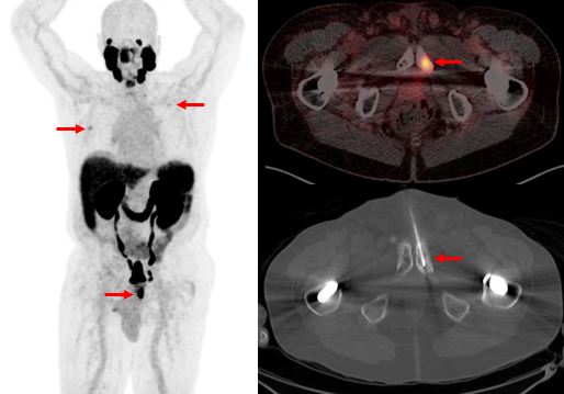
Results: To date, 78 subjects underwent piflufolastat F18 PET/CT (36 cohort 1; 42 cohort 2). In cohort 1, 15/36 (42%) demonstrated avid lesions outside the pelvis, including 9/36 (25%) subjects upstaged with unsuspected distant metastases (Figure), 3 (8%) upstaged with occult regional nodal metastases, 1 with unsuspected renal cell carcinoma metastases, 1 benign, and 1 pending confirmation. In cohort 2, 28/42 (67%) subjects demonstrated avid lesions (median PSA= 2.0 ng/mL), with 17 biopsy proven recurrences, 2 benign, and 9 pending confirmation. Across both cohorts, of 33 biopsies of piflufolastat F18 avid foci, 30 (91%) confirmed malignancy.
Conclusion: With pathology as the preferred standard of truth, piflufolastat F18 PET/CT detected unsuspected disease in patients with newly diagnosed high-risk and biochemically recurrent PCa with a PPV of >90%. Of note, 13/36% (13/36) of subjects with newly diagnosed high-risk PCa had pathologically-proven, previously occult, malignancy which would impact treatment strategies.
Figure: PYLARIFY PET/CT in a 72-year-old man with newly diagnosed Gleeson 8 prostate cancer, PSA 5.1. PET/CT demonstrates three avid osseous foci (arrows). The left pubic ramus focus was biopsy proven to represent metastatic prostate cancer.
Introduction
Solid organ transplant recipients may have elevated cancer risk due to immunosuppression. Large population cohort studies have shown that transplanted patients with low- or intermediate-risk prostate cancer (PCa) have similar outcomes to non-transplanted patients. However, there is limited data on the relationship between prior transplant exposure and risk for clinically aggressive PCa at presentation.
Methods
We queried the linked SEER-Medicare database to identify 123,261 screened patients, who were then diagnosed with prostate cancer. Criteria for screening included a PSA lab test or DRE exam in both the 12 month AND 13-36 month periods prior to diagnosis of PCa. History of solid organ transplant was identified using diagnosis or procedure codes in the 3 years preceding the diagnosis. Aggressive PCa phenotype was determined if patients had at least 3 of the following 5 characteristics: death from PCa within a year of diagnosis, de novo metastasis, regional lymph node metastasis, PSA >20, or Gleason Score 8-10 at presentation. Risk estimates of the likelihood of aggressive PCa at presentation for transplant vs. non-transplant cohorts were estimated in univariable and multivariable logistic regression models adjusting for age and race.
Results
We identified 292 transplant patients who went on to develop PCa. Table 1 shows the OR for risk of aggressive prostate cancer in both univariable and multivariable analyses. Both Univariable analysis and multivariable analyses demonstrated that prior transplant exposure was not associated with a significantly increased risk of aggressive PCa phenotype with ORs of .949 (95% CI 0.72-1.25, P = .71) and 1.187 (95% CI .896-1.572, p = .23), respectively.
Conclusions
Following solid organ transplantation, patients are not an increased likelihood of being diagnosed with aggressive prostate cancer. This data would suggest that increased screening and monitoring in this cohort is unnecessary as their risk is similar to the general population.
| Unadjusted | Adjusted | ||||
| Odds Ratio | 95% CI | P-value | Odds Ratio | 95% CI | P-value |
Transplant | 0.94 | 0.72-1.25 | 0.71 | 1.18 | 0.89-1.57 | 0.23 |
Age | 1.12 | 1.10-1.11 | <.0001 | 1.09 | 1.09-1.09 | <.0001 |
African American | 1.27 | 1.207-1.35 | <.0001 | 1.25 | 1.19-1.31 | <.0001 |
Table 1. Univariable and Multivariable OR of aggressive prostate cancer phenotype at diagnosis for transplant patients vs non-transplant controls.
Objectives:
International Continence Society (2009 guidelines) to consider neuromodulation in management of post-prostatectomy incontinence if the initial conservative therapy fails, as well as, in mixed incontinence and over-active bladder case. The objective of the present work is to study the role of Percutaneous Tibial Nerve Stimulation (PTNS) for men complaining of post-radical prostatectomy incontinence, mainly mixed incontinence and over-active bladder cases; prospective clinical trial.
Patients and methods
Post-radical prostatectomy men complaining of mixed incontinence and those with bladder over-activity were instructed to consider Percutaneous Tibial Nerve Stimulation (PTNS).
Eighteen men in the current study were subjected to 12 (weekly) PTNS sessions. They were asked to fill two days voiding diaries, as well as, the Expanded Prostate Cancer Index Composite for Clinical Practice (EPIC-CP), as it is a comprehensive instrument designed to evaluate patient function, bother and quality of life after prostate cancer treatment (0-60 points).
Results
After 12 PTNS sessions there is statistically decreased urinary frequency, decreased urgency and increased voiding volume. Moreover, EPIC-CP questionnaires decreased significantly (p=0.002) following 12 PTNS sessions. From the table, we reported 3 cases become completely dry and 7 men continent (<1 pad/day). Furthermore, there is 5 cases still have mild incontinence (1-2 pads/day), and only 3 men still consuming 2-3 pads daily.
| Pretreatment | Post-treatment |
Completely dry | 0 | 3 |
Continent < 1 pad / day | 0 | 7 |
Mild: 1-2 pads / day | 2 | 5 |
Moderate: 2-3 pads / day | 13 | 3 |
Severe: > 3 pads / day | 3 | 0 |
Continence progress among 18 Post-radical Prostatectomy incontinent men before and after 12 PTNS sessions
Conclusion:
Conclusion:
Percutaneous Tibial Nerve Stimulation as a minimally invasive neuromodulation tool seems to has a positive impact in post-radical prostatectomy incontinent men and their quality of life. It could fill the gap between failed conservative therapy and surgical intervention among those men.
Introduction:
Prolonged operative time (OT) is associated with a higher risk of surgical complications. One potentially modifiable risk factor for prolonged OT is resident involvement, as attending surgeons must balance OT with resident education. Herein, we apply machine learning to investigate how clinical factors, operative factors, and surgeon experience predict prolonged OT in robot-assisted radical prostatectomy (RARP).
Methods:
We conducted a retrospective study of RARPs at our institution from 2016 to 2017. Each case was broken down into 12 standardized steps. The operating surgeon of each step was identified as either expert (> 100 cases), training (<100 cases), or mixed. Classification and Regression Tree (CART®) identified the strongest predictors of prolonged OT from 15 clinical factors, task times of each step, and surgeon experience. Based on preliminary findings showing that the task times of two specific steps predicted prolonged OT, we used ElasticNet to further investigate how clinical factors and surgeon experience impact these two specific steps.
Results:
130 cases were included with median OT 122 minutes (IQR 104 – 147). Cases were performed by 14 experts (median 375 cases, IQR [150-1688]) and 14 training surgeons (30, [5-38]). CART® analysis selected two major predictors of prolonged OT: apical dissection and left lymph node (LN) dissection task times. Through 5-fold cross validation, CART® then identified 17 cases with prolonged OT predicted by task time > 16 minutes and > 20 minutes in the apical dissection and left LN dissection, respectively. ElasticNet analysis revealed non-nerve sparing procedure (p=.03) predicted longer apical dissection, while BMI (p=.03) and number of lymph nodes dissected (p<.01) predicted longer left LN dissection. Surgeon experience was not selected by CART® or ElasticNet as a statistically significant predictor of prolonged OT or task times of the two steps driving prolonged OT.
Conclusion:
Prolonged OT seems to be predicted by longer apical dissection and left LN dissection steps. The main factors driving task time of these two steps are patient and disease related factors. No strong evidence was found suggesting that surgeon experience is an independent and significant contributor to individual task times or total operative time.