Prostate Cancer 1

2:30 - 3:45pm Tuesday, 2nd November, 2021


28 Radiation-induced fistulas after pelvic radiation therapy: a systematic review and meta-analysis.

Michael J Sadighian MSc1, Nizar Hakam MBBS1, Gregory Amend MD1, Peggy Tahir MA MLIS2, Isabel E Allen PhD3, Behnam Nabavizadeh MD1, Jordan T Holler MPH1, William Shibley BA1, Kevin D. Li BS1, Behzad Abbasi MD1, Alexander Bell BA1, Osama Mohamad MD PhD4, Benjamin N. Breyer MD MAS1
1UCSF Department of Urology, San Francisco, CA, USA. 2UCSF Library Services, San Francisco, CA, USA. 3UCSF Department of Epidemiology and Biostatistics, San Francisco, CA, USA. 4UCSF Department of Radiation Oncology, San Francisco, CA, USA

Abstract

Objectives: Little is known about the prevalence of fistulas in patients with exposure to pelvic radiotherapy. The purpose of this systematic review and meta-analysis is to aggregate and summarize existing cohort study data on fistula prevalence among patients with a history of pelvic radiotherapy for pelvic malignancy.


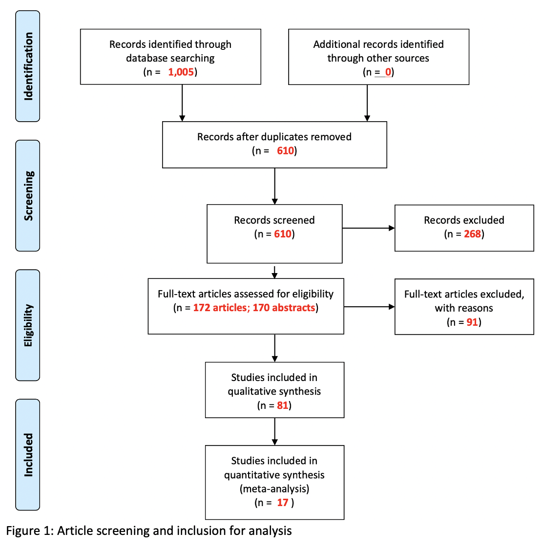
Materials and Methods: We queried PubMed, Embase, and Web of Science for studies pertaining to radiation induced fistulas in the pelvis. For this abstract, we included cohort studies reporting fistula prevalence among patients exposed to pelvic radiotherapy for the treatment of pelvic malignancy. We conducted meta-analysis using the fixed-effects model. We used I2 statistic to assess heterogeneity and the Newcastle-Ottawa Scale to assess risk of bias. PRISMA guidelines were followed, and our protocol was registered a priori on PROSPERO (CRD42020214451).


Results: We included 17 cohort studies with a total of 1,371 patients exposed to pelvic radiotherapy between 1983 and 2019. Mean patient age was 57.7 years and mean follow-up time was 32.5 months. The studies reported data on patients with cancer of the cervix (n=852), prostate (n=266), rectum (n=91), and multiple cancers (n=162). Pooled prevalence of radiation induced fistula was 3% (95% CI: 2% - 4%, I2 = 0%) and ranged between 1% and 34% in the included studies. Stratifying the pooled risk by year, cancer type, and fistula type demonstrated no significant heterogeneity between these substrata.


Conclusions: Patients undergoing pelvic radiotherapy for pelvic malignancy are at low risk for developing fistulas. The prevalence of fistulas has not changed over time and does not vary based on cancer type. Given that our mean follow-up time was 32.5 months, future studies should provide data on more long-term risk.

Uncaptioned visual

Uncaptioned visual

If funding provided, type in source company / entity name(s):

None

33 Race-Specific Trends in Prostate Cancer Screening and Presentation Before and After the 2012 United States Preventative Services Task Force Statement

Brandon Horton MPH1, Stacey Alexeeff PhD1, Stephanie Prausnitz MS1, Andrew Avins MD, MPH1, Joseph Presti, Jr. MD2
1Division of Research, Kaiser Permanente, Oakland, CA, USA. 2Department of Urology and Division of Research, Kaiser Permanente, Oakland, CA, USA

Abstract

Objective:  To assess temporal changes in the race-specific rates of PSA screening, prostate biopsy, incident prostate cancer detection, and metastatic cancer at presentation among screen-eligible men in Kaiser Permanente Northern California before and after the 2012 the United States Preventive Services Task Force Prostate Cancer Screening Statement.

Materials and Methods:  This was a retrospective study spanning the years 2006 to 2017, in screen-eligible Kaiser Permanente Northern California members (Black men ages 45-69, all other men ages 50-69) with no prior history of prostate cancer.  The number of screen-eligible men served as the denominator for all rate calculations.  We compared the race-specific biennial rates of PSA testing, prostate biopsy, incident prostate cancer detection, and metastatic cancer at presentation.

Results:  422,664 to 567,660 total men per biennial period were evaluated (72% White, 8% Black, 20% Asian).  Following the 2012 United States Preventive Services Task Force statement, all races experienced similar declines in screening (22-25%; Figure 1), biopsy (47-57%) and overall cancer detection (34-48%) rates. We found an increase in metastatic rates (39-105%) in all races (Figure 2). 

Uncaptioned visualUncaptioned visual

White are referent for all comparisons of rate ratios; tan colored numbers are statistically significantly different from referent, p<0.05

Conclusions: Following the 2012 Statement, in men under the age of 70, PSA screening, biopsy and incident prostate cancer-detection rates significantly decreased in a similar fashion across all races, while rates of metastatic disease significantly increased in all races.

If funding provided, type in source company / entity name(s):

The Permanente Medical Group Delivery Science and Physician Researcher Program

34 Prospective Validation of the Kaiser Permanente Prostate Cancer Risk Calculator in a Contemporary, Racially Diverse, Referral Population

Joseph Presti, Jr. MD1, Stacey Alexeeff PhD2, Brandon Horton MPH2, Stephanie Prausnitz MS2, Andrew Avins MD, MPH2
1Department of Urology and Division of Research, Kaiser Permanente, Oakland, CA, USA. 2Division of Research, Kaiser Permanente, Oakland, CA, USA

Abstract

Objective:   To prospectively validate a new prostate cancer risk calculator in a racially diverse population.

Materials and Methods:  We recently developed, internally validated and published the Kaiser Permanente Prostate Cancer Risk Calculator.  This study is a prospective validation of the calculator in a separate, referral population over a 21-month period.  All patients were tested with a uniform PSA assay and a standardized systematic, ultrasound-guided biopsy scheme.  We report on 3 calculator models:  Model 1 included age, race, PSA, prior biopsy status, body mass index, and family history of prostate cancer; Model 2 added digital rectal exam to Model 1 variables; Model 3 added prostate volume to Model 2 variables.  We considered three outcomes: high-grade disease (Gleason score >7), low-grade disease (Gleason score=6), and no cancer.  Predictive discrimination and calibration were calculated.  How each model might alter biopsy frequency and outcomes at various thresholds of risk was assessed.  We compared the performance of our calculator with two other calculators.

Results:  In 4178 patients (16.2% Asian, 11.3% African American, 13.5% Hispanic), cancer was found in 53%; 62% were Gleason score >7.  Using a high-grade risk threshold for biopsy of >10%, Model 2 predictions would result in 9% of men avoiding a biopsy, while only missing 2% of high-grade cancers.  At the same threshold, Model 3 predictions would result in 26% of men avoiding a biopsy, while only missing 5% of high-grade cancers.   The c-statistics for Models 1, 2, and 3 to predict high-grade disease vs. low-grade or no cancer were 0.76, 0.79 and 0.85, respectively.  The c-statistics for Models 1, 2, and 3 to predict any prostate cancer vs. no cancer were 0.70, 0.72 and 0.80, respectively.  All models were well calibrated for all outcomes.  Our Model 3 calculator had superior discrimination for high grade disease (c-statistic=0.85, 0.84-0.86) and any cancer (0.80, 0.79-0.82) compared to the PBCG calculator [(0.79, 0.78-0.80); 0.72 (0.70-0.73)] and the PCPT calculator [(0.75, 0.74-0.77); 0.69 (0.67-0.70)], respectively.  In the high-grade cancer predicted risk range of 0-30%, our Model 2 was better calibrated than the PCPT and PBCG calculators.  

Conclusions:  This validation of our calculator showed excellent performance characteristics.

If funding provided, type in source company / entity name(s):

The Permanente Medical Group Delivery Science and Physician Researcher Programs

108 The clinical cell-cycle risk score is associated with metastasis after radiation therapy and may identify men with prostate cancer who can forgo combined androgen deprivation therapy

Jonathan Tward MD, PhD1, Lauren Lenz MS2, Saradha Rajamani PhD2, Carl Olsson MD3, Deepak A. Kapoor MD3, Constantine Mantz MD4, Stanley L. Liauw MD5, Tatjana Antic MD5, Neal Shore MD6, Dan Albertson MD7, Jonathan Henderson MD8, Steve P. Lee MD9, Hiram A. Gay MD10, Jeff Michalski MD10, Arthur Hung MD11, David Raben MD12, Isla Garraway MD, PhD13, Michael S. Lewis MD13, Paul L. Nguyen MD14, David T. Marshall MD15
1Huntsman Cancer Institute, Salt Lake City, UT, USA. 2Myriad Genetics, Inc., Salt Lake City, UT, USA. 3Advanced Radiation Center of New York, New Hyde Park, NY, USA. 421st Century Oncology, Fort Meyers, FL, USA. 5University of Chicago Medical Center, Chicago, IL, USA. 6Carolina Urologic Research Center, Myrtle Beach, SC, USA. 7University of Utah Department of Anatomic Pathology and Molecular Oncology, Salt Lake City, UT, USA. 8Regional Urology, LLC, Shreveport, LA, USA. 9Long Beach VA Medical Center, Long Beach, CA, USA. 10Department of Radiation Oncology, Washington University, St. Louis, MO, USA. 11Oregon Health & Science University, Portland, OR, USA. 12University of Colorado, Aurora, CO, USA. 13Greater Los Angeles VA Medical Center, Los Angeles, CA, USA. 14Dana-Farber Cancer Institute, Boston, MA, USA. 15Medical University of South Carolina, Charleston, SC, USA

Abstract

Introduction: This study evaluated the ability of the combined clinical cell-cycle risk score (CCR) to prognosticate the risk of prostate cancer metastasis in men receiving dose-escalated radiation therapy (RT) with or without androgen deprivation therapy (ADT).


Methods: The CCR score is a validated model that combines the cell cycle progression score (CCP) with the UCSF Cancer of the Prostate Risk Assessment score (CAPRA). The CCR score and a CCR-based multimodality threshold score (2.112) were evaluated in a retrospective, multi-institutional cohort of men with National Comprehensive Cancer Center (NCCN) intermediate- or high-risk localized disease (N=741) who received single (RT)- or multimodality therapy (ADT with RT). Effects of prognostic variables were analyzed using Kaplan-Meier and Cox regression methods.


Results: Median follow-up was 5.6 years. CCR predicted metastasis [hazard ratio (HR) 2.21, 95% Confidence Interval (CI) 1.70-2.87, p<0.001]. The CCR score was a better prognosticator of metastasis (C-index 0.78) than either NCCN-risk group (C-index 0.72), CAPRA score (C-index 0.71), or CCP score (C-index 0.69) alone. In bivariate analyses, the CCR score remained highly prognostic for metastasis when comparing any ADT vs none (HR 2.19, 95% CI 1.62 to 2.97, p<0.001), ADT duration as a continuous variable (HR 2.11, 95% CI 1.59-2.79, p<0.001), or ADT use given as less than or at the recommended duration for each NCCN risk group (HR 2.19, 95% CI 1.68-2.84, p<0.001). Men with CCR scores either below or above the threshold (2.112) had a 10-year risk of metastasis of 4.1 % and 25.3%, respectively. For men below the threshold receiving RT alone versus RT+ADT, the 10-year risk of metastasis was 4.2% and 3.9%, respectively.


Conclusions: CCR is a highly precise and accurate predictor of metastasis in men undergoing dose-escalated RT, with or without ADT.   CCR adds clinically actionable information relative to guideline recommended therapies that are based on NCCN risk groups or CAPRA alone. Men with scores below the multimodality threshold may not significantly reduce their 10-year risk of metastasis with the addition of ADT.

If funding provided, type in source company / entity name(s):

Myriad Genetics, Inc.

46 Phase 1b/2 Study of Sabizabulin (VERU-111), an Androgen Receptor Transport Disruptor, in Men with Metastatic Castrate Resistant Prostate Cancer (mCRPC) Who Progressed on an Androgen Receptor Targeting Agent (ARTA)

Mark C Markowski MD PhD1, Mario A Eisenberger MD1, Christopher Pieczonka MD2, Robert H Getzenberg PhD3, Domingo Rodriguez MD3, K Gary Barnette PhD3, Mitchell S Steiner MD3, Daniel R Saltzstein MD4, Emmanuel S Antonarakis MD1, Ronald Tutrone MD5
1Sidney Kimmel Comprehensive Cancer Center, Johns Hopkins University School of Medicine, Baltimore, MD, USA. 2Associated Medical Professionals of NY, Syracuse, NY, USA. 3Veru Inc, Miami, FL, USA. 4Urology San Antonio, San Antonio, TX, USA. 5Chesapeake Urology Research Associates, Towson, MD, USA

Abstract

Objectives

Sabizabulin is a novel oral agent that targets microtubules to disrupt transport of AR.  A Phase 1b/2 clinical study was conducted to assess safety, maximum tolerated dose and efficacy in men with mCRPC that progressed on an ARTA.


Methods

A Phase 1b/2 study was conducted in men with mCRPC who have failed an ARTA. The Phase 1b portion (n=39) utilized a 3+3 design with escalating oral dosing of 4.5 mg to 81 mg  for 7 days on /14 days off drug per 21-day cycle, then was expanded to continuous daily dosing.  The Phase 2 portion (n=41) evaluated the recommended Phase 2 dose (63 mg PO daily).  A PK study evaluating the Phase 2 and Phase 3 trial dosage forms was also conducted.


Results 

Sabizabulin was well tolerated with the most common adverse events (>10% frequency) in patients that received 63 mg dose (n=54) including diarrhea, fatigue, ALT and AST increases which were mostly Grade 1 and 2. 


In the Phase 1b portion, in men who were treated with 63 mg (n=14), the median duration on study was 10.8 m (2.3-24.7m).  In the men with measurable disease (n=9), the ORR was 44% (n=4).  For men ³ 60mg dose (n=19), the estimated rPFS is 12.4 m (3 men still on study). The Phase 2 is ongoing with evidence of PSA responses and objective responses.  When combining Phase 1b/2 trials, in patients with measurable disease at baseline (n=29), the ORR (5PR +1CR observed) was 20.7%.  In patients that received ³ 63 mg (excluding baseline superscans)(n=55), the median rPFS is estimated to be 7.4 months with 10 men on study (Feb 2021).  


Conclusions

In the Phase 1b/2 clinical trial, oral 63mg daily dosing has a favorable safety profile and chronic dosing is feasible.  Efficacy was observed with PSA declines and long term durable responses.  The Phase 3 VERACITY study evaluating sabizabulin in chemotherapy naïve men with mCRPC who have failed an AR targeting agent is ongoing.  Sabazibulin appears to be in a similar class of other FDA approved targeted cytostatic drugs that have shown to significantly prolong progression and survival. 


If funding provided, type in source company / entity name(s):

Veru Inc

49 Development of an End-to-end Assessment of Suturing Expertise (EASE)

Taseen F Haque B.A.1, Monish Aron M.D.2, Justin W Collins M.D.3, Hooman Djaladat M.D.2, Ahmed Ghazi M.D.4, Kenneth A Yates Ed.D.5, Andre L Abreu M.D.2, Sia Danseshmand M.D.2, Mihir M Desai M.D.2, Alvin C Goh M.D.6, Jim C Hu M.D.7, Amir H Lebastchi M.D.2, Thomas S Lendvay M.D.8, James Porter M.D.9, Anne K Schuckman M.D.2, Rene Sotelo M.D.2, Chandru P Sundaram M.D.10, Jessica H Nguyen B.S.1, Inderbir Gill M.D.2, Andrew J Hung M.D.1
1Center for Robotic Simulation & Education, Catherine & Joseph Aresty Department of Urology, USC Institute of Urology, University of Southern California, Los Angeles, CA, USA. 2Department of Urology, University of Southern California, Los Angeles, CA, USA. 3University College London Hospital, Division of Uro-oncology, London, JS, United Kingdom. 4Simulation Innovation Laboratory, University of Rochester, Rochester, NY, USA. 5Department of Surgery, Keck School of Medicine, and Rossier School of Education, University of Southern California, Los Angeles, CA, USA. 6Urology Service, Department of Surgery, Memorial Sloan Kettering Cancer Center, Weill Cornell Medical College, New York, NY, USA. 7Department of Urology, Weill Cornell Medicine, New York, NY, USA, New York, NY, USA. 8Department of Urology, Seattle Children’s Hospital, Seattle, WA, USA. 9Swedish Medical Center, Seattle, WA, USA. 10Department of Urology, Indiana University, Indianapolis, IN, USA

Abstract

Objectives: Validated skills assessment tools have the potential to help trainees understand their progress. Herein, we aimed to exhaustively map out all relevant sub-skills of suturing and to define criteria around said sub-skills to differentiate performance.


Materials and Methods: The first step of this study was conducting a Cognitive Task Analysis (CTA), where 4 expert surgeons, moderated by an educational psychologist, systematically deconstructed the complete process of robotic suturing into its most basic sub-stitch maneuvers and described the accompanying skills to accomplish them at differing proficiency levels (“sub-skills”). We then utilized the well-established Delphi methodology to guide consensus on the CTA results from a panel of 16 leading surgical educators. Each sub-skill identified in the CTA underwent review and revision by using binary “agree”/“disagree” responses with free-text for commentary. This process was iterated until all sub-skills reached a content validity index (CVI), defined as proportion of participants who “agree” with each sub-skill, of at least 0.80.


Results: The 16 surgeons who served as panelists for the Delphi process had a median H-index of 23 (range 11-107). In Round 1 of the Delphi , 60/64 (94%) of proposed sub-skills met the CVI threshold. Three of the four (75%) sub-skills below the CVI threshold in Round 1 were within the “Pre-Planning” domain (Figure 1). The domains of EASE and their overall Round 1 CVIs are Pre-Planning (0.82), Needle Handling (0.90), Needle Entry (0.90), Needle Driving (0.97), Needle Withdrawal (1.0), Suture Placement and Management (0.92), and Knot Tying (0.90). In Round 2, the number of sub-skill descriptions was reduced to 61 as panelists suggested combining two sub-skills. These remaining sub-skills all reached CVI threshold. 


Conclusions: Our study defined the end-to-end process of suturing sub-skills and built consensus around them utilizing the expert opinions of leading surgical educators. The creation of EASE will set the foundation of future research goals, such as the full automation of skills assessment.


Uncaptioned visual

Figure: EASE Domains and Sub-Skills Schematic

If funding provided, type in source company / entity name(s):

Research reported in this publication was supported in part by the National Institute of Biomedical Imaging and Bioengineering of the National Institutes of Health under Award No. K23EB026493, and by the National Cancer Institute under Award No.1R01CA251579-01A1

115 Chronic Glucocorticoid Use and Risk for Advanced Prostate Cancer at Presentation: A SEER Population-based Cohort Study

Zorawar Singh MHS, Sarah K Holt PhD, John L Gore MD, MS, Jonathan L Wright MD, MS, George R Schade MD
University of Washington, Seattle, WA, USA

Abstract


Introduction

Oral glucocorticoids (steroids) are among the most commonly prescribed medications, with a large proportion of patients being chronic users. Steroids regulate diverse cellular functions and have cross-talk with androgen receptors, potentially propagating androgen independent prostate cancer (PCa). We aimed to examine the relationship between exposure to steroids and advanced PCa at presentation with a methodology to limit detection bias.

Methods

We queried the linked SEER-Medicare database to identify PSA screened patients diagnosed with PCa. Criteria for screening included a PSA lab test or DRE exam in both the 12 month AND 13-36 month periods prior to diagnosis of PCa. SEER-Medicare Part D prescription drug claims were used to identify steroid exposure. Steroid use was divided into exposure groups based on duration of use in the 3 years prior to diagnosis: controls with no exposure, <30 days, 30d-1yr, 1-2yrs, and >2+ years. Advanced PCa was defined as de novo systemic metastases or regional lymph node metastasis at presentation. Risk estimates of the likelihood of advanced PCa at presentation for steroid exposure groups vs. controls were estimated in univariate and multivariate (MV) logistic regression models adjusting for age, race, and comorbidities. Dose response was evaluated in analogous MV regression analysis with duration of steroid use as a continuous variable (days).

Results

We identified 22,920 PSA screened patients diagnosed with PCa of which 28.96% used glucocorticoids in the exposure period.  Both univariate and MV analyses found a significantly increased risk for advanced PCa in patients with >2yrs of steroid exposure with OR of 2.06 (95% CI 1.35-3.14) and 1.74 (1.12-2.69), respectively (Table 1). Analogous MV regression analysis demonstrated a dose response relationship between duration of steroid use and risk for advanced PCa at presentation (p=0.01).

Conclusions

In this population-based PSA screened cohort, prolonged steroid use was associated with increased risk of de novo advanced PCa. With their widespread use, it is important to consider the role steroids may play in PCa pathogenesis.

Uncaptioned visual


120 The Diagnostic Performance of Piflufolastat F 18-PET/CT in High-Risk and Recurrent Prostate Cancer: OSPREY and CONDOR Study Results

David Josephson MD1, Bela Denes MD2
1Tower Urology, Cedars Sinai Medical Center, Los Angeles, California, USA. 2Lantheus Medical Imaging, Inc., Billerica, Massachusetts, USA

Abstract

Background:  PSMA-targeted PET/CT is a new imaging modality for prostate cancer (PCa) detection and localization.  Piflufolastat F 18 (PYLARIFY®; previously known as 18F-DCFPyL) is a novel PSMA targeted radiotracer recently approved by the FDA for imaging men at risk for metastasis prior to initial treatment and recurrence following prior therapy.  OSPREY and CONDOR were prospective multicenter clinical trials designed to determine the safety and diagnostic performance of piflufolastat-PET/CT in the initial and recurrent disease setting respectively.

Methods:  In OSPREY, piflufolastat was evaluated in men with high-risk PCa who were planned for radical prostatectomy with lymphadenectomy (Cohort A, n=268).  Co-primary endpoints were specificity and sensitivity for detecting metastases in pelvic lymph nodes (PLNs).  The CONDOR primary endpoint was correct localization rate (CLR) defined as PPV with correct anatomic lesion co-localization between piflufolastat and the standard of truth in men with BCR and uninformative conventional imaging.  For both studies, 9 mCi (333 MBq) of piflufolastat was administered 1-2 hours prior to PET/CT.

Results:  For OSPREY Cohort A (n=252 evaluable), piflufolastat-PET/CT demonstrated a sensitivity among the three readers ranging from 30.6-41.9% (lower bound of 95% CI: 19.2-29.7%), specificity of 96.3-98.9% (lower bound of 95% CI: 93.6-96.0%), and PPV and NPV ranging from 78.1-90.5% (lower bound of 95% CI: 63.8-69.9%) and 81.4-83.8% (lower bound of 95% CI: 76.4-78.9%), respectively.  For CONDOR, 208 men (median PSA 0.8 [0.2-98.4] ng/mL) underwent piflufolastat-PET/CT.   The study achieved its primary endpoint: CLR of 84.8% to 87.0% among three readers; the lower bound of 95% CI for CLR by all three reviewers was >77%.  Most frequently reported adverse reactions were headaches, dysgeusia and fatigue, occurring at rate of ≤2% between both clinical studies.

Conclusions:  Piflufolastat demonstrated high PPV, NPV, and specificity as well as acceptable sensitivity for PLN metastases in a high-risk PCa cohort.  In patients with biochemically recurrent PCa and uninformative imaging, high CLRs and PPVs across three independent readers were reported.  Piflufolastat was safe and well tolerated.  NCT02981368 and NCT03739684.


Source of Funding:  Progenics Pharmaceuticals, Inc., a Lantheus company


If funding provided, type in source company / entity name(s):

Source of Funding: Progenics Pharmaceuticals, Inc., a Lantheus company