Abstract Text
Expand your capabilities
Interested in imaging with far-red fluorescent probes? Expand your multiplex capabilities with near-infrared (NIR) modules for the Olympus FLUOVIEW FV3000 microscope, including a 730 nm or 785 nm diode laser and GaAs PMTs for high-sensitivity detection up to 890 nm.
This workshop will focus on the advantages of fluorescence multiplexing and deep tissue imaging using near infrared (NIR) laser light.
NIR laser sources can help in visualizing biological structures more clearly and at higher resolution deep within the specimen. NIR excitation can also enable more gentler live-cell imaging conditions.
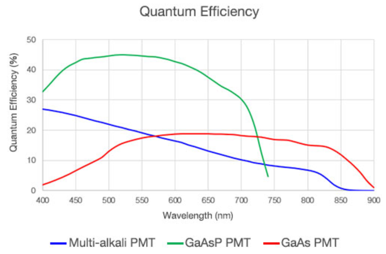
Using a selection of cooled gallium arsenide PMTs significantly extends the detection range to up to 890 nm. The free choice of combination of Multi-alkali, GaAsP and GaAs detectors enables users to configure the best fit for their application.
Optics which support multi color with NIR imaging
Transmission values and chromatic corrections are the other part of the game, which needs to be taken care for meaningful NIR imaging. We would like to highlight these points by showing the individual elements which contribute to excellent confocal NIR imaging.