On-target/off-tumor toxicity has been one of the challenges for adoptive cell therapy against solid tumors. Even though carcinoembryonic antigen (CEA) is overexpressed in a variety of solid tumor types, including cancers of the lung, colon, and pancreas, and is an attractive tumor-associated antigen, CEA-targeting immunotherapies have caused severe toxicity in the clinical trials due to CEA expression on normal cells. To overcome this hurdle, CEA Tmod, a NOT logic-gated T cell therapy, was developed by pairing a CEA specific CAR (activator) with an HLA-A*02 specific blocker. CEA Tmod cells are designed to kill tumor cells while protecting normal cells by exploiting loss of heterozygosity (LOH) at the HLA locus on tumor cells. Here we demonstrate selective cytotoxicity against tumor cells by CEA Tmod cells in a series of in vitro and in vivo experiments. In addition, functional assays allow assessment of off-target activity of the activator and blocker in Tmod cells. Finally, an NGS-based strategy to identify HLA-A*02 LOH patients, supporting clinical success of Tmod therapy, is described.
Introduction: Currently, it is well known that immune cells play major role against cancer. However, malignant cells are able to evade immune cells. Usually, tumor microenvironment is replete of regulatory molecules that are capable of changing function and phenotype of infiltrating immune cells. In the present work, we aimed to characterize infiltrating cells and subsets of T lymphocytes within metastatic melanoma lymph nodes.
First, lymph nodes obtained during surgical resection were confirmed as metastatic by anatomopathological analysis. Out of 13 samples, 9 were confirmed for metastatic disease. The lymph node tissue was then shredded and washed in culture media. Next, we determined cells subpopulations, subsets of T lymphocytes and molecules expressed by them by flow cytometry in LSR II Fortessa equipment. The results were analyzed using the software FlowJo. Our findings indicated that the main type of infiltrating cells were B and T lymphocytes, compatible with the usual populations present in lymph nodes, with prevalence of CD4 (68±2%) over CD8 (26±6%). Regarding the subsets of T lymphocytes, we observed a prevalence of memory T CD4 (61±18%) or T CD8 (51±17%) lymphocytes compared to transition or naïve phenotype. Amongst the subsets of memory cells, there was prevalence of effector memory for CD8 cells and no differences between effector or central memory for CD4. Yet, we observed a high percentage of T lymphocytes expressing CCR10, followed by CXCR2, both very important chemokine receptors in melanoma context. Finally, we also have observed higher expression of PD-1 (76±14%), followed by TIM3 (49±19%) and LAG-3 (34±11%), which molecules can down regulate the cytotoxic activity of these lymphocytes, and should be closely monitored during in vitro lymphocyte expansion for further autologous therapy development. Characterization of immune cells within metastatic melanoma microenvironment can provide relevant information prior to lymphocyte expansion.
Loss of heterozygosity (LOH) is commonly observed in many solid tumor types. To exploit such irreversible genetic difference between tumor and normal cells, we have developed a cell therapy that utilizes a combination of tumor-specific target and HLA deletion to focus cytotoxicity on cancer cells. The engineered T cells contain a dual-receptor system (Tmod): (i)an activating CAR that binds tumor-specific targets and (ii) an inhibitory LIR-1 based receptor (blocker) that binds HLA-A*02. Tmod cells have comparable potency to CAR-T cells but have striking selectivity for A*02(-) tumor over A*02(+) normal cells in vitro and in vivo. Moreover, the blocker module is able to block the activation of endogenous TCRs triggered by HLA mismatches, consistent with the use of the Tmod system to control graft-versus-host disease (GvHD) for allogeneic cell therapy. In mixed lymphocyte reaction with allogeneic stimulators, Tmod cells exhibit minimal allo-response—as low as the autologous background level. To further develop the system into an off-the-shelf therapy, we have added a B2M-targeting shRNA to knock down HLA class I and mitigate rejection of Tmod T cells from host T cells. Such engineered Tmod cells possess the key properties of on-target potency, tumor selectivity, blocking of GvHD, and escape from allo-rejection.
Fractionated radiotherapy is a mainstay of head and neck squamous cell carcinoma (HNSCC) treatment but fails in two-thirds of cases due, in large part, to development of radioresistance. New insights into the predominant pathways determining radiotherapy response could help develop mechanism-informed therapies to reduce the emergence of radioresistance and improving cure rates. Here we report that radioresistant HNSCC cells exhibit increased tumor aggressiveness. Using unbiased proteome profiler antibody arrays, we identify that upregulation of c-Met phosphorylation is one of the critical mechanisms for radioresistance in HNSCC cells. We further uncover that radioresistance-associated HNSCC aggressiveness is effectively exacerbated by c-Met but is suppressed by its genetic knockdown and pharmacological inactivation. Mechanistically, the resulting upregulation of c-Met signaling increases the expression of plexin domain containing 2 (PLXDC2), which in turn modulates cancer cell plasticity by epithelial-mesenchymal transition (EMT) induction and enrichment of the cancer stem cell (CSC) subpopulation, leading to resistance of HNSCC cells to radiotherapy. Depletion of PLXDC2 overcomes c-Met-mediated radioresistance through reversing the EMT progress and blunting the self-renewal capacity of CSCs. Therapeutically, the combination of c-Met inhibitor SU11274 and radiation induce tumor remission and limit lymph node metastasis in an orthotopic mouse model. Collectively, these significant findings not only demonstrate a novel mechanistic underpinning of radioresistance-associated aggressiveness but also provide a possible therapeutic strategy to target radioresistance in HNSCC patients.
Tumor immune surveillance is a critical process in inhibiting tumorigenesis. By stimulating patients’ immune systems to selectively kill cancer cells, immunotherapy can substantially improve the treatment outcomes for cancer patients. Accumulating evidence has shown that long non-coding RNAs (lncRNAs) play critical roles in the development of various diseases such as cancer. Although these findings have demonstrated that lncRNAs could modulate the human immune response in the tumor microenvironment, most lncRNAs’ function and their roles in tumor immunity remain unknown. High-throughput CRISPR screenings have enabled the functional characterization of novel genes involved in the regulation of cancer phenotypes. Recently developed CRISPR-SAM technology can activate gene expression by targeting their promoter. The activated lncRNAs will go over similar endogenous post-transcriptional modification and lead to a “physiological” level of overexpression. The CRISPR-SAM technology provides a unique opportunity to systematically characterize novel lncRNA regulators in tumor immune response. Here, we performed a CRISPR activation screening of 9744 lncRNAs in melanoma cells co-cultured with human TCR-engineered T cells which previously were used as adoptive T cell therapy. Further integrative analysis using tumor immunogenomics data revealed that IL10RB-DT and LINC01198 are significantly correlated with tumor immune response and survival in melanoma and breast cancer. Specifically, IL10RB-DT suppresses T cells’ activation via inhibiting IFN-γ-JAK-STAT1 signaling and antigen presentation in melanoma and breast cancer cells. On the other hand, LINC01198’s upregulation sensitizes the killing of tumor cells by T cells. Mechanistically, LINC01198 interacts and activates NF-κB component p65 which enhances p65’s transcription activity and promotes its downstream targets including type I and II interferon responses. These results demonstrate the feasibility of high-throughput in vitro genetic screening for potential lncRNAs which relate to the adoptive T cell therapy. Meanwhile, our study systematically characterizes novel lncRNAs participating in tumor immune response and suggests novel targets for cancer immunotherapies.
Acute myeloid leukemia (AML) is a highly lethal cancer for which effective immunotherapies are actively sought. Recently, a novel group of putative AML tumor-specific antigens (TSA) was characterized. Distinct from neoantigens derived from mutated genes and tumor-associated antigens (TAA), MHC class I peptides arising from allegedly non-coding regions of the genome, specifically in AML cells, have been discovered. These aberrantly expressed TSA (aeTSAs) are shared between patients which makes them interesting targets for immunotherapies. Knowing that T cell receptor (TCR) confers specificity to T lymphocytes, the goal is to discover and characterize aeTSA-specific TCRs in order to redirect T cells toward leukemic cells.
To this end, we expanded aeTSA-specific T cells from naive CD8+ T cells obtained from healthy donors through co-culture with autologous dendritic cells loaded with the relevant aeTSA. The aeTSA-specific CD8+ T cells identified by MHC-multimer staining were sorted for RNA extraction TCR sequencing. Amplicon sequencing reveals that the expanded anti-aeTSA TCR repertoire is markedly oligoclonal, facilitating the identification of 1 to 10 dominant TCR α and β chains. In contrast, TCR repertoire specific for the viral antigen LMP-2426-434 and the TAA WT-137-45 were more diverse. In addition, functional avidity tests of T cells targeting these antigens (aeTSA, LMP-2426-434 and WT-137-45) were performed using ELISpot with decreasing concentration of peptides and revealed that functional avidity of T cells recognizing TSA (aeTSA and viral TSA) are similar, suggesting that aeTSAs stimulate high-avidity responses. In parallel, two TCRs recognizing two HLA-A*0201 associated aeTSA have been fully characterized. Using virus-free, CRISPR-Cas9 based, approaches, we have performed endogenous TCR deletion and TRAC-directed aeTSA-TCR insertion in primary human T cells for further characterization, paving the way for the use of aeTSA-specific immunotherapy in AML.
Chimeric antigen receptor (CAR) T cell therapy targeting CD19 has achieved remarkable success in treating B-cell malignancies, however some patients fail to respond due to poor fitness of the adoptively transferred T cells. To potentially increase the number of patients that could benefit from CART19 therapy, we investigated whether CRISPR-Cas9-mediated disruption of PDCD1 and/or CTLA-4 in patient T cells could enhance efficacy. Indeed, we demonstrate, in both an in vitro model of CART dysfunction and xenograft mouse models, that disruption of CTLA-4, but not PDCD1 or PDCD1/CTLA-4 results in superior CART cell performance evidenced by increased proliferation, cytokine production and enhanced anti-tumor response. Furthermore, CART19 cells manufactured from T cells depleted of CTLA-4 maintain CAR expression on the T cell surface under conditions of chronic antigen exposure, which is a driver of CAR-dysfunction. To understand mechanistically how CTLA-4 disruption contributes to the increased anti-tumor efficacy of CART19 cells we perform transcriptomic analyses and identify upregulated pathways, aligning with the observed therapeutic superiority, including granzyme signaling, cell cycle regulation and cytokine signaling. Moreover, CTLA-4 disrupted CART19 cells display unopposed CD28 signaling, which is observed by IPA and GSEA analyses and supported by xenograft mouse studies where increased CD28 protein expression is detected in the peripheral blood of mice treated with CTLA-4 KO compared to WT CLL CART19 cells. Importantly we confirm the clinical relevance of these results, establishing that depletion of CTLA-4 enables the manufacture of potent CART19 products from CLL patient T cells that previously produced an ineffective product. Our findings demonstrate that disruption of CTLA-4 can invigorate dysfunctional patient T cells, providing a strategy for increasing the number of patients that respond to CART19 therapy.
Although tumor infiltrating Vγ9Vδ2T cells often have a good prognostic value, their role in cancer immune surveillance remains to be defined. We employed a human genetically engineered step-wise mutagenesis model of colorectal cancer and demonstrate that a single oncogenic mutation introduced into healthy organoids derived from the colon is sufficient to activate T cells through a Vγ9Vδ2TCR. During early mutagenesis we observed changes in spatial dynamics of BTN2A1 which allowed, after transformation binding of soluble Vγ9Vδ2TCR and re-localization and an increased expression of the small GTPase RhoB. Using a spatial protein screening technology platform we identified a novel protein network that is responsible for modulating spatial orchestration during early transformation by acting directly on BTN3A1 and indirectly on BTN2A1. This mode of action depended on phosphorylation of BTN3A1 and allowed tumor control in vitro and in vivo emphasizing the crucial role of these molecules from early mutagenesis to advanced cancer stages and the therapeutic potential of a Vγ9Vδ2TCR.
Infiltration of T cells to the tumor microenvironment is up to now one of the major bottlenecks of adoptive cellular immunotherapy and considered as an important hurdle of succesfull CART therapy especially against solid tumors. In this work we developed an in vitro 3D tumor model pipeline consisiting of both malignant and healthy tumor components embedded in an extracellular matrix that enabled the precise quantification of tumor specific T cell behavour with respect to tumor targeting and specific migration to the tumor site. We found that the amount of CD8+ T cells migrated to the tumor site is the major determinant of successful tumor killing however this migration also depended on the presence of CD4+ T cells. We have identified a chemokine as one of the most important soluble factors that limits CD8+ T cell migration to the tumor microenvironment. Using its major receptor that is naturally low or non-expressed by CD8+ T cells, as an arming strategy for CART cells, we were able to significantly improve CD8+ T cell migration to the tumor site and therefore induce a more robust tumor killing. This phenomenon was demonstrated for a great panel of patient-derived hematological malignancies, and more importantly, also for various patient-derived solid tumor organoid models. This work demonstrates that arming CD8+ CART cells with chemokine receptors may provide a valuable tool to increase therapy success of adoptive T cells especially in solid tumors.
Introduction
Whilst the majority of metastatic cancers remain resistant to curative drug treatment, a number of rarer malignancies, comprising gestational choriocarcinoma, testicular cancer and ovarian germ cell tumors, acute lymphoblastic leukemia, high grade lymphomas, Hodgkin’s disease and some the childhood malignancies have been routinely curable with cytotoxic chemotherapy drugs for more than 50 years
The explanation for this dramatic divergence in curability between these relatively rare malignancies and the more common incurable metastatic malignancies has long been a subject of great interest and scientific debate
Hypothesis
Recently we have suggested that a key central component determines heightened chemotherapy sensitivity and cancer curability. This is the persistence in the malignant cells of the naturally heightened apoptotic sensitivity that their usually transient cells of origin hold at the time of their malignant transformation
Whilst the genetic event of mitosis is common to all human cells, the chemotherapy curable malignancies each appear to have a very close temporal association between their cells of origin and the specialized unique genetic events.
These events are nuclear fusion (trophoblast tumours), immunoglobulin VDJ gene recombination (acute leukaemia), immunoglobulin gene somatic hypermutation and class switching (high grade NHL), meiosis (germ cell tumours) or gastrulation (childhood malignancies).
Discussion
We will discuss the key biological events occurring in the cells of origin at the time of malignant transformation and how these unique pro-apoptotic phenotypes are preserved in these malignancies. These processes plus the absence of any hierarchical cancer stem cells makes these malignancies highly sensitive to DNA damaging chemotherapy.
Further information (PMID: 32582272)
Over the course of an individual’s lifetime, genomic alterations accumulate in somatic cells. Retrotranspositions of long interspersed nuclear element-1 (L1), a widespread mobile element in the human genome, contribute to tumor initiation by disruption of tumor suppressor genes through L1 insertion and/or L1-mediated complex genomic rearrangements. However, the retrotransposition activity of L1 in normal human cells is poorly understood. Here, we explored the whole-genome sequences of 892 single-cell clones established from various tissues collected from 28 individuals. Remarkably, 88% of colorectal epithelial cells acquired somatic L1 retrotranspositions, carrying ~3 events per cell on average with substantial intra- and inter-individual variances, which was accelerated at least 10-fold during tumourigenesis. Breakpoints of retrotranspositions suggested that a few variant mechanisms can be involved in the L1 retrotransposition processes. Fingerprinting of donor L1s using source-specific unique sequences revealed 34 hot L1s, 44% of which were newly discovered in this study, and many ultra-rare hot L1s in the human population showed higher retrotransposition potential in somatic lineages than common sources. Multi-dimensional analysis of somatic retrotranspositions events with early embryonic developmental relationships, genome-wide methylation, and gene expression profiles of the clones demonstrated that (1) retrotranspositions occur from early embryogenesis at a substantial rate, (2) epigenetic activation of hot L1s is stochastically acquired during the wave of early global epigenomic reprogramming, rather than by the sporadic loss-of-methylation at the late stage, and (3) most L1 transcripts in the cytoplasm do not generate retrotranspositions in somatic lineages. In summary, this study provides insights into the retrotransposition dynamics of L1s in the human genome and the resultant somatic mosaicism in normal human cells.
Dysregulation of the DNA damage response (DDR) is a key inducer of genomic instability that promotes cancer initiation and progression which may be exploited to enhance sensitivity to cytotoxic drugs and DNA repair inhibitors. We previously reported that silencing of Lactate Dehydrogenase C (LDHC) perturbs DDR signaling in breast cancer cells, resulting in impaired long-term survival and sensitization to DNA damage inducers and DNA repair inhibitors. These findings indicate that LDHC could be used as a novel target in cancer precision medicine to improve treatment response to anti-cancer drugs. LDHC is an attractive target for therapeutic intervention with limited off-target effects as it features a highly tumor-specific expression and has been implicated in several cancer hallmarks. Hence, we sought out to explore the mechanistic networks and key molecules that contribute to LDHC-associated DDR dysregulation. Differential expression and network analysis of LDHC-silenced cells identified 55 down- and 47 up-regulated genes that were enriched in pathways related to cell cycle checkpoint control, BRCA1-mediated DNA damage response and NF-kb signaling. Upstream regulator analysis revealed mTOR and CASP3 as potential regulators of the LDHC-associated transcriptome network. Further analysis of the mTOR pathway confirmed a downregulation in protein expression of the pro-tumorigenic molecules STAT1, STAT3 and SMAD3 following LDHC silencing. Similarly, we observed a decrease in protein expression of the STAT3 downstream anti-apoptotic molecule Bcl2 in LDHC-silenced cells. Functional mTOR and STAT3 inhibitor studies are ongoing to assess the extent to which these pathways contribute to the LDHC-silencing cellular phenotype. Our findings highlight functional networks and molecular mediators in breast cancer that may play a role in DNA damage response dysregulation and loss-of-survival following LDHC therapeutic targeting.
Metastasis and therapeutic resistance are a major clinical challenge, responsible for the vast majority of cancer deaths. A subpopulation of tumor cells known to have intrinsic resistance to standard therapies and contribute to metastasis function as “cancer stem”, or tumor-initiating, cells (TICs). Enriched populations of TICs are typically identified by markers such as the cell surface marker combination of CD44+/CD24- or fluorescent reporters for signaling pathways that regulate TIC function, such as STAT3. However, marker expression can be unstable and there is no established method to lineage trace long-lived TICs or to follow them as they undergo cell state changes.
We developed a two component STAT3 signaling-specific lentiviral lineage tracing (LT) system. The first component is a vector that labels cells with active STAT3 signaling (EGFP+), followed by a self-cleaving peptide and tamoxifen-inducible Cre-recombinase (4M67-EGFP-P2A-CreERT2). The second component is a constitutively expressed dual-color switching Cre-dependent reporter vector (EFS-loxPdsRedloxP-mNeptune2). Addition of tamoxifen drives color switching from dsRed to mNeptune2 via CreERT2 recombination in STAT3 signaling cells. Sum159 cells were transduced with the LT system and reporter activity was validated both in vitro and in vivo using confocal microscopy and flow cytometry. Four LT cell populations (EGFP+/mNeptune2+, EGFP+/dsRed+, EGFP-/mNeptune2+, and EGFP-/dsRed+) were enriched using fluorescence activated cell sorting, then analyzed by single cell RNA sequencing (scRNA seq).
Our results confirm the STAT3 LT reporter identifies STAT3 signaling cells (EGFP+), which can be labelled (mNeptune2+) upon addition of tamoxifen. We conducted a tamoxifen dose response curve to identify the optimal TAM dose for labeling STAT3 signaling cells. scRNA seq uncovered gene expression patterns within the TIC compartment, revealing similarities and differences in gene expression between the TIC compartment and the remaining reporter populations.
Acute erythroid leukemia (AEL) is a disease of erythroid lineage commonly with TP53 mutations, complex karyotype, and poor prognosis irrespective of currently available therapies. We aimed to identify therapeutically targetable dependencies in AEL using a genome-wide CRISPR/Cas9 knockout screen in two Trp53/Bcor-mutant mouse AEL cell lines that were previously established (Iacobucci I et al. Blood 2021). Analysis of the screens revealed the Urod gene encoding uroporphyrinogen decarboxylase as a critical dependency in both cell lines. UROD is a cytosolic enzyme in the heme biosynthesis pathway involved in converting cytosolic uroporphyrinogen III into the key heme biosynthetic intermediate, coproporphyrinogen III. Interestingly, UROD was significantly overexpressed and active by data-driven network inference algorithm in AEL compared to other leukemia subtypes in an analysis of RNA-seq data from over 2,000 acute leukemia samples. We validated UROD as a dependency in a targeted fashion using electroporation of Cas9 and synthetic sgRNA ribonucleoprotein complex in the two screened cell lines, and additional mouse and commercially available human cell lines (TF-1 and HEL). Targeted knockout of UROD resulted in decreased tumor cell growth over time in culture and an increase in necrotic cells compared to non-targeting guide control cells. Sequencing analysis revealed high editing efficiency (>85%) preceding the wave of necrotic cells followed by a recovery of cells expressing wild-type UROD, indicating that UROD is a dependency in these cells. These results were corroborated with an increase in ALAS1 expression, which is negatively regulated by cellular heme, and shows that UROD knockout cells are unable to generate cellular heme, and this is leading to their loss of fitness. Moreover, mitochondrial function assays showed significant decreases in basal respiration, ATP production, maximal respiration, and spare capacity in a time-dependent manner, followed by the rescue of mitochondrial function after loss of editing efficiency and re-expression of UROD.
To overcome the poor outcomes of head and neck cancer (HNC), we have advanced our study from the perspective that HNC is a symbiotic evolving system, addicted to the transcriptional reprograming triggered by microenvironmental stresses. In this context, we developed an ultra-rapid mouse carcinogenesis model (4W) induced by a YAP1 transcriptional coactivator (Omori et al, Sci Adv 2020) and are conducting integrative epigenetic analyses on YAP1-induced transcriptional reprogramming. WES, RNA-seq, Chip-seq, and WGBS on YAP1-induced mouse tumors and cell lines demonstrated that YAP1 epigenetically causes carcinogenesis without affecting genome-wide chromatin confirmation, but inducing hypomethylation on the super enhancers (SE), related to tissue regeneration (i.e., recapitulation of wounds that don’t heal condition). Poor prognosis was associated with YAP1-induced carcinogenesis gene module in the TCGA data (p = 0.00033) and with the level of YAP1nuclear protein in the 119 HNC samples (p = 0.0116). RNA-seq, Chip-seq with HNC cell lines showed that YAP1 is essential for the assembly of SE and YAP1 SE module including IL6 was associated with unfavorable survival in the TCGA data (p = 0.031) and the existence of IL6-YAP1 feed-forward loop was confirmed through in vitro assays. EEM and motif assays revealed that YAP1, collaborating with PITX2 transcriptional factor (TF), regulates TGF-beta-induced EMT and CAF, consistent with the recent HNC single-cell RNA-seq study (Puram et.al, Cell 2017). Thus, the involvement of YAP1 in the metastasis was suggested. Bioinformatic analyses indicated YAP1-iduced immune evasion. In vitro assays and chip-seq on human HNC samples support the significance of PITX2 as a YAP1 partner TF in SE. Co-expression of YAP1 plus PITX2 or BRD4 further worsened the prognosis than the individual factor alone. We are confirming above- results by spatial transcriptomics and multiplex IHC. Collectively, our data indicate the essential role of YAP1 in the symbiotic evolution of HNC.
TP53 is the most frequently mutated gene in human cancer. Around 10% of TP53 mutations in human cancer are nonsense mutations. Given the high frequency of TP53 mutations overall, tumors with TP53 nonsense mutations represent a substantial number of cancer patients worldwide. The most common TP53 nonsense mutation is p.R213X, which is also the 7th most common of all cancer-associated TP53 mutations. Previous studies have demonstrated a high incidence of spontaneous tumors in Trp53-null and Trp53R172H knock-in mice, with development of lymphomas and sarcomas from 3-6 months of age in homozygous mice and from 10-12 months in heterozygotes. Here we have examined a possible tumor phenotype of Trp53R210X mice. We have used CRISPR/Cas9 genome editing to generate mice harboring the nonsense mutation p.R210X, corresponding to human p.R213X. Our results so far indicate that Trp53R210X mice are initially phenotypically normal. However, the proportion of female Trp53R210X/R210X mice is dramatically reduced. Based on our current numbers, female homozygous mice are very poor breeders, and significantly smaller than female heterozygous and wildtype littermates. Trp53R210X/R210X mice start developing tumors at 2.5 months of age, and the current maximal lifespan in this ongoing study is <8 months. The most common tumor type in homozygous mice thus far is lymphoma. This tumor phenotype is in agreement with previous data on the tumor phenotypes of Trp53-null and Trp53R172H homozygous mice. Our new mouse model will allow further studies of the effects of Trp53 nonsense mutation in a multi-organ system. This model will also be a valuable asset in preclinical evaluation of novel therapeutic compounds targeting nonsense mutations. Our long-term goal is to develop more efficient treatment for tumors carrying nonsense mutant TP53. This may also be relevant for treatment of tumors harboring nonsense mutations in other tumor suppressor genes, such as APC and PTEN.
The Heat Shock Protein 27 (Hsp27) has emerged as a principal factor of the Castration-Resistant Prostate Cancer (CRPC) progression, also, an Antisense Oligonucleotide (ASO) against Hsp27 (OGX-427 or Apatorsen) has been assessed in different clinical trials. Here, we illustrate that Hsp27 highly regulates the expression of the human DEAD-box protein 5 (DDX5), and define DDX5 as a novel therapeutic target for CRPC treatment. DDX5 overexpression is strongly correlated with aggressive tumor features, and notably with CRPC. DDX5 downregulation using a specific ASO-based inhibitor which acts on DDX5 mRNAs inhibits cell proliferation in pre-clinical models, and particularly restores the treatment sensitivity of CRPC. Interestingly, through the identification and analysis of DDX5 protein interaction networks, we have identified some specific functions of DDX5 in CRPC that could contribute actively to tumor progression and therapeutic resistance. We first present the interactions of DDX5 and the Ku70/80 heterodimer and the transcription factor IIH (TFIIH), thereby uncovering DDX5 roles in different DNA repair pathways. Collectively, our study highlights critical functions of DDX5 contributing to CRPC progression and provides preclinical proof-of-concept that a combination of ASO-directed DDX5 inhibition with a DNA damage-inducing therapy can serve as a highly potential novel strategy to treat CRPC.
Humans develop carcinomas (epithelial malignancies) at a high rate compared to closely related primates. New biomarkers and potential therapeutic targets are being developed to monitor and combat malignancies. Siglecs are cell surface receptors on most white blood cells of the immune system that recognize ligands bearing sialic acids (Sias), a family of glycans found at the terminal end of the glycan chains on cell surface and extracellular glycoconjugates. In particular, Siglec-XII (encoded by the gene SIGLEC12), although on macrophages, is surprisingly prominent on some epithelial cells and has several uniquely human features: a fixed homozygous missense mutation (absent in primates) inactivating its natural ligand recognition property; and a polymorphic frameshift mutation eliminating full-length protein expression in ~60%–70% of worldwide human populations. Despite the loss of canonical sialic acid binding, Siglec-XII still recruits Shp2 and accelerates tumor growth in a mouse model. Although Siglec-XII is only present in about 30% of the normal human population, the expression of Siglec-XII was found in about 80% of aggressive human epithelial carcinomas, such as colorectal cancer (CRC). We hypothesized that dysfunctional Siglec-XII facilitates human carcinoma progression. In this work we show that transfection of the human cDNA in a SIGLEC12 null carcinoma cell line provoked a reduction in cell attachment as well as accelerates spheroid growth and invasion. Siglec-XII expressing cells expressed higher pERK/ERK protein expression levels compared to controls. Furthermore, we generated a novel transgenic mouse which allows conditional expression of Siglec-XII in villi and crypts of the small and large intestine (SIGLEC12-Villin1-Cre-ERT2). In the azoxymethane-dextran sodium sulfate model of colitis-associated CRC, Siglec-XII expressing mice showed more and significantly larger tumors than controls. This work will contribute to a better understanding of the impact of Siglec-XII on cancer progression.
The most important problems in cancer therapy are death due to acquired therapeutic resistance and therapeutic toxicity. The aim of this study was to test adaptive therapy as an evolutionary strategy in preclinical models with MCF7 cancer cell lines resistant to palbociclib and fulvestrant. MCF7 resistant breast cancer cells were injected in the mammary fat pads of NSG mice and grew orthotopically. We treated these mice with gemcitabine or capecitabine either intermittently or by adjusting their doses. We also used a combination of drugs that we applied in tandem or switched the drugs in every application (ping-pong) either intermittently or by adjusting their doses. In this experiment, first, we wanted to see if gemcitabine and/or capecitabine can control the resistant MCF7 cells and, second, compare adaptive therapy with standard therapy. We adjusted the drug dosage based on the tumor burden as observed via bioluminescence imaging twice per week. We found that in the ping-pong combination, in both intermittent and dose adjustment, the survival of mice increased significantly. In tandem combination therapy, the intermittent treatment was less successful compared to dose adjustment, however it was better than standard therapy in increasing survival. In single drug adaptive therapy, there was a great increase in survival in capecitabine dose adjustment treatment compared to both intermittent and standard therapy. In addition, there were no differences between adaptive strategies and standard therapy using gemcitabine.Overall, adaptive therapy strategies could prolong progression-free survival besides reducing the tumor burden. We often observed that tumors continued to shrink even after we stopped therapy and we had several weeks off-treatment. This suggests that once a tumor is under control using adaptive strategies, lower drug doses could be used to increase progression-free survival.
The MET receptor, a receptor tyrosine kinase (RTK) activated by its single ligand – hepatocyte growth factor (HGF), plays essential roles in the tumorigenicity, angiogenesis and invasiveness of glioblastoma (GBM). Nevertheless, trials of HGF/MET-targeted therapy in GBM patients has failed to show clinical benefits, suggesting undiscovered mechanisms underlying the aberrant activation of MET signalling in GBM. Here, we discovered a 404 amino acid novel MET variant (MET404), encoded by circular MET RNA (circMET) with the aid of the N6-methyladenosine (m6A) reader YTHDF2, serves as an independent ligand of MET receptor in GBM. Genetic ablation of circMET in mice led to reduced MET404 expression and mitigated MET signalling. Conversely, CRISPR-Cas9 mediated conditional MET404 knock-in plus P53knock-out in mouse astrocytes initiated robust GBM tumorigenesis and significantly shortened overall survival. MET404 is a secreted protein and directly interacts with the MET β subunit on a different domain compared to HGF, and thus potentiates constitutive activation of MET signalling even in the absence of HGF stimulation. High MET404 expression is associated with poor prognosis in GBM patients, indicating its clinical relevance. Targeting MET404 with a neutralizing antibody or genetic ablation impaired GBM tumorigenicity both in vitro and in vivo, with synergistic benefits when combined with a traditional MET inhibitor. Overall, we identified a novel independent MET variant that promotes GBM tumorigenicity, offering a potential new targeted strategy for GBM treatment.
Long noncoding RNAs (lncRNAs) have emerged as crucial players in cancer biology and other prevalent diseases. Unlike protein coding mRNAs, the majority of lncRNAs are poorly conserved, and are not evolutionarily neutral. LncRNAs regulate biological processes as functional RNAs. Recent studies, however, suggest that a subset of lncRNAs produce functional proteins. In this study, we have discovered a short (132 amino acid) polypeptide, which we named Tumor-Specific Polypeptide 1 (TSP1), encoded by a pancreas- and testis-specific RNA annotated as a putative long noncoding RNA by Sun and Gadad et al. (lncRNA1456, aka LOC105373323) transcribed from the X chromosome. TSP1 is upregulated in luminal A, luminal B, and HER2 molecular subtypes of breast cancer. We show that TSP1 modulates estrogen-dependent, as well as estrogen-independent growth, of MCF-7 cells by regulating cancer pathways in xenograft models. We have found that TSP1 shares homology with homeodomain containing transcription factors. TSP1 interacts with PHF8, a histone demethylase which is also encoded by an X-linked gene, that demethylates H3K9me2 and other sites to regulate transcription. Mechanistically, TSP1 positively regulates histone demethylase activity of PHF8 in vitro and regulates gene expression in breast cancer cells. These results identify TSP1 as a coregulator of transcription and emphasize the need to interrogate the functional roles of lncRNA-derived peptides.
Endometrial cancer (EC) is the 4th most common cancer in women in the developed world. Close to 20% of women still succumb to recurrent cancers of the uterus. The most overrepresented subset contributing to lethal uterine cancers has been demonstrated to be SWI/SNF deficient dedifferentiated endometrial carcinoma (DDEC). DDEC tumors possess both well-differentiated and undifferentiated regions. Examination of DDEC lesions has revealed that over 70% of the undifferentiated regions within these neoplasms lack the expression of core chromatin remodeling proteins, like SMARCA4. To ascertain how loss of SMARCA4 may drive dedifferentiation and metastasis in DDEC, SMARCA4-deficient EC cell lines were generated by CRISPR. EC cell lines lacking SMARCA4 expression exhibited partial EMT yet were less pro-tumorigenic than SMARCA4 intact EC cells when functionally evaluated in vitro. Serial passaging SMARCA4 knockout (KO) EC cells in vivo led to the formation of tumors that closely recapitulate clinical DDEC both in histology and gene expression changes. Prior to cellular dedifferentiation being evident in vivo, EC cell lines that are SMARCA4 KO exhibit a senescent-like intermediary state characterized by an increased secretion phenotype and condensed chromatin. Experimentally evolving our cell line model of DDEC with SMARCA4 deficiency through successive serial passaging also illuminated that cellular dedifferentiation in the context of the endometrium likely follows a sequence of epigenetically driven events. It appears that a terminally dedifferentiated cell cluster emerges over time, and this cluster can be identified by dramatic alterations in chromatin occupancy. Determining the extent to which loss of SMARCA4 contributes to the acquisition of DDEC is a critical step towards improving treatment practices. Models of SWI/SNF deficient DDEC most importantly though, contribute substantially to our understanding of cancer evolution and perhaps how tumors of all tissue types that contain undifferentiated cells hijack biology through epigenetic mechanisms and achieve clinical aggressiveness.
Melanoma is associated with poor outcomes and the rapid acquisition of drug resistance. Common therapies include inhibitors of the MAPK pathway. The cAMP pathway, and the transcription factors involved in this pathway have been highlighted in resistance to RAF/MEK inhibitors. ICER, as a repressor of gene expression (an antagonist of CREB) of the cAMP pathway, might be involved in this observed resistance. In order to test this hypothesis we developed transgenic zebrafish lines expressing three different forms of ICER in the zebrafish model for melanoma, Tg(mitfa:BRAFV600E); mitfa(lf); p53(lf). The three different forms of zebrafish ICER-Iɣ were as follows: wild-type- (wt-), no-lysine- and S35&41A-ICER. No-lysine-ICER is a an “un-ubiquitinatable form of ICER where all ten lysines are replaced by arginines and in S35&41A-ICER serines 35 and 41 were substituted for alanines. We have shown before that phosphorylation on serines 41 by ERK1 was a prerequisite for polyubiquitination and proteasomal degradation, whereas phosphorylation on a serine 35 by the mitotic kinase cdk1 was necessary for monoubiquitination an alter subcellular localization to the cytosol. Tumor incidence curves showed that two forms of ICER (wt- and no-lysine-ICER) significantly accelerated melanoma onset compared to EGFP control. Both zebrafish expressing wt-ICER and no-lysine-ICER succumbed to malignancies much faster than control EGFP and S35&41A-ICER expressing fish. In contrast, the zebrafish expressing S35&41A phosphorylation mutant ICER have a significantly longer life than EGFP control. Melanocytes expressing both wt- and no-lysine-ICER grew in confluent patches in zebrafish, unlike melanocytes in the EGFP- control and S35&41A-ICER expressing zebrafish, which grew in a wild-type stripe pattern. A pathological and molecular analysis will be presented investigating the hypothesis that this presumed paradox is related to the development of a refractory melanoma of the ICER-expressing melanocytes.
Activation of PI3K/AKT/mTOR signalling, most commonly by activating mutations of PI3K/AKT family members or loss of PTEN phosphatase function, contributes to carcinogenesis of many malignancies.
BTK (Bruton’s tyrosine kinase) is a non-receptor tyrosine kinase of the Tec kinase family, which is critical for B-cell development, differentiation and malignancies. Resistance frequently follows BTKi treatment. Reduced levels of BTK and upregulated PI3K/AKT signalling are hallmarks of these ibrutinib-resistant cells. Upregulation of PI3K-β expression is demonstrated to drive resistance in ibrutinib-resistant cells1.
The tumour suppressor PTEN is the major negative regulator of the PI3K/AKT signal. Loss of PTEN function is common in a number of cancers, including prostate. Preclinical studies have indicated that the PI3Kβ isoform is the critical lipid kinase that drives primarily PI3K pathway activation, cell growth, and survival in PTEN-deficient tumor cells2-6.
In this work, we explore the potential of PI3K-β/δ inhibitor CVL237(KA2237) in overcoming BTK-resistant B-cell lymphoma and PTEN-deficient prostate. CVL237 significantly inhibits the tumour growth in the BTK-resistant and PTETN-deficient xenograft model, suggesting that PI3K-β and PI3K-δ are the key nodes underlying AKT/mTOR axis signalling and cell survival in ibrutinib-resistant DLBCL and PTEN-null prostate.
CVL237, an oral dual selective PI3K-β/δ inhibitor, can reverse the resistance and inhibit tumour growth in the BTK-resistant xenograft mouse model. The effect is enhanced when combined with a BTK inhibitor.
Meanwhile, CVL237 exhibits antitumor activity in the PTEN-deficient CDX model and shows a synergistic effect with PARPi.
Some studies1 have demonstrated that reduced levels of BTK and compensatory upregulated PI3K/AKT pathway are potentially responsible for the emergence of ibrutinib-related resistance in DLBCL cells. Targeting the PI3K/AKT/mTOR axis with a PI3K-β/δ selective dual inhibitor reduced both tumorigenic properties and survival-based PI3K/AKT/mTOR signalling of ibrutinib-resistant cells.
Therefore, target inhibition of PI3K-β/δ is a potential therapy for BTK-resistant lymphomas and PTEN-deficient solid tumours.
The most common cancer is colorectal cancer(CRC). One of the colorectal cancer risk factors is bile acids. Bile acids are made by the liver, functioning digestion of dietary lipids, and are toxic to cells as detergents. Several studies have shown that continuous administration of bile acids causes cancer, and colorectal cancer is correlated with FXR expression. FXR (farnesoid X receptor) is a superfamily of nuclear receptors. Intestinal cancer stem cells and cancer progression are down-regulated by activation of FXR, but the mechanism remains unclear. We confirmed from RNA sequencing that the expression of 1C(one-carbon) metabolism genes is upregulated by FXR activation. 1C metabolism regulates some biochemical pathways and is also interrelated with some cancers. Some papers have shown that 1C metabolic genes play a crucial role in cancer proliferation because it is persisting cellular functions. We identified the clinical data of COAD (Colon adenocarcinoma) patients. We confirmed with the TCGA-COAD patient data that the expression direction of a specific 1C metabolic gene was the same according to the FXR activation. Patients with high expression in both the ATF4 and 1C metabolic genes have a poor prognosis. We hypothesized that colorectal cancer cells whose proliferation was reduced by FXR activation could survive through the 1C metabolic pathway. We suggest novel therapeutic strategies for the treatment of colorectal cancer.
Diffuse-type gastric adenocarcinoma is the most aggressive subtype of gastric cancer (GC) and known to resistant to chemotherapy. Meanwhile, several studies suggested that steroid-hormonal signaling affects the progression of gastric cancer. Steroid hormone receptor-mediated signaling plays an important role in cancer proliferation and tumorigenesis by recruiting cofactors to its response elements, however, the mechanisms in GC are still unclear. Here, we showed that an epigenetic reader, well-known transcriptional co-activator, was up-regulated in EMT-type gastric cancer cells and inhibition of the protein suppressed the proliferation by regulating steroid-hormonal signaling. We identified inhibition of the protein can downregulate the activation of endogenous steroid hormone receptor using luciferase reporter assay. To investigate differentially expressed genes (DEGs) through inhibition of the protein, RNA-sequencing analysis was also performed with EMT-type GC cells. We found several steroid hormonal signaling target genes were downregulated in the epigenetic reader inhibited group. Consistently, the regulation of steroid hormonal receptor signaling pathway was associated with Gene Ontology (GO) analysis of down-regulated DEGs. Collectively, our results indicate that an epigenetic regulator promotes cell proliferation by regulating steroid hormonal receptor signals and suggest it as a therapeutic target for diffuse-type GC.
Ubiquitin-Proteasome System (UPS) is a crucial protein degradation system consisting of several factors such as E1, E2, and E3 enzymes, ubiquitin, and proteasome. Bortezomib, a proteasome inhibitor, has been commercialized as anti-hematologic cancer therapy for Multiple Myeloma targeting UPS. Interestingly, recent reports suggest that chemicals target UPS also have the potential for solid malignancies therapy. In cancer cells, the cell cycle rate is so fast that proteotoxic stress occurs frequently, especially in KRAS mutant patients, selective addiction to proteasome function is frequent. In pancreatic cancer patients, KRAS mutations occur at a very high rate, and we found that certain deubiquitinase genes are up-regulated in pancreatic cancer tissue via TCGA and GTEx databases. In addition, it was analyzed that the mortality rate of patients with high levels of expression of these deubiquitinase genes is high. Then GSEA screening results showed that their expression is positively correlated with cell cycle terms such as E2F and G2M checkpoints. Here we hypothesize that these genes are important keystones in controlling the cell cycle of the pancreatic cancer cell. To conduct in-depth analysis, mRNA bulk sequencing, Proteomics, and single-cell RNA sequencing were conducted. Through these Multiomics analyses, it was re-confirmed that these genes are related to E2F targets and G2M checkpoints as we predicted. What's interesting is that they also have a big correlation with Mitochondrial function. In this poster, we present how these deubiquitinase genes are analyzed through Multiomics and which mechanisms affect pancreatic cancer cells.
Cell-free chromatin particles (cfChPs) that circulate in blood are known to readily enter into healthy cells, integrate into their genomes and damage their DNA (doi: 10.1007/s12038-015-9508-6). We show here that treatment of NIH3T3 mouse fibroblast cells with nanogram quantities of cfChPs isolated from sera of cancer patients led to euchromatinization of histones by 6h which was accompanied by marked activation of gene expression networks of DNA damage, inflammation, metabolism, cancer, metastasis, stemness and stress. The cells became aneuploid by 96 h, and displayed extensive centrosome abnormalities, chromosomal aberrations including large scale translocations and formation of centric micronuclei. Also detected were numerous extrachromosomal DNA and double minutes, ostensibly as consequence of chromothripsis. Activation of these oncogenic pathologies resulted in acquisition of phenotypic capabilities represented by the 10 hallmarks of cancer, including the immune check-point PD-L1. The cells became visibly transformed by 96 hours, which could be prevented by concurrent treatment with anti-histone antibody complexed nanoparticles. Independent experiments conducted with cfChPs isolated from 70 cancer patients and 70 healthy volunteers at concentrations of 5ng-10ng showed transformation in 54/70 experiments in case of the former and 0/70 in the latter. However, transformation occurred in 41/70 experiments when the concentration cfChPs from healthy volunteers was increased 4-5 fold. Inoculation of the treated cells into SCID mice induced tumours with high efficiency and which contained abundant human DNA. These results suggest that cancer is caused by extraneous oncogenic agents in the form of cfChPs, and that they may be involved in both cancer initiation and metastasis. The wide spectrum of cellular and genetic alterations induced by cfChPs may help to reconcile the conflicting theories of carcinogenesis and provide a unified mechanism for cancer hallmark activation.
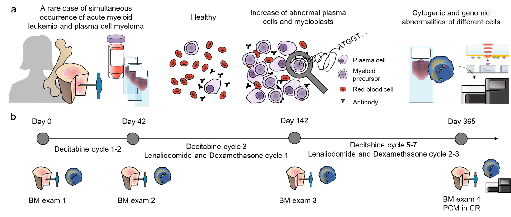
Co-occurrence of Multiple myeloma and acute myelogenous leukemia is extremely rare, so that five cases are reported globally. Two neoplastic cells commonly originate form multipotent hematopoietic stem cell, but depending clonal acquisition or loss at lower level, they part way with each other. We identified the clonal changes in heterogeneous hematopoietic lineages by integrating whole genome sequencing, copy number variations, cell morphology, and cytogenetic aberrations at single-cell level. We selected each single cells from Wright stained slide or fluorescence in situ hybridized slide, with subsequent whole genome sequencing, using novel Hema-seq technique. By integrating results in each cell lineage, we inferred the sequential clonal changes along hematopoietic tree, revealing the decisive changes leading to myeloma cell and leukemic cell.
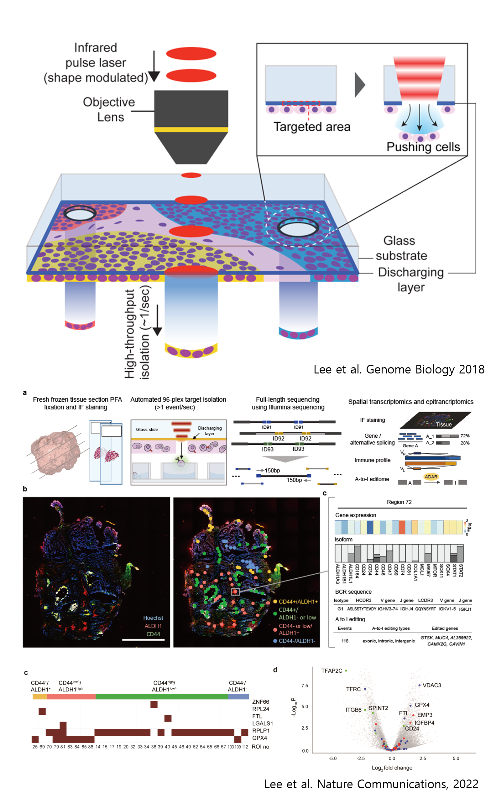
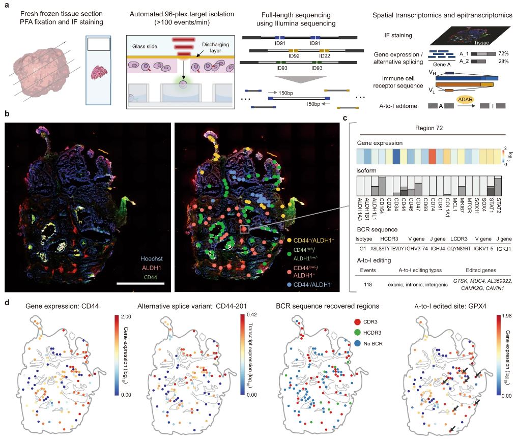
Cancer is spatially heterogeneous in terms of genetic molecules. Revealing genomic, epigenomic, transcriptomic, and epitranscriptomic features that are unique to the malignant cell populations within cancer lead to cancer biomarker discoveries that can be translated into diagnostic or therapeutic tools. Here, we introduce spatially-resolved laser-activated cell sorting (SLACS) technology coupled to next generation sequencing and mass spectrometry that analyzes genetic molecules from regions of interest within spatial context. Specifically, we demonstrate application of SLACS in various cancers such as breast cancer, glioblastoma, glioma, meningioma, multiple myeloma, and leukemia. Examples of discovering cancer biomarker and their mechanism of action within spatial context are also provided. Bridging spatial technologies to omics technologies, the discovery of specific markers within spatial context will provide insights into the next generation diagnostics and cancer therapeutics such as cancer vaccines.
We have previously found that a safe and controlled manipulation of endocytosis in vivo may have disruptive therapeutic potential for antibody-dependent cell cytotoxicity in the clinical setting. Here data is presented to show how the delivery of antibody-drug conjugates can be improved by increasing tumour cell "loading" and endosomal delivery. In analysis of the tumour cell / immune cell synapse we additionally present a central mechanism in the biogenesis of the active immune synapse. In immune cells this mechanism is a potential target for pharmaceutical regulation of human immune response. In healthy humans this cell biological pathway is present in immune privileged cells and immune cells. Our data shows that expression of components of this pathway in tumour cells can lead to biogenesis of a pseudo-immune synapse with concomitant expression of proteins normally detected in immune privileged cells. Pathology analysis of human tumours suggests this may be a tumour inherent change which leads to tumour evasion of immuno-surveillance.
Defects in pathways governing genomic fidelity have been linked to improved response to immune checkpoint blockade therapy (ICB). Pathogenic POLE/POLD1 mutations can cause hypermutation, yet how diverse mutations in POLE/POLD1 influence anti-tumor immunity following ICB is unclear. Here, we comprehensively determined the effect of POLE/POLD1 mutations in ICB and elucidated the mechanistic impact of these mutations on tumor immunity. Murine syngeneic tumors harboring Pole/Pold1 functional mutations displayed enhanced anti-tumor immunity and were sensitive to ICB. Patients with POLE/POLD1 mutated tumors harboring telltale mutational signatures respond better to ICB than patients harboring wild-type or signature-negative tumors. A mutant POLE/D1 function-associated signature-based model out-performed several traditional approaches for identifying POLE/POLD1 mutated patients that benefit from ICB. Strikingly, the spectrum of mutational signatures correlates with the biochemical features of neoantigens. Alterations that cause POLE/POLD1 function-associated signatures generate TCR-contact residues with increased hydrophobicity, potentially facilitating T-cell recognition. Altogether, the functional landscapes of POLE/POLD1 mutations shape immunotherapy efficacy.
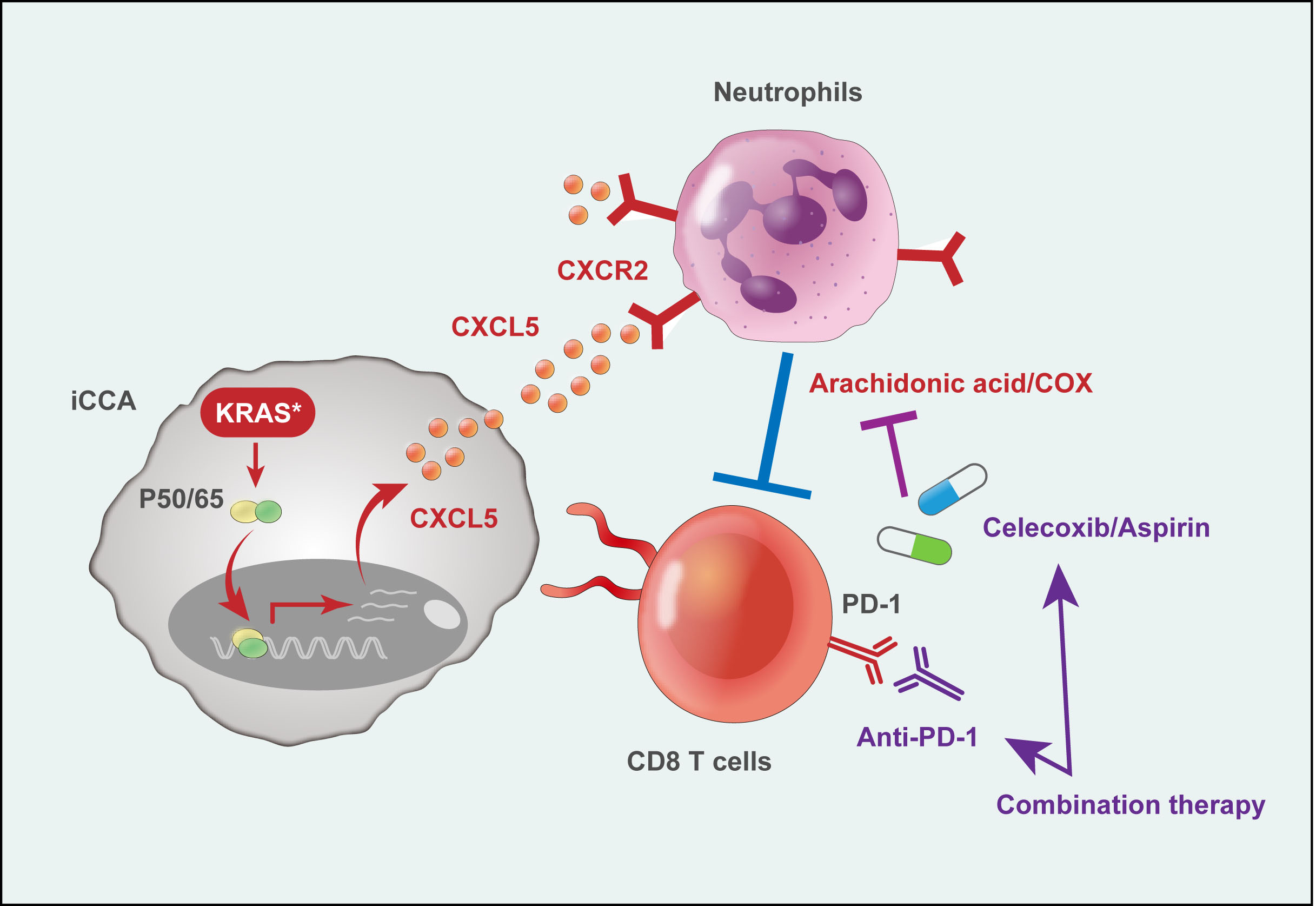
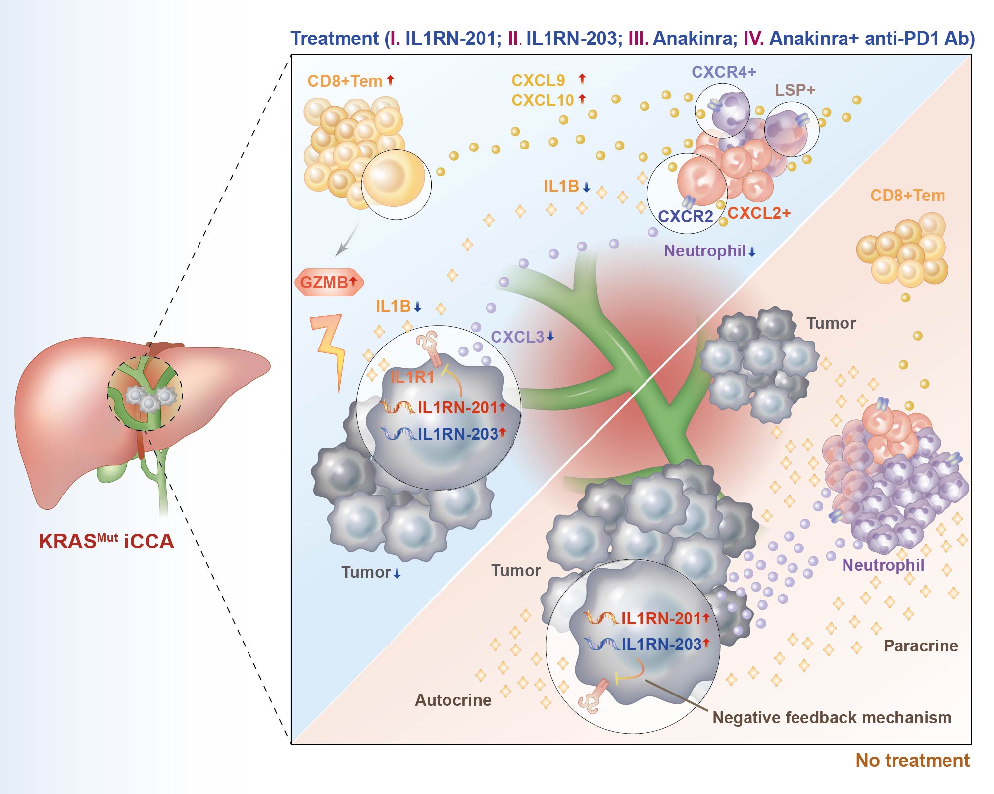
Metabolic reprogramming in the tumor microenvironment retards antitumor immunity and the efficacy of immune checkpoint blockade (ICB). Here, we subdivide intrahepatic cholangiocarcinoma (iCCA) into three metabolic subtypes with distinct prognostic significances according to Protein-Scale-Based Classification. Specifically, the highest enrichment scores for the worst prognostic subtype converge on Cyclooxygenase/Arachidonic Acid (COX/AA) metabolic pathway, accompanied by the enrichment of KRAS mutations. In iCCA mouse models, KRAS mutation promotes the upregulation of COX/AA signature and the accumulation of arachidonic acid metabolites. Single-cell RNA sequencing nominates neutrophil as the main resource of COX/AA metabolism. Mechanistically, mutated KRAS activated NF-κB pathway to upregulate CXCL5 expression, which promotes neutrophil infiltration through the CXCL5-CXCR2 axis to suppress the anti-tumor activity of cytolytic T cells. Anti-inflammatory drugs, such as COX2 selective inhibitor celecoxib or aspirin, in combination with anti-PD-1, show remarkably synergistic effects on the treatment of KRAS-mutated iCCA. Combined anti-inflammatory drugs and anti-PD-1 treatment of patient-derived tumor fragments from KRAS-mutated iCCA ex vivo reinvigorates anti-tumor activity and predicts clinical response. Moreover, the expression levels of CXCL5 and COX/AA signature correlate with patient survival and response to ICB in several patient datasets. Our findings propose a readily translatable adjuvant strategy for the treatment of KRAS-mutated iCCA that is refractory to ICB monotherapy.
Immunotherapy is used worldwide to treat cancer by activating host immune cells. However, the response rate of immunotherapy is still low and the mechanism by which cancer cells evade T cell attack is still unclear. Here, we show that inhibiting glucocorticoid receptor-mediated signaling pathway can enhance the efficacy of immunotherapy. Inhibition of glucocorticoid receptor-mediated signaling increases NF-κB signaling pathway, which is the representative pathway of regulating inflammation signals. Inhibition of signaling in vivo carried out delay in tumor growth and increased the number of cytotoxic CD8 T cells residing in tumors. Also, stimulation of GR-mediated signaling pathway led to lower response rate of immunotherapy using anti-PD1 in vivo. Thus, this study provides the insight on cancer cell surveillance against T cell through glucocorticoid receptor-mediated signaling pathway. Together, these results suggest that inhibiting GR signaling pathway should be considered as a therapeutic target in cancer immunotherapy.
Introduction
The response rate of immune checkpoint blockade (ICB) is still low. With the accumulation of large-scale ICB data, efforts to use these data to build machine learning predictors of ICB response are rising. However, as most datasets are still quite small, pertaining machine learning models may often ‘overfit’ the data, i.e., have a much weaker performance on independent test data than on the data they were learned upon.
Methods
Here we analyzed a cohort of 1479 samples across 16 cancer types (Chowell et al. 2022). Based on 16 genomic/clinical features, we built 20 different machine learning models, comparing their performance in terms of the Area Under ROC Curve (AUC) and importantly, in terms of the AUC difference between training vs test sets, using a standard cross validation procedure.
Key Results
Conclusions
The new LR computational model identifies key features predicting pan-cancer ICB response and survival. The use of combined genomic and clinical features holds potential to further facilitate clinical ICB patient stratification beyond TMB.
Abstract
The FDA has recently approved a high tumor mutational burden (TMB-high) biomarker, defined by ≥10 mutations/Mb, for the treatment of solid tumors with pembrolizumab, an immune checkpoint inhibitor (ICI) that targets PD1. However, recent studies have shown that this TMB-high biomarker is only able to stratify ICI responders in a subset of cancer types, and the mechanisms underlying this observation have remained unknown. The tumor immune microenvironment (TME) may modulate the stratification power of TMB (termed TMB power), determining if it will be predictive of ICI response in a given cancer type. To systematically study this hypothesis, we inferred the levels of 31 immune-related factors characteristic of the TME of different cancer types in The Cancer Genome Atlas (TCGA). Integration of this information with TMB and response data of 2,277 patients treated with anti-PD1 identified key immune factors that determine TMB power across 14 different cancer types. We find that high levels of M1 macrophages and low resting dendritic cells in the TME characterized cancer types with high TMB power. A model based on these two immune factors strongly predicted TMB power in a given cancer type during cross-validation and testing (Spearman Rho=0.76 & 1, respectively). Using this model, we predicted the TMB power in nine additional cancer types, including rare cancers, for which TMB and ICI response data are not yet publicly available. Our analysis indicates that TMB-high may be highly predictive of ICI response in cervical squamous cell carcinoma, suggesting that such a study should be prioritized.
Background:
Cytotoxic T lymphocyte (CTL) plays a crucial role in anti-cancer immunity. Progression of CTL to exhausted T lymphocyte (ETL) cells that overexpress inhibitory receptors can substantially decrease effector cytokines production and diminish the cytolytic activity in tumor microenvironment (TME). However, while the activity levels of CTL and ETL are considered important determinants of Immune checkpoint inhibitor (ICI) response, it has been repeatedly observed that their predictive power of the latter is quite limited. Studying this conundrum on a large scale across the TCGA cohort, we find that ETL and CTL activity (estimated based on conventional gene signatures in the bulk tumor expression) is strongly positively correlated in most cancer types. We thus hypothesized that their limited predictive power may arise due to their high concordance in the bulk expression, such that their opposing associations with response effectively cancel each other.
Methods & Results:
Aiming to better characterize these two CD8+ immune states as ICI response biomarkers, we analyzed several melanoma single cell expression datasets via an interaction linear regression model and identified 13 genes whose expression state decouples the CTLs and ETLs. We further tested and validate this decoupling signature in several melanoma bulk expression ICI cohorts by first demonstrating that in high-decoupling-score patient groups, the correlation between ETL and CTL activities is indeed markedly lower than in the high-decoupling-score patient groups. Second, the performance of CTL activity in predicting ICI response is significantly better in the high-decoupling-score than that in the low one. Finally, importantly, in the high decoupling score, CTL activity is a better predictor of melanoma patients ICI response than state-of-art ICI prediction methods.
Significance:
These results demonstrate the utility of a new decoupling score for boosting the power of CTL activity in predicting ICI response in melanoma.
Immune checkpoint blockade (ICB) therapy has revolutionized cancer treatment. However, only a fraction of the patients responds to ICB therapy. Accurate prediction of patients to likely respond to ICB would maximize the efficacy of ICB therapy. The tumor microenvironment (TME) dictates tumor progression and therapy outcome. Here, we classified TME by analyzing the transcriptome from 11,069 cancer patients based on angiogenesis and T-cell activity. We found three distinct angio-immune TME subtypes conserved across 30 non-hematological cancers. There was a clear inverse relationship between angiogenesis and anti-tumor immunity in TME. Remarkably, patients displaying TME with low angiogenesis with strong anti-tumor immunity showed the most significant responses to ICB therapy in four cancers. Re-evaluation of the renal cell carcinoma clinical trials provided compelling evidence that the baseline angio-immune state is highly predictive of ICB responses. This study offers a rationale for incorporating baseline angio-immune scores for future ICB treatment strategies.
CD8 T cells eliminate infected and malignant cells. However, during chronic infection and cancer, CD8 T cells are constantly stimulated, leading to exhaustion and loss of killing potential. Inhibitory receptors on CD8 T cells such as PD1 and Tim3 contribute to this dysfunctional state, and as a result, have become immunotherapeutic prospects to reinvigorate exhausted CD8 T cells in cancer. Nevertheless, some patients remain unresponsive, highlighting the need to identify novel inhibitory molecules. We asked if phosphatidylserine (PS), a lipid metabolite, functions as a metabolic inhibitory checkpoint on exhausted CD8 T cells. PS localizes to the inner plasma membrane but is best known to be externalized during apoptosis where it can exert immunosuppressive actions. Less understood is the exposure of PS on live cells in vivo. Using an LCMV chronic infection model and three different mouse tumor models we find that antigen specific exhausted CD8 T cells expose PS in vivo, independent of cell death. Transcriptomics and metabolomics suggest an upregulated PS metabolic circuit within exhausted CD8 T cells potentially contributes to its exposure. Therapeutically, blocking exposed PS in vivo with aPS antibodies during chronic infection reinvigorated the CD8 T cell response and worked synergistically with current cancer treatments such as aPDL1. RNA-seq of exhausted CD8 T cells isolated from aPS treated mice suggest that blocking exposed PS induced an increased proliferative state within these cells. Lastly, CD8 T cells from human renal cell carcinoma and non-small cell lung carcinoma also expose PS and upregulate a PS metabolic circuit, highlighting the translational potential of targeting exposed PS. Overall, we demonstrate that CD8 T cells externalize PS as a potential ‘non-classical’ inhibitory molecule in mice and humans and begin to uncover an interesting aspect of exhausted CD8 T cell biology.
Cellular heterogeneity in the tumor immune microenvironment (TIME) of gastric cancer (GC) is progressively molded in pre- and early-malignant lesions and has a profound impact on disease progression and responses to therapeutics, in particular those based on immune checkpoint blockade (ICB). Here, we generated for the first time single-cell RNA sequencing (scRNA-seq) and TCR sequencing (scTCR-seq) profiles of ICB-treated primary GC tumors, as well as liver and ovary metastases. When integrated with public scRNA-seq datasets, our data capture a full trajectory of GC malignancy comprising more than 400,000 cells derived from close to 100 samples. Built upon this atlas of GC cell states, we identified remarkable transformation in the abundance of distinct cell-lineage populations and population-specific transcriptional programs, including 1) enrichment of NK-like CD8+ T cells in metastases independent of destinations and 2) rejuvenation of exhausted CD8+ T cells and enhanced immunogenicity of gastric cancer cells induced by anti-PD-1 treatment. Finally, our spatiotemporal GC landscape as a molecular reference also enables high-resolution interpretation of future single-cell omics data that may be masked by de novo analysis.
Metabolic reprogramming of tumor cells providing metabolic energy and biological macromolecules as well as microenvironment necessary for tumor growth, metastasis, and resistance to immune checkpoint therapies has emerged as an attractive cancer-specific target for developing safer and more effective new therapeutics. RXRα, a unique member of the nuclear receptor superfamily, regulates different metabolic pathways by serving as an obligate heterodimer partner for many nutrient-sensing nuclear receptors and binding to a variety of dietary lipids. We found that RXRα is frequently modified and/or proteolytically cleaved in different types of tumor cells, during tumorigenesis in animals, and in clinical tumor specimens. Modified RXRα proteins play a crucial role in the development and maintenance of the tumor metabolic reprogramming by nongenomically regulating AMPKα, GSK-3β, mTOR, TGFβ, and mitochondrial activities. We have also identified NM6603, a new small molecule compound that selectively binds to the modified RXRα via a unique mechanism. In EMT6 triple-negative breast cancer mouse model, NM6603 effectively suppressed the growth and metastasis of EMT6 tumor cells in a CD8+ T cell dependent manner. Mechanistic studies revealed a potent effect of NM6603 on recruiting CD8+ T cells to EMT6 tumor through its inhibition of TGFβ signaling known to serve as a determinant of immune exclusion. NM6603 inhibition of TGFβ signaling is accompanied with its activation of AMPKα and GSK-3β. Together, our results revealed a critical role of RXRα-mediated tumor metabolic reprogramming in regulating immune checkpoint therapy and identified a promising new RXRα compound that can overcome immune exclusion. NucMito recently received clearance from FDA to proceed with phase I clinical trial of NM6603 in patients with advanced solid tumors.
The gut microbiome is emerging as a critical modulator of tumor initiation, progression, immune evasion and therapy resistance, while the molecular mechanisms of microbes that drive cancer evolution, phenotypic plasticity and metastasis remain largely unknown. This is due to a lack of good experimental model systems to study the interaction between primary human normal and cancer cells with the microbiota. We need new models for organotypic co-culture that can maintain pathogenic bacteria and patient-derived organoids in their corresponding microenvironments. Modeling epithelial-microbiome interactions in colorectal cancer poses a particular challenge because of the profound and often uncharacterized gradients of oxygen as well as gradients in signaling ligands and other nutrients that spatially organize distinct microbes, epithelial differentiated and stem cell states along a luminal-basal axis in the intestine. We are developing a platform to reproduce these key aspects of the in vivo microenvironment that allow the anaerobic microbiome to thrive in a strictly hypoxic environment while allowing it to directly interact with the epithelium supported by physiologic conditions. Here we will present several important advancements: First, we defined the oxygen gradient in the gut of mice by measuring oxygen levels along the intestine to estimate the consumption of bacteria and the scavenging function of mucus. With this newly acquired in vivo information, we constructed a system that allows precise and real-time monitoring of oxygen gradients necessary for allowing intestine-microbiome coculture to occur under physiologically accurate chemical microenvironment. This system enabled us to construct a microbiome-organoid coculture and directly observe the process of biofilm formation on intestinal epithelium. Finally, we used our platform to investigate the role of oxygen gradients, shear stress, and metabolites in facilitating the establishment and maintenance of intestinal stem/differentiated cell hierarchies with patient-derived organoids. Our platform will enable robust mechanistic interrogation of microbial modulators of intestinal cancer.
Fusobacterium nucleatum is an oral anaerobe that could be described as an opportunistic pathogen. Outside of the oral cavity the pathogenic potential of F. nucleatum is especially apparent in colorectal cancer, where it is thought to drive tumor progression. However, a complete mechanistic understanding of how F. nucleatum contributes to the development or progression of tumors is still lacking. Recently, ALPK1 has been identified as a novel pattern recognition receptor that is able to recognize bacterial ADP-heptose and subsequently induce pro-inflammatory NF-κB responses. Here, we show that sterile culture supernatant derived from Fusobacterium spp. induces an NF-κB response and subsequent IL-8 release in HeLa-57a cells, HT-29 cells and colorectal cancer organoids. This NF-κB response is absent after stimulation of ALPK1-/- HeLa-57a or HT-29 cells. Additionally, Fusobacterium nucleatum supernatant causes formation of DNA double stranded breaks in HT-29 cells, as shown by yH2AX western blot staining and 53BP1 confocal microscopy staining. Lastly, chemical and molecular weight analysis revealed that factor with the F. nucleatum that is driving DNA damage and NF-κB induction is smaller than 3 kDa and not of proteinaceous nature. Thus, a small, unknown factor within the culture supernatant of Fusobacterium spp. induces DNA damage, and drives NF-κB response in an ALPK1-dependent manner. These findings could prove pivotal in understanding the pathogenesis of F. nucleatum and pave the road for new therapeutics that inhibit F. nucleatum-mediated development of colorectal cancer.
Gastric (GC) and colorectal cancers (CRC) are due to over time accumulation of genetic and epigenetic alterations resulting from complex interplay between host, bacterial and environmental factors. Helicobacter pylori (Hp) is a major risk factor for GC. Its presence in the stomach modifies the ecological niche allowing colonization by other bacteria. Hp-induced microbiota remodeling have shown an increase of Escherichia genus both in the gastric and fecal microbiota of GC patients, leading to emerging explorations of the contribution of such dysbiotic bacteria to GC etiology. In contrast, CRC development has long been associated to many different bacteria, such as pathogenic strains of Escherichia coli (Ec). Our hypothesis is that GC likely involves synergistic or sequential impacts of several pathobionts, rather than only one pathogen. Such combined impact could in turn also play a role in CRC onset, supporting epidemiological studies pointing to Hp as a risk factor for CRC. Based on molecular tracks establishing links between bacteria and host pathways, we aim to understand how Hp and Ec co-inhabitation in the same host can modulate the pro-tumoral potency of each. To investigate this, we i) develop a human gastric epithelial cells chronic infection model to study underlying mechanisms that could promote tumorigenesis, focusing on the consequences on DNA methylation and expression of key target genes, together with DNA damages and cell proliferation. First data suggest a more pronounced impact of co-infection on several readouts, particularly on DNA methyltransferase activity; ii) develop a mouse model of long-term co-infection to study consequences on stomach and gut colonization efficiency, inflammation, and tumorigenesis. Co-infection in mice leads to higher gastric inflammatory lesions compared to mice only infected by Hp. Experiments are in progress to further investigate these aspects.
Colorectal cancer (CRC) is the second cause of cancer-related deaths worldwide. Its etiology is multifactorial and comprises dysbiosis and the chronic exposure to resident pathobionts, such as AIEC and extraintestinal pathogenic Escherichia coli.
The polyketide synthase (pks) is a pathogenicity island encoding colibactin (Clb), a genotoxin alkylating DNA and causing double-strand breaks. Clb is mainly expressed by E. coli from the phylogenetic group B2. Strikingly, a Clb mutational signature is enriched in tumors of the gastrointestinal tract, and especially in tumor suppressor genes such as APC. Most pks+ E. coli also express the cytotoxic necrotizing factor 1 (CNF1) cyclomodulin, a toxin that deamidates a specific amino acid in the Rho small GTPases (Rac1, CDC42 and RhoA), leading to their permanent activation.
In this study, we investigated the carriage of pks and cnf1 in CRC patients. We screened 120 control, 24 adenoma, and 108 CRC patients (from stages I to IV) using qPCR on DNA extracted from stool. We observed significantly more pks positive bacteria in patients suffering from CRC compared with controls. A similar tendency was observed for cnf1. We confirmed these observations upon development of a bioinformatic approach on previously published metagenomic data from worldwide cohorts, showing that pks and cnf1 producing bacteria are obviously part of the CRC microbiome. We also performed qPCR on DNA from CRC tissue biopsies to investigate the presence of pks and/or cnf1-producing bacteria in the tumor microenvironment. In a complementary approach, we explored whether the CNF1 toxin manipulates the main signaling pathways deregulated in CRC, using a mouse intestinal organoid model.
Methionine is a key component of dietary proteins. However, the role of methionine in cancer progression remains inconclusive: dietary methionine restriction represses cancer growth and improve cancer therapy in xenografted tumors; methionine is also critical for T cell activation and differentiation, making it a potential tumor suppression nutrient. To investigate the impact of dietary methionine on tumor progression, we fed a genetic intestinal tumor model, Apcmin+/- mice, with either a methionine-restricted diet (MR diet) containing 0.172% DL-methionine or a control diet (CTRL diet) containing 0.86% DL-methionine. We were surprised to find that MR diet feeding reduces T cell activation, exacerbates tumor growth and dramatically shortened symptom-free survival. Apcmin+/- mice are immunocompetent in contrast with immunodeficient mice utilized in previous xenograft and PDX studies. Next, we compared the effects of MR diet feeding on the growth of allograft CT26.WT cells in immunocompetent Balb/c and immunocompromised NSG mice. MR diet feeding suppress tumor growth in NSG mice but not in Balb/c mice. Anti-PD1 antibody significantly suppressed the growth of tumors in control diet-fed but not MR diet-fed Balb/c mice, suggested MR reduces the efficacy of ICI. Mechanistically, the impact of MR diet on tumor growth and anti-tumor immunity is partially dependent on gut microbiota-mediated non-cell autonomous activation of immune cells. MR alters composition of gut microbiota and reduces microbial production of hydrogen sulfide. Fecal transplantation from methionine-restricted tumor-free animals can suppress T cell activation and enhance tumor growth in tumor-bearing recipient mice. Conversely, dietary supplementation of hydrogen sulfide donor or methionine stimulates anti-tumor immunity and suppresses tumor progression. In summary, our findings reveal a vital role of gut microbiota in mediating methionine restriction-induced suppression of anti-tumor immunity and suggest that any possible anti-cancer benefits of the methionine restriction require careful considerations of both the microbiota and the immune system.
Immunotherapies have demonstrated remarkable success on some melanoma patients, but, for most patients, the efficacy is mild and temporary due to primary and acquired resistance. Overcoming the immune resistance is an unmet clinical needs for melanoma therapies. Melanoma are derived from melanocytes, which develop from neural crest and share a neuroectodermal origin with the nervous system. We investigated whether immune-privilege of nervous system is exploited by melanoma, and identified that both melanoma cell -intrinsic and -extrinsic immunosuppression are carried out by nerve growth factor (NGF). To melanoma cells, through an autocrine loop, NGF signals through its TrkA receptor to desensitize IFN-g response and excludes T and NK cells out of tumor cell vicinity. To effector T cells, which upregulates surface TrkA expression upon TCR activation, the paracrine NGF signaling recruits SHP-1 to TCR complex and directly dampens TCR signaling and effector function, especially for those TCRs with moderate affinity against antigen. Targeting NGF genetically or pharmacologically with Larotrectinib reshapes the melanoma microenvironment and sensitized immunotherapy for tumor eradication. This superior efficacy is associated with protective memory carried by T cells with moderate affinity TCRs. Our finding reveals a comprehensive mechanism through which the NGF-TrkA axis suppresses CD8+ T cell infiltration and function. Due to the prevalent expression of NGF and TrkA in melanoma patient samples, this also provides a novel mode of action for Larotrectinib to be repurposed as an immune sensitization reagent.
An in-depth understanding of epithelial breast cell response to growth-promoting ligands is required to elucidate how signals from the microenvironment affect cell-intrinsic regulatory networks and their resultant cellular phenotypes, such as cell growth, progression, and differentiation. Understanding the cellular response to these signals is particularly important in understanding the mechanisms of breast cancer initiation and progression. There is increasing evidence that aberrant epigenetic marks are present in cells of the breast tumor microenvironment and are known to affect primary cellular processes such as proliferation, differentiation, and apoptosis. However, the mechanisms by which epigenetic microenvironment signals influence these cellular phenotypes are complex and currently not well established. To deconvolute the complexity of the epigenetic microenvironment signals in breast cancer we developed a novel correlation model: HOCMO (Higher-Order Correlation Model) using proteomics data to reveal the regulatory dynamics among signaling proteins, histones, and growth-promoting ligands in the breast epithelial cells. In the proposed model, the protein-ligand and histone-ligand correlations at multiple time points are first encoded in two three-way tensors. Then, a non-negative tensor factorization model is used to capture and quantify the protein-ligand and histone-ligand correlations spanning all time points, followed by a correlation score to reveal the 3-way correlations among ligands, histones, and proteins. Our method revealed the onset of specific protein-histone signatures in response to growth ligands contributing to distinct cellular phenotypes that are indicative of breast cancer initiation and progression. Furthermore, the differential and enrichment analyses of transcriptomic data corroborated with the correlations found between ligands and signaling proteins at a specific time point by the HOCMO.
An information-based modality for the treatment of cancer is proposed that is based on the manipulation of the tumor microenvironment. Instead of directly changing the internal state of the cancer cells, an information-based modality alters the signals being passed between the cells to instruct the cells to perform an action. This modality is based on a theory of Structure Encoding in DNA, where information about body part structure controls the epigenetic state of cells in the process of development from pluripotent cells to fully differentiated cells. It has been noted that cancer is often due to errors in morphogenetic differentiation accompanied by associated epigenetic processes. This implies a model of cancer called the Epigenetic Differentiation Model. A major feature of the Structure Encoding Theory is that the characteristics of the differentiated cell are affected by intercellular information passed in the tissue microenvironment, which specifies the exact location of a cell in a body part structure. This is done by exosomes that carry fragments of long non–coding RNA and transposons which convey structure information. In the normal process of epigenetic differentiation, the information passed may lead to apoptosis due to the constraints of a particular body part structure. The proposed treatment involves determining what structure information is being passed in the microenvironment of a particular tumor, then adding artificial exosomes that overwhelm the current information with commands for the cells to go into apoptosis.
The limited efficacy of the current anti-tumor microenvironment (TAM) strategies is due in part to the poor understanding of the roles and relative contributions of the various tumor stromal cells to tumor development. Here, we describe a versatile in vivo anthrax-toxin-protein delivery system allowing for the unambiguous genetic evaluation of individual tumor stromal elements in cancer progression. Our reengineered tumor-selective anthrax toxin exhibits potent anti-proliferative activity by disrupting ERK signaling in sensitive cells. Since this activity requires the surface expression of the CMG2 toxin receptor, genetic manipulation of CMG2 expression using our cell-type-specific CMG2 transgenic mice allows us to specifically define the role of individual tumor stromal cell-types in tumor development. Here, we validated this powerful approach by assessing the contribution of tumor stromal endothelial cells. Our results demonstrate that disruption of ERK signaling only within tumor endothelial cells is sufficient to halt tumor growth. Our results further implicate that c-Myc is a downstream effector of ERK signaling, and that the MEK-ERK-c-Myc-central metabolic axis in tumor endothelial cells is essential for tumor progression.
Checkpoint blockade immunotherapy (CBI) through antibody-mediated inhibition of PD-1 with or without CTLA-4 prolongs the survival of ~40% of patients with metastatic melanoma but ultimately fails in most patients and tumor types. To extend the benefits of CBI to more patients, it is crucial to understand how anticancer immune responses develop and are regulated within the tumor microenvironment. We recently showed that CTLA-4 CBI triggers CD28-dependent proliferation of tumor-associated T regulatory cells (Tregs), limiting therapeutic efficacy (Marangoni Cell 2021). We now use intravital multiphoton microscopy, genetic reporters of T cell activation, and transgenic mice to investigate how PD-1 blockade modulates Treg responses in immunogenic melanoma.
Previous reports (Kamada PNAS 2019, Kumagai Nat Immunol 2020) suggested that PD-1 blockade increases tumor Treg proliferation and accumulation by enhancing TCR signaling. In apparent agreement, we found that PD-1 antibodies greatly expanded Treg numbers in the immunogenic D4M-pOVA melanoma model. However, intravital multiphoton microscopy combined with the intracellular calcium reporter Salsa6f (Dong, Othy eLife 2017) revealed that PD-1 blockade does not enhance TCR triggering in tumor Tregs. Also, inducible deletion of PD-1 in melanoma-associated Tregs did not increase their numbers or activation yet allowed Treg accumulation after administration of PD-1 antibodies. Thus, indirect mechanisms are the primary driver of Treg accumulation in immunogenic melanoma treated with PD-1 CBI.
Mechanistically, CD8+ T cells were the main source of IL-2 within melanoma, PD-1 blockade increased IL-2 production by CD8s, and IL-2 led to intratumor Treg accumulation via the upregulation of ICOS. Decreasing Tregs to pre-immunotherapy levels in Foxp3-DTR mice or through anti-ICOS antibodies improved the efficacy of PD-1 CBI. Thus, PD-1 antibodies do not act directly on Tregs but trigger their expansion by increasing IL-2 production in intratumor CD8s. PD-1 antibody-mediated Treg accumulation limits the efficacy of PD-1 CBI against immunogenic melanoma.
Recent studies have identified a unique cancer-associated fibroblast (CAF) population termed antigen-presenting CAFs (apCAFs), characterized by the expression of major histocompatibility complex class II molecules, suggesting a function in regulating tumor immunity. Here, by integrating multiple single-cell RNA-sequencing studies and performing robust lineage-tracing assays, we find that apCAFs are derived from mesothelial cells. During pancreatic cancer progression, mesothelial cells form apCAFs by downregulating mesothelial features and gaining fibroblastic features, a process induced by interleukin-1 and transforming growth factor β. apCAFs directly ligate and induce naive CD4+ T cells into regulatory T cells (Tregs) in an antigen-specific manner. Moreover, treatment with an antibody targeting the mesothelial cell marker mesothelin can effectively inhibit mesothelial cell to apCAF transition and Treg formation induced by apCAFs. Taken together, our study elucidates how mesothelial cells may contribute to immune evasion in pancreatic cancer and provides insight on strategies to enhance cancer immune therapy.
Epitranscriptomic features, such as single-base RNA editing, are sources of transcript diversity in cancer, but little is understood in terms of their spatial context in the tumour microenvironment. Here, we introduce spatial-histopathological examination-linked epitranscriptomics converged to transcriptomics with sequencing (Select-seq), which isolates regions of interest from immunofluorescence-stained tissue and obtains transcriptomic and epitranscriptomic data. With Select-seq, we analyse the cancer stem cell-like microniches in relation to the tumour microenvironment of triple-negative breast cancer patients. We identify alternative splice variants, perform complementarity-determining region analysis of infiltrating T cells and B cells, and assess adenosine-to-inosine base editing in tumour tissue sections. Especially, in triple-negative breast cancer microniches, adenosine-to-inosine editome specific to different microniche groups is identified.
Protein glycosylation, the attachment of carbohydrates onto proteins, is a fundamental process that alters the biological activity of proteins. Changes to glycosylation states are associated with many forms of cancer including breast cancer. Through immunohistological analysis of breast cancer patient tumors, we have discovered the expression of an atypical glycan- polysialic acid (polySia)- in breast cancer. Notably, we have identified polySia expression in not only tumor cells but also on tumor infiltrating lymphocytes (TILs) and our study reveals ST8Sia4 as the predominant polysialyltransferase expressed in breast tumors. Evaluation of ST8Sia4 expression in tumor cells identified an association between high expression levels and poor patient outcomes whereas ST8Sia4 expression in infiltrating stromal cells was associated with good patient outcomes. Investigation into CD56, a known carrier of polySia chains, found CD56 and polySia expression on breast tumor cells and TILs. CD56 tumor cell expression was found to significantly associate with HER2 expression. In a HER2 expressing breast cancer cohort, CD56 tumor cell expression was found to positively correlate with polySia expression. Evaluation of CD56 tumor cell expression identified a significant association between CD56 expression and poor patient outcomes. By contrast, CD56 expression on TILs was significantly associated with good clinical outcomes. CD56 TIL positive tumors were also consistently polySia TIL positive. Interestingly, we identified the presence of polySia+ lymphocytes in tumors lacking CD56 positive TILs. The presence of these polySia+/CD56- TILs was a poor prognostic indicator. Overall, this study provides the first detailed report of polySia and CD56 in breast cancer and demonstrates that the prognostic significance is dependent on the cell type expression within the tumor.
Osteosarcoma (OS) is the most common primary bone malignancy, and like many forms of malignant bone disease (MBD), progression is accompanied by tumor growth, dissemination, and destruction of bone. The current standard for treating OS is by chemotherapy followed by surgical removal of the primary tumor. Chemotherapy has the capacity to increase 5-year survival from 20–40% (surgery alone) to 55–90%. Survival rates within the chemotherapeutic responder category can be stratified into those with greater than 90% tumor necrosis after therapy versus the remainder, who have 5-year survival rates of 90% and 55%, respectively. The effectiveness of chemotherapy can be negatively influenced by tumor-induced osteolytic bone lesions (OLs) that provide a protective environment for tumor propagation and survival. The canonical Wnt (cWnt) inhibitor Dickkopf-1 (Dkk-1) has been shown to drive bone destruction and tumor survival in several forms of MBD, including OS.
Herein, we describe an experimental vivo morpholino antisense therapy directed against Dkk-1, termed DkkMo, with the capacity to slow tumor expansion, increase tumor necrosis and preserve the shape and volume of bone in a cell-based and a patient derived xenograft (pdx)-based murine model of orthotopic OS. In these models, we demonstrate that as a single-agent or when co-administered with doxorubicin, DkkMo reduces expression of key tumor survival factors known to modulate responses to nutritional and chemotherapeutic stresses resulting in slowed tumor growth and increased necrosis. Furthermore, DkkMo re-initiates local cWnt signaling thereby inhibiting destruction of bone. Systemic administration at therapeutic doses did not cause weight loss or other gross indicators of toxicity in mice.
These preclinical findings indicate that DkkMo has the potential to safely target tumor growth, survival and local bone tissue destruction, and therefore may represent a powerful means to improve treatment of OS and other MBDs.
Neutrophils, first responders of the innate immune system, have been implicated in lung cancer progression and metastasis to secondary organs. Homeostatic trafficking and neutrophil arming are controlled by the C-X-C chemokine receptor type 2 (CXCR2) and the circadian rhythm. These factors regulate peripheral compartmentalization and neutrophil extracellular trap (NET) formation in a time-of-day-dependent manner. However, it remains unknown whether the CXCR2 axis can be temporally targeted to prevent neutrophil induction during lung cancer progression, while at the same time avoiding neutropenia. Here, we show that the expression of CXCR2 ligands and NETosis accurately predict adverse oncological outcomes in lung adenocarcinoma patients. Using pre-clinical models of primary and metastatic lung cancers, we found that tumor cell-supplied CXCR2 ligands dictate neutrophil recruitment and activation during disease progression. In these models, unsupervised immunophenotyping of peripheral leukocytes revealed that the host’s systemic response to lung cancer is subject to robust time-dependent oscillations that modulate CXCR2 expression and NET formation. Consistently, we found that metastasis of lung cancer to the liver followed a diurnal pattern that coordinated with circadian fluctuations in circulating NETs. Finally, quantitative systems modelling of CXCR2 inhibitor pharmacodynamics identified optimal posological schedules to curb diurnal surges in neutrophil trafficking. Our results suggest that metastatic events that are mediated by NETs may occur at specific times during the day-night cycle and provide a rationale for the temporal targeting of neutrophil functions in lung cancer. The finindings reported here bring new and clinically relevant insights into the intersection between innate immunity and the circadian rhythm in lung malignancies, which support the need for chronopharmacology of immune-based cancer treatments.
Cell competition is a social cellular phenomenon whereby unfit cells are selectively eliminated by surrounding cells. For example, although loss of a cell polarity regulator, Scribble drives tumorigenesis, these Scribble mutant cells are selectively eliminated when surrounded by WT cells through cell competition. This tumor-suppressive cell competition has been extensively studied in Drosophila, but its molecular mechanism remains largely unknown in mammals.
We recently found that FGF21 triggers this cell competition, using MDCK cells depleted of Scribble in a tetracycline-inducible manner (scribKD cells). FGF21 was upregulated via the ASK1-p38 signaling in scribKD cells. FGF21 secreted from scribKD cells induced cell competition through FGFR1 of WT cells. Interestingly, FGF21-overexpressing cells attracted WT cells, suggesting that FGF21 attracts surrounding WT cells to compress and eliminate scribKD cells. More recently, we found that nitric oxide produced by NOS3 activated ASK1 via S-nitrosylation. NOS3 was activated by AKT1 phosphorylation, and both NOS3 and AKT1 were required for this cell competition. Thus, we identified the S-nitrosylation of ASK1 as a key post-translational modification that induces a “kick-me-out” signal, FGF21 in a tumor-suppressive cell competition.
The presence of tumor-infiltrating lymphocytes (TIL) correlates with a better outcome in patients with breast cancer. Importantly, their spatial distribution is also a prognostic marker for the outcome of breast cancer patients and their response to systemic therapy, highlighting the importance of an intact tissue structure for the characterization of patients’ tumors. Here, we developed a robust method for RNA extraction and exome-capture RNA-sequencing of laser-capture microdissected tumor compartments from formalin-fixed paraffin-embedded (FFPE) samples. We applied this spatial transcriptomics approach to study immune cell infiltration within different tumor compartments (stromal versus intraepithelial) of triple-negative breast cancer. We found a highly variable spatial distribution of immune cell subsets among tumors. This transcriptomic analysis revealed that the immune repertoires of intra-epithelial T and B cells were consistently less diverse than those of stromal T and B cells. T-cell receptor (TCR) sequencing confirmed a reduced diversity and an increased clonality of intra-epithelial T cells as compared to matched stromal T cells. Most clonotypes were present in both intra-epithelial and stromal T cell repertoires, however the cumulative frequency of shared clonotypes was higher in intra-epithelial versus stromal TIL, likely reflecting the expansion of tumor antigen-specific T cells capable of migrating into tumor cell nests. Our method is widely applicable and can elucidate intratumoral immune cell dynamics that may lead to improved anti-cancer immunotherapy.
Members of the integrin family play a critical role in prostate cancer (PCa) metastasis and the development of castration-resistant PCa. Expression of αv integrins, including αvβ3, and αvβ6, positively correlates with angiogenesis and migration, adhesion, epithelial-mesenchymal transition of prostate tumor cells. However, there is no demonstrated mechanism for the elevated delivery and retention of integrins on the plasma membrane (PM) of PCa cells. Our group introduced the novel concept of "onco-Golgi," postulating that the fragmented phenotype of this organelle and resulting endoplasmic reticulum stress (ER stress) are associated with formation of pro-metastatic glycosyl epitopes. We found the ATF6-mediated branch of ER stress plays a significant role in cellular adaptation of tumor cells. ATF6 also promotes formation of ER-PM junctions, which serve as an avenue for cell surface delivery of high-mannose Integrin αv. Depletion of ATF6 reduces Integrin αv trafficked through these junctions. After Golgi fragmentation, Integrin αv is abnormally glycosylated, and its representation is enhanced at the PM by increased interaction with Galectin-3 (Gal-3). For the first time, we found that progression of PCa is associated with enhanced Integrin αv expression and colocalization with Gal-3 at the PM. In high-grade prostate tumors, PM expression of Integrin αv correlates positively with ATF6 activation but inversely with nuclear androgen receptor (AR). This implies that the androgen refractory state is associated with overexpression of integrins. We found that treatment of androgen-refractory PCa cells with autophagy inhibitor hydroxychloroquine (HCQ) converts their fragmented Golgi to a more compact phenotype, restores AR expression, and reduces PM Integrin αv via its deposition in early endosomes. HCQ treatment also reduces tumor size and metastasis of PC-3 cell-derived orthotopic tumors in nude mice. In these tumors, the effect of HCQ is especially prominent when combined with ATF6 depletion, suggesting a novel therapeutic option to impede integrin-mediated progression of PCa.
Glioblastoma is the most common and lethal form of intracranial tumor, with no effective treatment available. Despite the growing awareness that extensive molecular and cellular heterogeneity in glioblastoma is a major impediment to successful therapy, how to overcome the heterogeneity remains unexplored. To comprehensively characterize such glioblastoma heterogeneity, we previously used an integrative approach, combining single-cell RNA-seq (scRNA-seq) of 28 patient samples and patient-derived xenografts (PDX), with analysis of 401 TCGA bulk glioblastoma specimens. We found that glioblastoma is dominated by a limited set of four malignant subpopulations that recapitulate (1) neural progenitor-like (NPC-like), (2) oligodendrocyte-progenitor-like (OPC-like), (3) astrocyte-like (AC-like) and (4) mesenchymal-like (MES-like) states. Among the four recurrent cellular states, three states recapitulate neurodevelopment, while the origin of the fourth state—resembling mesenchymal cells—remains poorly understood.
Here we dissect glioblastoma-to-microenvironment interactions by scRNA-seq and spatial analysis of human tumors and model systems, combined with functional experiments. We observe that macrophages are co-segregated with MES-like subpopulations and induce a transition of glioblastoma cells into MES-like states. Mechanistically, macrophage-released Oncostatin M (OSM) signals through OSMR/GP130 and LIFR/GP130 receptor heterodimers on glioblastoma cells to activate transcription factor STAT3. Our functional analyses further reveal that glioblastoma MES-like states are associated with increased T cell cytotoxicity and exhibit vulnerabilities to T-cell mediated killing of cancer cells. The ability to induce MES-like states by OSM or other treatments may present a new therapeutic option when coupled with immunotherapies.
1. Neftel C*, Laffy J*, Filbin MG*, Hara T*, et al. An Integrative Model of Cellular States, Plasticity, and Genetics for Glioblastoma. Cell. 2019 Aug 8;178(4):835-849
2. Hara T*, Chanoch-Myers R*, et al. Interactions between cancer cells and immune cells drive transitions to mesenchymal-like states in glioblastoma. Cancer Cell. 2021 Jun 14;39(6):779-792
Defining drivers of cancer stemness can provide opportunities to control cancer progression. Here, we report that the receptor LPAR4/GPR23/P2Y9 is specifically expressed on cells exposed to environmental stress or cancer drugs where it promotes stress tolerance, self-renewal, and tumor initiation. Pancreatic cancer cells sense cellular stress via loss of the tumor suppressor miR-139-5p resulting in LPAR4 expression. LPAR4-expressing tumors display enrichment of key extracellular matrix (ECM)-related genes that are established drivers of cancer stemness, and surprisingly do not require stimulation with the canonical LPAR4 ligand, lysophosphatidic acid. Specifically, upregulation of fibronectin via an LPAR4/AKT/CREB axis is indispensable for LPAR4-induced tumor initiation in vitro or in vivo, and fibronectin deposited by LPAR4-expressing cells signal through integrin α5β1 or αVβ3 transferring stress tolerance to LPAR4-negative cells. Therefore, stress- or drug-induced LPAR4 enhances cell-autonomous production of a fibronectin-rich ECM, allowing cells to survive “isolation stress” and compensate for the absence of stromal-derived factors by creating their own tumor-initiating niche.
Glioblastoma (GBM) is a fast-growing primary brain tumor characterized by high invasiveness and resistance. This results in poor patient survival. Resistance is caused by many factors, including cell-extracellular matrix (ECM) interactions. Here, we addressed the role of the adhesion protein integrin α2, which we identified in a high throughput screen for novel potential targets in GBM cells treated with standard therapy consisting of temozolomide (TMZ) and radiation. In our study, we used a range of primary/stem-like and established GBM cell models in vitro and in vivo. To identify regulatory mechanisms, we employed high-throughput kinome profiling, Western blotting, immunofluorescence staining, reporter and activity assays. Our data show that integrin α2 is overexpressed in GBM compared with normal brain and, that its deletion causes radiochemosensitization. Similarly, invasion and adhesion are significantly reduced in TMZ-irradiated GBM cell models. Furthermore, we found that knockdown of integrin α2 impairs proliferation of GBM cells without affecting DNA damage repair. At the mechanistic level, we found that integrin α2 affects the activity of activating transcription factor 1 (ATF1) and modulates the expression of kinase 1 (ERK1) regulated by extracellular signals. Finally, we demonstrated that deficiency of integrin α2 inhibits tumor growth and thereby prolongs survival of mice with orthotopically growing GBM xenografts. Taken together, our data suggest that integrin α2 may be a promising target to overcome GBM resistance to radio- and chemotherapy. Thus, it would be worth evaluating how efficient and safe the adjuvant use of integrin α2 inhibitors is to standard radio(chemo)therapy in GBM.
Introduction
Most cancer deaths are caused by metastasis yet understanding how metastatic cancers adapt from their origin tissues to their target tissues remains a fundamental challenge. Here, we ask if the overall gene expression of primary and metastatic tumors is more similar to their tissue of origin (TOR) or to their target tissue. We then aim to identify the key pathways in metastatic tumors whose expression becomes significantly closer to their target tissue than to the TOR of their primary tumors.
Methods
We analyzed the expression profiles of three pertaining cohorts, including: (a) primary tumors of 9 cancer types, where their metastatic target is known (liver, lung, or brain) (TCGA), (b) metastases of 12 cancer types (MET500), and (c) their corresponding origin and target normal tissue samples (GTEx). To quantify the similarities between tumor samples and their origin and targeted tissues, we computed the Euclidean distance between their expression profiles, termed their transcriptomic distances.
Results
Conclusions
This first of its kind systematic analysis of the expression landscape of primary tumors and metastases with respect to their TOR and target tissues provides a transcriptomic-wide view of cancer tumors adaptations to their metastatic niches.
Integrin αvβ3 is a marker of cancer progression, promoting drug resistance and metastasis in a range of epithelial cancers. Hence, a humanized anti-αvβ3 antibody (Etaracizumab/Abegrin) was developed in the 1990s, designed to eliminate αvβ3+ cancer cells via NK cell mediated antibody-dependent cellular-cytotoxicity (ADCC). Etaracizumab/Abegrin has been proven to be safe with efficacy in a subset of patients in clinical trials. Our recent study of the immune microenvironment of αvβ3+ human epithelial tumors showed increased tumor-associated macrophages (TAMs) but minimal accumulation of NK or T cells. Furthermore, we have found that αvβ3+ cells are sufficient to secrete factors that enrich TAMs in their microenvironment.
Therefore, we re-engineered Etaracizumab to favor TAM engagement over NK cells. This new humanized anti-αvβ3 antibody, ABT101, binds to integrin αvβ3 with the same affinity as Etaracizumab, but eliminates cancer cells via macrophage mediated ADCC and promotes increased anti-tumor activity in vivo compared to Etaracizumab. However, ABT101 does not induce antibody-dependent cellular phagocytosis (ADCP) possibly due to the high expression of CD47, “don’t eat me” signal, on cancer cells. Thus, ABT101 was re-designed to block CD47 (ABT103). ABT103 demonstrates higher target cell killing than ABT101 as it induces both macrophage mediated ADCC and ADCP. These findings underline an innovative principle of “antigen-effector cell matching” to design antibodies that exploit TAM enrichment in the microenvironment. Furthermore, by triggering both ADCC and ADCP, therapeutic antibodies may reach their optimal ability in eradicating these aggressive cancers.
Glioblastoma (GBM) is the most aggressive primary brain tumour and is resistant to nearly all available treatments. Emerging results show that natural killer (NK) cells are at the frontline of antitumor response and can be modified and leveraged in immunotherapy against various kinds of solid tumors, including GBM. The advantages of NK cells over T cells in therapeutic setting have been reported recently, such as broader reactivity and lower toxicity. However, the immune-hostile nature of GBM allow a substantial proportion of intractable cells, glioblastoma stem cells (GSCs) in particular, to evade the attack from NK cells, impeding the further application of this leading immunotherapy. Here, we discovered that EZH2-92aa, a novel protein encoded by circular EZH2 (circEZH2) upregulated in GBM, induces the resistance of glioblastoma stem cells (GSCs) to NK cells via dysregulating NK group 2D ligands (NKG2DLs, one of the most critical NK activators). EZH2-92aa translation is positively driven by DEAD-box polypeptide 3 (DDX3) in GBM. It not only directly binds the promoters of major histocompatibility complex class I chain-related gene A/B (MICA/B) and hampers their transcription, but also indirectly represses UL16-binding protein (ULBP) transcription by stabilizing EZH2. Downregulation of NKG2DLs (MICA/B and ULBPs) in GSCs mediated NK cell resistance. Stable EZH2-92aa knockdown enhanced NK cell-mediated GSC eradication in vitro and in vivo and synergized with anti-PD1 therapy. Our results signify the immunosuppressive role of EZH2-92aa in paralyzing the NK cell response and the clinical potential of combining EZH2-92aa inhibition and adoptive transfer NK cell therapy in GBM.
Peritoneal metastasis (PM), a terminal malignant disease, often arises from gastric or colorectal cancer. Majority of PM patients are treated with standard systemic chemotherapies. However, selected PM patients are also treated with the combination of cytoreductive surgery (CRS) and local hyperthermic intraperitoneal chemotherapy (HIPEC). Studies suggest that compared to systemic chemotherapy CRS/HIPEC enhances survival of the patients; but frequent recurrence of the disease limits overall 5-years survival. Therefore, new effective treatments are needed to improve survival of PM patients. Limited evidences suggest that CRS/HIPEC treatment approach seems to activate the immune components leading to better survival of selected PM patients. For that reason, we are interested in investigating novel combination treatments that are able to induce protective immunity against PM lesions.
To identify novel drug combinations and to select drugs combinations for in-vivo experiments, we treated human colorectal cancer cells (HT-29 and HCT-8) with either oxaliplatin (a common chemotherapy to treat PM) alone or in combination with DNA damage inhibitors (ATRi) or cell proliferation inhibitors (MEKi) or multi-kinase inhibitors (Sorafenib). We noticed enhanced cytotoxicity when cells were treated with oxaliplatin + ATRi. Using our well-established syngeneic PM mouse model, we showed that oxaliplatin or ATRi had minimal effects on tumor growth. Whereas the combination of oxaliplatin + ATRi significantly reduced growth of PM lesions, which was due to accumulation of functional CD8+T cells within the lesions. Furthermore, depletion of CD8+ T cells abrogated the protective effects of the combination therapy suggesting that CD8+ T cells are crucial to control tumor growth. Overall, these results suggest that optimal priming of the immune system with novel drug combinations may provide long-term control of PM lesions.
The treatment of metastatic melanoma has been revolutionized with a variety of immune checkpoint blockade (ICB) therapies available in the last decade. Despite durable responses exhibited by a subgroup of patients, most of patients develop acquired resistance within five years. Molecular mechanisms of primary and acquired resistance to ICB are still widely unknown, which warrant further mechanistic investigation.
Towards that goal, we used a holistic approach to molecularly profile 35 longitudinal samples derived from 7 patients with metastatic melanoma (3 responders and 4 non-responders) who progressed on sequential ICB therapies that were procured at pre-, on-, and post-treatment time points. We integrated analyses of data generated from Whole-exome Sequencing (WES), RNA sequencing (RNAseq), NanoString nCounter vantage 3D which were on the bulky tumor level, and Cyclic Immunofluorescence (CyCIF), NanoString Digital Spatial Profiling (DSP) technology and Multiplex Immunohistochemistry (mIHC) platforms which are on the single cell resolution to elucidate evolutionary trajectories that occurred in both tumor and tumor immune microenvironment (TiME). Integrated analyses of genomic, transcriptomic and proteomic data identified the activation of the MAPK pathway as a potential mechanism of resistance to ICB therapies. Spatially-resolved, image-based immune phenotyping at single cell resolution revealed that response to ICB is associated with infiltration of immune cells accompanied by induced activities of myeloid, T cells and macrophages in the TiME
In summary, our study not only reveals the dynamic co-revolution of both tumor and immune cells residing in TiME following sequential ICB therapies that may have orchestrated response and resistance to ICB therapies but also provides a general framework to unravel mechanisms of response and resistance to ICB therapies. A deeper understanding and elucidation of evolving mechanisms of response and resistance to ICB therapies will point us to generate hypothesis-driven salvage therapeutic strategies that will potentially overcome resistance to ICB therapies.
KRAS mutations have been causally linked to tumor-promoting inflammation and defined as a crucial factor driving carcinogenesis. We identified a negative feedback mechanism in KRAS mutated intrahepatic cholangiocarcinoma (iCCA) that abrogate aberrant RAS/MAPK signaling. Specifically, based on high-throughput data gathered from massive human and murine samples, we pioneered a feedback mechanism, which is the upregulation of interleukin 1 receptor antagonist (IL1RN)-201/203 induced by mRNA splicing, played a vital anti-inflammatory role in KRAS mutated iCCA. In murine models, both IL1RN-201/203 up-regulation and Anakinra treatment could significantly promote antitumor immune response by manipulating the recruitment and phenotypes of neutrophils. Consequently, the tumor inflammatory environment was remodeled and then KRAS mutation-mediated iCCA progression was inhibited. Furthermore, we observed that Anakinra treatment worked synergistically with anti-PD1 Ab to further increase intratumoral infiltration of GZMB+ CD8+ T cells. Interestingly, the IL1RN splicing mediated inflammation checkpoint was prevalent in various human cancers with activated RAS/MAPK signaling. A similar shift in the tumor inflammatory milieu was discovered by the treatment of patient-derived tumor fragments from multiple types of cancer with KRAS mutation. In conclusion, our work described a novel inflammation checkpoint derived from IL1RN mRNA splicing, which could be a promising therapeutic target for KRAS mutated iCCA and other cancers.
Introduction
The tumor immune microenvironment (TIME) state can be characterized by the cell abundance and gene expression of the different immune cell-types it inhabits. As it is not easily accessible, here, we ask if the latter may be predicted, at least to some extent, from matched blood scRNA-seq data of cancer patients.
Methods
We analyzed 26 tumor-blood matched scRNA-seq data (together with the patients’ clinical information including HPV infection status, alcohol use, tobacco use, age and sex) of head and neck squamous cell carcinoma (HNSCC). We developed a machine learning framework to predict cell abundance and gene expression levels of TIME immune cells from an individual patient’s blood scRNA-seq data and her/his clinical information.
Key Results
Conclusions
This is the first of its kind expression based "liquid biopsy" analysis to predict cell type abundance and gene expression patterns of TIME immune state from the blood. The use of such scRNA-seq liquid biopsy to study the TIME is a promising way to further facilitate personalized cancer precision therapy.
Despite advances in cancer therapy, the clinical outcome of gastric cancer (GC) patients is still poor, largely due to tumor heterogeneity. Thus, it is important to find a hidden vulnerability of clinically refractory subtype of GC. Here, we report that aggressive GC cells rely heavily on endocytosis, facilitated by caveolin-1 (CAV1), for survival. Via analyzing both bulk and single-cell RNA-seq data, we found that CAV1 was highly up-regulated in the most aggressive SEM (stem-like, genome stable, EMT, and mesenchymal)-type GC cells, thereby enabling CAV1-mediated endocytosis and further utilization of extracellular proteins via lysosomal degradation. Down-regulation of CAV1 alone was sufficient to induce cell death in SEM-type GC cells, emphasizing that the endocytosis is a critical survival mechanism in aggressive GC cells. Consistently, chloroquine, the lysosomal inhibitor, successfully blocked CAV1-mediated EM-type GC, resulting in the marked suppression of tumor growth in chemo-resistant GC cells in vitro including patient-derived tumoroids, and in vivo. Together, these findings suggest that CAV1-mediated endocytosis is a metabolic pathway responsible for the survival and aggressiveness of gastric cancer and thus a potential therapeutic target
High-grade serous ovarian carcinoma (HGSOC) is the most common histologic type of epithelial ovarian cancer (EOC). Owing to its poor survival outcomes, it is essential to identify novel biomarkers and therapeutic targets. The hippo pathway is critical in various gynaecological cancers. Herein, we examined the clinicopathological significance of key genes in the hippo pathway. TCGA and GEO data were curated to analyse the mRNA expression and the clinicopathological association in HGSOC. The protein levels of significant genes in HGSOC tissue were analysed using TMA-based immunohistochemistry. VGLL3 expression was significantly correlated with advanced tumour stage and poor overall survival (p=0.037), while VGLL4, TEAD3, TEAD4, YAP, and TAZ showed no significant association with survival and tumour stage. VGLL3 expression was associated with 8,995 gene expressions (p-value <0.001), and four known and six novel cancer-related signalling pathways, implying that VGLL3 is involved in the deregulation of many genes and pathways. Furthermore, VGLL3 expression was consistently associated with abundant macrophages, and VGLL3 expression and macrophages were independent prognostic factors (p=0.003 and p=0.023, respectively). Our study revealed that VGLL3 may have a distinct role in clinical outcomes and immune cell infiltration in patients with HGSOC and may be EOC’s potential prognostic marker.
We aimed to study the impact of stromal components on the modulation of the antitumoral immune response and therapeutics in solid cancer. We performed studies with pancreatic and ovarian cancer mouse models as well as tissues from tumor bearing patients. Tissue stiffness was measured by atomic force microscopy. Single cell RNAseq of tumor tissues from pancreas was used to define a stiffness signature of cancer-associated fibroblasts (CAF) associated with immunosuppression. We identified βig-h3 stromal protein as a key actor of the immune paracrine interactions in pancreatic and ovarian cancer. This protein acts directly on tumor-specific CD61 expressing CD8+ T cells in pancreatic cancer and unconventional T cells in ovarian cancer as well as F4/80 macrophages.
We further report the identification of a cell population that shares pericyte, stromal and stemness features (Pericyte Stem Cells -PeSCs), does not harbour the KrasG12D mutation and drives tumoral growth in vitro and in vivo. This population induced resistance to anti-PD-1 immunotherapy. The use of anti-βig-h3 therapy was able to unleash anti-PD-1 immunotherapy.
We revealed a unique stiffness signature of Cancer-Associated Fibroblasts (stiffness promoting- spCAFs). This population loses PDGFRα surface expression in vivo in stiff areas. The PDGF-AA ligand trap strategy in vivo reprogramed the spCAFs population leading to diminished tissue rigidity and enhanced immune response.
Overall our results point out a key role for the stromal components as "partners in crime" in solid cancers. We show an important translational potential targeting βig-h3 in stromal cancers. We identified a novel cell population, PeSCs, that instruct immunosuppressive myeloid cell responses to bypass PD-1 immunotherapy. Moreover, we deciphered the role of PDFG signaling in controlling solid tumor mechanical immune escape.
Neutrophil extracellular traps (NETs) are extracellular web-like structures of cytosolic and granule proteins assembled on de-condensed chromatin. NETs trap and kill bacteria, fungi, viruses, and parasites. Although some studies implicate NETs in tumor metastasis, whether NETs can kill cancer cells is largely unexplored. We previously showed that PIK3CA mutations render colorectal cancers (CRCs) more dependent on glutamine and that a combination of CB-839, a glutaminase inhibitor, and 5-FU induced tumor regression of PIK3CA mutant CRCs. Here, we report the combination of CB-839 and 5-FU induced NETs in PIK3CA mutant CRCs in xenograft, syngeneic, and genetically engineered mouse models. Disruption of NETs by either DNase I treatment or depletion of neutrophils in CRC tumors attenuated the efficacy of the drug combination. Mechanistically, the drug combination induced the expression of IL-8 preferentially in PIK3CA mutant CRCs to attract neutrophils into the tumors. Moreover, the drug combination increased the levels of reactive oxygen species in neutrophils, thereby inducing NETs. Cathepsin G, a serine protease localized in NETs, enters CRC cells by binding to a cell surface protein RAGE. The internalized cathepsin G cleaves 14-3-3 proteins, releases Bax, and triggers apoptosis in CRC cells (Fig. 1). Lastly, we have completed a phase II clinical trial of the combination of CB-839 and capecitabine, an oral prodrug of 5-FU, in PIK3CA mutant metastatic CRC patients who were refractory to prior 5-FU treatment. Among thirty patients treated with the drug combination, two patients had a partial response (tumors shrunk over 30%), and fifteen patients had stable diseases. An analysis of tumor biopsies from these patients showed that NETs were significantly up-regulated in post-treatment biopsies compared to their pre-treatment counterparts. In summary, our studies illuminated a paradigm-shifting concept that drug treatment-induced NETs kill cancer cells through a previously unrecognized molecular mechanism.
Obesity is linked to greater cancer incidence and death across multiple tumor types, including breast cancer. due, in part, to inflammatory perturbations within adipose tissue that can disrupt homeostasis locally and systemically. We have previously shown that obesity enhances breast cancer metastasis to lung using preclinical models of obesity. This effect was dependent on obesity-associated lung neutrophilia, and was evident as early as 48-hours post-tail vein injection of breast cancer cells, suggesting that obesity influences extravasation. Thus, this study examines how obesity-associated inflammation affects breast cancer extravasation to lung. We show that obesity enhances cancer cell extravasation in association with increased vascular permeability, through loss of endothelial adhesions. This is dependent on neutrophils, such that neutrophil depletion strengthens endothelial integrity and reduces extravasation. Mechanistically, neutrophil-produced reactive oxygen species (ROS) in obese mice increase neutrophil extracellular traps (NETs) and weaken endothelial junctions, facilitating tumor cell extravasation. In vivo treatment with catalase (a ROS scavenger), or NET inhibitors, reverses this effect. Confirming the translational relevance of our findings, we performed imaging mass cytometry on lung metastasis patient samples and observed greater neutrophils with low catalase levels correlating with elevated body mass index. Given that obesity is increasing on a global scale, understanding the mechanistic relationship between obesity and cancer is crucial.
Brain metastases (BrMs) are the most common form of brain tumors in adults, and frequently originate from lung and breast primary cancers. BrMs are associated with a high mortality, emphasizing the need for more effective therapies. Genetic profiling of primary tumors is increasingly used as part of the effort to guide targeted therapies against BrMs, and immune- based strategies for the treatment of metastatic cancer are gaining momentum. However, the tumor immune microenvironment (TIME) of BrM is extremely heterogeneous and whether specific genetic profiles are associated with distinct immune states remains unclear. Here, we performed an extensive characterization of the immunogenomic landscape of human BrMs by combining whole-exome/whole-genome sequencing, RNA-sequencing of immune cell populations, flow cytometry, immunofluorescence staining and tissue imaging analyses. This revealed unique TIME phenotypes in genetically distinct lung- and breast-BrMs, thereby indicating new perspectives for the development of personalized immunotherapies tailored by the genetic makeup of the tumors.
Leukemic stem cell-specific targeting may improve the survival of patients with acute myeloid leukemia (AML) by avoiding the ablative effects of standard regimens on normal hematopoiesis. Herein, we performed an unbiased screening of compounds targeting cell surface proteins and identified clinically used DPP4 inhibitors as strong suppressors of AML development in both murine AML model and primary human AML cells xenograft model. We found in retrovirus-induced AML mouse models that DPP4-deficient AML cells-transplanted mice exhibit delay and reversal of AML development, whereas deletion of DPP4 has no significant effect on normal hematopoiesis. DPP4 activates and sustains survival of AML stem cells that are critical for AML development in both human and animal models via binding with Src kinase and activation of NF-kB signaling. Thus, inhibition of DPP4 is a potential therapeutic strategy against AML through suppression the survival and stemness of AML cells.
It has been proven that intracellular lactate drives a new type of post-translational modification (PTM), lysine lactylation (Kla). We performed the first global lactylome profiling on a prospectively collected hepatitis B virus (HBV)-related hepatocellular carcinoma (HCC) cohort. Integrative lactylome and proteome analysis of the patient-derived tissue samples identified 9,275 Kla sites, 9,256 (99.80%) of which are on non-histone proteins, indicating the prevalent occurrence of Kla on whole proteome of HCC. We also scouted the upstream mechanisms and found that EP300 and class IIB HDACs (HDAC6, HDAC10) may play potential regulatory roles in non-histone Kla. Specifically, Kla preferentially affects the enzymes involved in vital metabolic pathways, including carbohydrate metabolism, tricarboxylic acid (TCA) cycle, amino acid metabolism, fatty acid metabolism and nucleotide metabolism. Higher Kla levels on the proteins in these metabolism pathways are closely linked to aggressive clinical features (higher level of AFP, larger tumor diameter and observed tumor thrombus) and the presence of driver mutations (e.g., TP53 and CTNNB1) in HCC. Furthermore, we verified that lactylation at K28 inhibited the function of adenylate kinase 2 (AK2), the key enzyme catalyzing the conversion of ATP to ADP, thus leading to energy disorders and facilitating the proliferation, invasion and metastasis of HCC cells in vitro and in vivo. In conclusion, our work therefore reveals that Kla can be the ‘accelerator’ of HCC progression stepped on by aberrant tumor metabolism.
A key step in metastasis is the systemic dissemination of circulating tumor cells (CTCs). Clinically, CTC abundance is dynamic; rising numbers of CTCs predict metastatic progression and early mortality. However, our understanding of the temporal dynamics of CTC abundance and molecular factors that regulate CTC dissemination is limited. One barrier to studying CTC dynamics and regulatory mechanisms is the rarity of CTCs in model organisms which can be experimentally perturbed. Here we transplanted mouse tumor cells into mice or rats and found that rats are a more efficient in vivo model organism to study CTC dissemination compared to a traditional mouse model, producing 10 times more CTCs than mice. Using this mouse-to-rat transplant model, we uncover an acute 50-fold rise in breast tumor CTC abundance occurring late in tumor progression. Further, low-to-high CTC transition is accompanied by marked increase in intratumoral necrosis. These findings are corroborated by longitudinal study of CTC abundance and tissue necrosis markers in blood plasma from patients with metastatic breast cancer. Bulk RNA sequencing of mouse-to-rat xenograft tumor necrotic core compared to the non-necrotic rim revealed distinct tumor-specific and host-specific transcripts in the necrotic core. We confirm that one of the highest enriched genes in the necrotic core, angiopoietin-like 7 (Angptl7), is a tumor-derived secreted factor localized in the peri-necrotic zone immediately surrounding the necrotic region. Subsequently, we show that Angptl7 suppression causes vascular changes and markedly reduces tumor necrosis, single CTCs, CTC clusters, and metastasis. Our findings uncover a non-linear transition in CTC abundance governed by tumor-derived factors in the peri-necrotic zone and identify Angptl7 as a therapeutic target for CTC and metastasis suppression.
Pancreatic ductal adenocarcinoma (PDAC) cells derive their resistance to therapy and aggressive clinical course from the symbiotic signaling and metabolic interactions with cancer-associated fibroblasts (CAFs). Trogocytosis is a process of “nibbling” of plasma membranes between two cells. Here, we demonstrate CAFs are the primary recipients of exogenous lipids which they transfer to metabolically “parasitic” PDAC cells via a contact-mediated trogocytosis. Whereas trogocytosis has been described normal development, the biochemical and signaling regulators of trogocytosis between CAFs and PDAC cells have not been defined.
We determine that trogocytosis occurs through heterotypic CAF-PDAC interactions whereby membrane blebbing (exaggerated by release of Ca2+ from the ER) results in bleb “uptake” by the PDAC cells. The blebs express externalized phosphatidylserine (PtdSer), and blockade of PtdSer in vitro transiently deters trogocytic uptake of CAF membranes. Probing a short list of candidate targets involved in regulation of cholesterol trafficking, membrane fusion and protrusions using CRISPRi, indicates that CAFs deficient in TMEM16F exhibit markedly reduced ability to support the viability of cholesterol-auxotrophic PDAC cells in lipid-poor media. As a promising therapy target, TMEM16F is a Ca2+-regulated scramblase increasing PtdSer on the outer leaflet of the plasma membrane. TMEM16F is highly expressed in CAFs compared to fibroblasts isolated from the adjacent tumor. The TMEM16F-null CAFs are unable to sustain of cholesterol-auxotrophic PDAC cells and do not support the growth of PDAC in an orthotopic co-implantation model. Furthermore, several clinically antibiotics are selective TMEM16F inhibitors. One of the TMEM16F inhibitors, niclozamide, is effective in blocking cholesterol transfer from CAFs to PDAC cells in vivo.
Our overall model is that activated CAFs initiate trogocytosis by expressing PtdSer as “eat me” signals. This process is regulated by Ca2+-dependent phospholipid scramblase TMEM16F. Re-purposing of clinically available TMEM16 inhibitors could make a tangible impact on treatment of PDAC patients in the near term.
Brain metastasis is the development of secondary tumors within brain tissue. To reduce brain metastasis incidence and cancer mortality, rationally-designed therapeutic approaches targeting the mechanistic underpinnings of brain metastasis progression is imperative. Immune cells within the brain metastatic niche have indisputable and ubiquitous roles in regulating brain tumor progression. Yet, the regulation of CNS immunity by peripheral and systemic factors during early brain metastatic colonization through late-stage outgrowth are not completely understood. The gut microbiota composition plays a crucial role in regulating the host’s peripheral immune system, correlates with anti-cancer immunotherapy efficacy and has profound influence on brain behavior and function by reshaping the brain immune niche. Here, we aim to identify how gut microbiota modulation influences the dynamic brain immune landscape throughout brain metastasis outgrowth. Antibiotic-induced gut microbiota dysbiosis led to a significant increase in brain metastasis burden in contrast to vehicle-treated controls, suggesting that gut dysbiosis remodeled the brain metastatic niche to a tumor-promoting environment. Using single-cell analysis, we analyzed the evolving brain immune landscape prior and at three timepoints during brain metastasis progression in mice with or without gut dysbiosis. Vehicle and antibiotics treated mice exhibited dissimilar gene expression changes over the time course mainly in neutrophils and Ly6C+ monocytes but also in CD8+ T cells and recently defined, innate-like T cell killers. Additionally, gut dysbiosis associated with a significant change in relative frequency of γδ T cells but minimally with most other cell types. By dissecting these differing immune responses, we aim to identify immune components that ultimately control brain metastasis outgrowth and reveal the protective effects maintained by a diverse host gut microbiota. Understanding the constituents and host-intrinsic regulators of the brain metastatic niche shaped through gut-brain communication will guide the development of novel and feasible brain metastasis prevention strategies through gut microbiota modulation.
B cells located in tumor-associated tertiary lymphoid structures (TLSs) participate in anti-tumor immunity1. However, the contribution of B cells to anti-tumor immunity needs to be further investigated. The transcription factor Blimp-1 (encoded by Prdm1) is necessary for the generation of plasma cells, the antibody-producing cells2. Here we show that Blimp-1 deficiency in B cells promoted antitumor immunity. We found that Blimp-1-deficient B cells increased the expression of costimulatory molecules CD80 and CD86 to enhance effector T cell function. Major histocompatibility complex class II (MHC II) was required for the anti-tumor efficacy of B cell-specific Blimp-1 knockout (BcKO) mice. Experimental maneuvers that impaired antigen recognition or presentation by B cells led to more aggressive tumor growth, whereas growth was inhibited when B cells recognized tumor-specific antigens. Antibodies derived from tumor-bearing mice showed no activity in tumor control. The Blimp-1 inhibitor valproic acid suppressed tumor growth. We demonstrate that inhibition of plasma cell differentiation and antibody secretion results in enhanced tumor-associated antigen presentation by B cells and thereby tumor resistance. Our results suggest that B cells could be a potential source of immunotherapy against cancer.
Although immunotherapy has revolutionized cancer treatment, many immunogenic tumours remain refractory to treatment. This can be largely attributed to an immunologically “cold” tumour microenvironment characterized by an accumulation of immunosuppressive myeloid cells and exclusion of activated T cells.
Here, we demonstrate that genetic ablation or therapeutic inhibition of the myeloid-specific hematopoietic cell kinase (HCK) in mice enables activity of antagonistic anti-PD1, anti-CTLA4, or agonistic anti-CD40 immunotherapies in otherwise refractory pancreatic ductal adenocarcinomas, and augments response in treatment-susceptible colon tumours. Accordingly, HCK-deficient hosts develop fewer metastatic lesions in breast and pancreatic tumour models and the latter show reduced reduced desmoplastic reactions with fewer cancer-associated fibroblasts. Mechanistically, HCK ablation reprograms tumour-associated macrophages and dendritic cells in NF-kB-independent manner towards an inflammatory endotype. When treated with immune checkpoint inhibitors, tumours in HCK-deficient hosts display enhanced CD8+ T cell recruitment and improve anti-tumour immune responses in a CXCR3- and IL12-signaling dependent manner. Likewise, therapeutic inhibition of HCK in humanized mice engrafted with patient-derived xenografts counteracts tumour immunosuppression, improves T cell recruitment, and impairs tumour growth. This observation is consistent with reduced overall survival of HCK-high expressing patient cohorts with colon, pancreatic or other solid malignancies.
Collectively, our results suggest HCK activity as a negative determinant of anti-tumour immune responses. Accordingly, therapeutic targeting of HCK activity enhances response to immune- and chemotherapy by simultaneously stimulating immune cell activation and inhibiting the immunosuppressive tumour microenvironment.
Despite intensive basic and translational research, the complex biology of pancreatic ductal adenocarcinoma (PDAC) remains a significant challenge and limits overcoming its dismal prognosis. The tumor microenvironment (TME) actively contributes to PDAC pathogenesis via a dynamic bidirectional tumor-stroma dialog and cancer-associated fibroblast (CAF) subpopulations orchestrate TME architecture, modulate disease progression, and also prognosis. Although our understanding of tumor-stroma crosstalk is continuously growing, the impact of certain gene mutations, occurring in the tumor epithelium, on the programming of a cancerous TME remains largely unknown. Homologous recombination deficiency (HRD), caused by mutations such as ATM, promotes PDAC aggressiveness, a mesenchymal phenotype, and desmoplastic reaction, but also sensitizes towards platinum-based therapies and PARP1 inhibition. To investigate whether and how HRDness impacts TME biology, we investigated murine ATM- and/or P53-knockout PDACs, as well as human counterpart tissues. Both human and murine systems revealed that ATM status significantly impacts TME composition in both P53-proficient and deficient contexts, with an imbalanced myCAF/iCAF ratio in favor of an αSMA+ CAF enrichment in HRD tumors. Additionally, secretomics and proteomics investigations demonstrated a greater TGFβ release capacity, and further in vivo and in vitro analyses a downstream activation of canonical TGFβ signaling in CAFs. Strikingly, we finally demonstrate in several ex vivo and in vivo models that a combinatorial therapy involving a TGFβRI inhibitor reverses cancer-promoting TME remodeling and exacerbates FOLFIRINOX cytotoxic effects in ATM-deficient HRD PDAC. These findings suggest that ATM-deficient HRD malignant cells mediate a TGFβ-dependent CAF fate switch towards myCAF differentiation, in turn fostering tumor cell invasion and chemoresistance. Overall, our study conceptualizes how genotype-specific tumor-stroma feedback and -forward signaling axes redraft TME towards a cancer-promoting fate and introduces exploiting HRDness-specific vulnerabilities via multiple interference strategies.
Glioblastoma (GBM) is the most common primary tumor arising in the central nervous system. The currently approved standard of care has transient clinical benefit as GBM tends to be exceedingly aggressive. Despite pronounced efforts to identify novel therapies, curative options for GBM do not exist, and the survival rate of diagnosed patients is very low. Therefore, there is an urgent need for new treatment strategies. One approach would be to co-target and thereby disrupt distinct hallmarks of cancer, aiming to elicit sustained therapeutic responses. We have assessed this concept by combining a tricyclic antidepressant -imipramine - with drugs targeting VEGF-A ligand or VEGF-Receptor in mice bearing de novo GBM. All monotherapies were ineffective. In notable contradistinction, we found that combinatorial regimens significantly increased survival benefit and regressed established tumors. Investigation of the basis for the therapeutic efficacy revealed that combining the VEGF pathway inhibitor with imipramine accentuated autophagy while modifying the angiogenic tumor vasculature to be more normal-like with induction of high endothelial venules. In addition, imipramine downregulated an M2-like phenotype of tumor-associated macrophages via histamine receptor and reprogramed them to express chemokines attracting otherwise rare CD8 T cells, which demonstrably contributed to the observed efficacy. As such, these hallmark co-targeting combinations reprogram the GBM microenvironment from immunosuppressive to pro-inflammatory, thereby rendering it immunogenic and sensitizing the tumors to immune checkpoint blockade, as evidenced by enhanced responses when an anti-PD-L1 therapy was included in the mix. The results to be presented will elaborate on a provocative new therapeutic approach for glioblastoma that has the prospect of motivating clinical evaluation in this daunting form of human cancer.
Cutaneous melanoma is a highly aggressive cancer capable of distant and lethal metastatic spread. Recent breakthroughs have come from understanding oncogenic signaling and cancer immunobiology. Targeted therapies successfully block MAPK signaling in BRAFV600e mutant melanoma with remarkably high clinical responses followed by rapid relapse, whereas checkpoint inhibitors activating the immune response induce long-lasting responses, albeit only in a subset of patients. These limitations have driven interest in understanding innate and acquired resistance.
Using an immunocompetent genetically-engineered mouse model of BRAF-driven melanoma, which phenocopies the human disease in its development, histopathology, and response to therapy, we focused on the tumor microenvironment (TME), seeking to elucidate resistance mechanisms.
Our investigations have revealed that tumor-associated macrophages (TAMs) are a critical component of the TME, predominantly polarized toward a pro-tumoral “M2-like” phenotype, producing immunosuppressive factors and exhibiting extensive immuno-suppressive capabilities suggesting that they might significantly hinder immune responses in the melanoma TME.
Seeking to assess their functional importance, we have found that combining conventional strategies with TAMs-reprogramming agents stimulates anti-tumor immune responses, leading to improved survival and responsiveness to standard-of-care therapies. These data highlight the central role played by macrophages in melanoma progression and demonstrate that pharmacologic reprogramming of macrophages represents a new therapeutic modality with the potential to elicit more effective anti-tumor immune responses against this devastating disease.
Background
Ovarian cancer (OC) metastases occur by directly seeding into the peritoneum as well as by lymphatic and haematogenous spread. Risk factors for the development of distant metastases are stage, grade, and lymph node involvement. The proliferation of lymphatic vessels seems to be implicated in tumor progression and metastasis dissemination. Cancer associated omental fat contains a multitude of cells affecting tumor growth, especially in inflamed tissue. The impact of an adipose-rich microenvironment containing these cells on the dissemination of metastasis via lymphatic vessels has not been investigated yet.
Materials and Methods
To examine the effect of omental fat on lymphangiogenesis in OC we used a cohort of 101 human specimens. We analyzed lymphatic vessels histologically with D2-40 and Lyve-1 markers. We also analyzed the interaction of immune cells within this tumor environment. Cytokine profiling has been performed by multiplex immune-assays.
Results
We observed a higher density of tumor-associated vessels, especially lymphatic vessels in OC in contact with the omentum; mainly localized along the adipose tissue. We also found an increase of CD20 cells in tissues with fat and especially in contact with the omentum. We also measured a higher secretion of VEGF-C and bFGF in tissues with fat compared to tissues without fat.
Discussion
In conclusion, omental fat in OC seems to have an impact on lymphangiogenesis. The close contact of immune cells with omental fat may promote the growth of new lymphatic vessels via higher VEGF-C and bFGF concentrations. Further investigations must be performed to understand the exact mechanisms underlying this phenomenon.
ETP-ALL (Early T cell Progenitor Acute Lymphoblastic Leukemia) represents a high-risk subtype of T cell acute lymphocytic leukemia (T-ALL). Therapeutically, ETP-ALL patients frequently relapse after conventional chemotherapy highlighting the need for alternative therapeutic approaches. Using our ZEB2Tg ETP-ALL mouse model we previously documented the potential utility of the catalytic LSD1 inhibitor (GSK2879552) for treating mouse/human ETP-ALL. However, this approach proved to be inefficient, especially in killing human ETP-ALL cell xenografts in vivo due to enhanced IL7 mediated signalling from the tumor microenvironment. Here we have revealed the novel involvement of ZEB2/LSD1 complexes in repressing the intrinsic apoptosis pathway by inhibiting the expression of several pro-apoptotic proteins such as BIM (BCL2L11) as a major driver for ETP-ALL survival. Treatment with LSD1i (particularly with the steric inhibitor SP2509) restored the expression of ZEB2/LSD1 pro-apoptotic BIM (BCL2L11) target. In combination with downstream IL7-R pathway component inhibitors such as the JAK/STAT pathway inhibitor (JAKi, Ruxolitinib) or with a direct inhibitor of the anti-apoptotic BCL2 protein (BCL2i, ABT-199) resistance of human and mouse ETP-ALL to LSD1i was reversed. This new combination approach efficiently inhibited the growth of human and mouse ETP-ALL cells in vivo by enhancing their differentiation and triggering an apoptotic response. These results set the stage for novel combination therapies to be used in clinical trials to treat ETP-ALL patients.
Non-small cell lung cancer (NSCLC) is characterized by molecular heterogeneity with diverse immune cell infiltration patterns, which has been linked to both, therapy sensitivity and resistance. However, full understanding of how immune cell phenotypes vary across different patient and tumor subgroups is lacking. Here, we dissect the NSCLC tumor microenvironment at high resolution by integrating 1,212,463 single-cells from 538 samples and 309 patients across 29 datasets, including our own dataset capturing cells with low mRNA content. Based on the cellular composition we stratified patients into immune deserted, B cell, T cell, and myeloid cell subtypes. Using bulk samples with genomic and clinical information, we identified specific cellular components associated with tumor histology and genotypes. Analysis of cells with low mRNA content uncovered distinct subpopulations of tissue-resident neutrophils (TRNs) that acquire new functional properties in the tissue microenvironment, providing evidence for the plasticity of TRNs. TRN-derived gene signature was associated with anti-PD-L1 treatment failure in a large NSCLC cohort.
Mitochondria is an important organelle supplying energy and regulating cellular homeostasis. In cancer biology, the proper regulation of mitochondrial function has been attracting attention as one of the important topic. Dysregulation of mitochondria is associated with diverse cellular events such as metabolic shifts including the Warburg effect, redox status, and stress responses. Recently, mitoregulin (MTLN), a micro protein encoded by LINC00116 has been reported to control mitochondrial functions although the role of MTLN in cancer cells remains unclear. Here, we found that MTLN regulates overall mitochondrial functions such as membrane potential, reactive oxygen species generation, and shape control in breast cancer cells. Interestingly, these changes in mitochondrial function resulted in abnormal mitochondria-associated ER membranes (MAMs) formation, which is crucial for the stress adaptation response during early phase of ER-stress. Indeed, MTLN deficient breast cancer cells showed the improper induction of the stress relieving genes including chaperone proteins, indicating those cells could not relieve ER stress. Consequently, the vulnerability to bortezomib, a clinically available ER-stress-inducing anti-cancer agent, was significantly enhanced by the downregulation of MTLN. In conclusion, MTLN controls stress adaptation responses in breast cancer cells, as a key regulator of mitochondria-ER harmonization, and thereby the expression level of MTLN may serve as an indicator to assess the responsiveness of cancer cells to proteasome inhibitors.
Gastric cancer is the third leading cause of cancer-related deaths worldwide. Although the progression model toward to intestinal-type gastric cancer is well established by Correa et al., the pathway leading to other type of cancer, such as diffuse-type cancer, is still obscure. Likewise, recent case-study shows that the patients with hypertrophic gastropathy has higher risk of gastric cancer, suggesting other progression pathway exists.
Rab25 small GTPase plays dual roles in epithelial carcinogenesis. Rab25 acts as a tumor suppressor by modulating integrin trafficking in the skin and gastrointestinal tract. By contrast, Rab25 serves as oncogenic function in ovarian cancer and triple negative cancer. Since the varying targets were controlled by Rab25, understanding the cellular localization and target molecules of Rab25 is important to study the putative role of Rab25 in cancer progression.
In present study, we found that Rab25 was strongly expressed in gastric parietal cells responsible for gastric acid secretion and EGF ligand production. To assess the pathophysiology of Rab25 knock-out (KO) mice, we sacrificed mice at 6-, 9-, 12- month after birth. While distinct lesion was not observed in early-time point, 12-month-old Rab25 KO mice developed spontaneous dysplasia, along with massive foveolar hyperplasia, in corpus region. Interestingly, it seemed that disease progression did not follow Correa’s cascade characterized by oxyntic atrophy and emergence of metaplasia from gland base. Using in vitro parietal cell culture, we elucidated that Rab25 could control Transforming growth factor-alpha (TGFA) secretion and Rab25 deficiency promoted oversecretion of TGFA, leading to Rab25-related gastric disease. Notably, oversecreted TGFA activated EGFR signaling in foveolar cells. While gastric tumor rarely observed in Rab25 KO mice alone, the mice crossed with the hypertrophic gastropathy model induced gastric tumor within 12 months. Collectively, we provide new insights into the role of Rab25 in gastric environment and cancer progression.
Mitochondrial hyperpolarization is a hallmarker of solid tumors, including the most aggressive primary brain tumor glioblastoma, which can suppress the efflux of proapoptotic mediators through the mitochondrial transition pore to mark cancer cells an apoptosis resistance state and malignant statue. Here we uncovered that mitochondria homeostasis in glioblastoma tissue prone to maintain higher mitochondrial quality control activity than paired normal brain tissues so as to maintain higher mitochondrial potential. Mitochondrial fission followed by mitophagy constitute the key steps of mitochondrial quality control. MP31, a mitochondrial located uORF translated micro-peptide reported by our group previously, can function as a tumor suppressor by limiting lactate oxidation in mitochondria and inducing mitochondrial potential loss. Here, we describe that MP31 induced mitochondrial potential loss can trigger mitochondrial fission but block mitophagic flux, which will disrupt the mitochondrial homeostasis in glioblastoma cells. Thus damaged mitochondria can not be degraded and accumulated in cancer cells, which will induce ROS production and DNA damage. MP31 blocks mitophagic flux by suppressing the fusion of autophagosome with lysosome and inhibiting lysosomal acidification. MP31 inhibits lysosomal acidification by competitive binding LDHB with V-ATPase, the major contributor of lysosomal acidification. We have also tested the protein expression of key regulators govern the fusion of autophagosome with lysosome, but none of the tested gene changed. We speculate that MP31 may target certain new regulators of this step that have not yet been discovered. Moreover, it was found that the mitophagic flux blockage of GBM cells can enhance the sensitivity of GBM cells to TMZ treatment by suppressing protective mitophagic activity in vitro and in vivo. Collectively, MP31 can also function as a tumor suppressive peptide by blocking mitophagic flux, disrupting mitochondrial homeostasis and sensitizing GBM cell to chemotherapy.
Immunotherapy has dramatically impacted cancer therapy, but it has been challenging to apply immunotherapy to most solid tumors that do not display neoantigens. One way to target these tumors is to induce necrosis, which robustly activates immune cells, inducing immunogenic cell death. However, anticancer therapy-induced necrosis was primarily characterized by morphological changes, and the molecular drivers of necrosis were largely obscure. To probe necrosis, we used our necrosis inducing anticancer agents, the small molecules BHPI and second-generation ErSO, which kill cancer cells by hyperactivating the anticipatory unfolded protein response (a-UPR). In orthotopic xenografts, ErSO eradicates primary and metastatic therapy-resistant ER+ breast cancer, induces complete or near-complete regression of ovarian and endometrial cancer and elicits robust responses in breast and ovarian cancer PDOs. Using genome wide CRISPR-Cas9 screens with negative selection against our necrosis-inducing a-UPR hyperactivators, BHPI and ErSO, we identified the calcium-activated, ATP-inhibited, plasma membrane sodium channel, TRPM4, as critical for anticancer therapy induced necrosis. TRPM4 knockout in multiple models abolished ErSO-induced ATP depletion, sustained UPR activation, cell swelling, necrotic cell death and increased migration of immune cells. Notably, knockout of TRPM4 completely abolished the ability of ErSO to induce regression of ER+ breast tumors in mice. Supporting a broad role for the TRPM4 pathway in anticancer therapy induced necrosis, rapid cancer cell death induced by four necrosis-inducing cancer therapies unrelated to ErSO, that range from FDA-approved to preclinical, is strongly reversed by TRPM4 knockout. ErSO treatment induces migration of macrophage into regressing tumors. Medium from cancer cells killed by necrosis-inducing ErSO, but not by an apoptosis inducer, robustly activates mouse and human macrophages and dramatically increases macrophage migration and activation, as shown by induction of pro-inflammatory cytokines. This work identifies a protein that plays a pivotal role in the action of diverse anticancer therapies inducing immunogenic necrosis.
Ewing sarcoma is the second most common bone tumor in childhood and adolescence with an unsatisfactory survival rate. The EWS-FLI1 fusion oncogene is found in ~85% Ewing sarcoma patients as a unique driver and ideal drug target. Currently the first-line therapy in clinic to treat Ewing sarcoma includes chemotherapy, radiotherapy and surgery with no available targeted therapies. Although extensive efforts have been devoted to determine EWS-FLI1 transcriptional targets and binding partners, the mechanisms regulating EWS-FLI1 protein stability remain elusive. To this end, we identify OTUD7A as a major physiological deubiquitinase that stabilizes EWS-FLI1 proteins in Ewing sarcoma. Our data suggest that genetic depletion of OTUD7A in various Ewing sarcoma cell lines reduces EWS-FLI1 protein levels and subsequently reduces Ewing sarcoma cell growth in vitro and xenografted Ewing sarcoma tumor growth in nude mice. Confirming expression of OTUD7A in Ewing sarcoma cell lines and patient tumors, collaborating with Atomwise, Inc, we performed an AI-driven virtual drug screen of a 4-million small molecule compounds, and identified one hit we named 7Ai as a potential OTUD7A catalytic inhibitor. Excitingly, treatment with 7Ai reduces EWS-FLI1 protein abundance in multiple Ewing sarcoma cell line in vitro, and subsequently decreases Ewing sarcoma cell growth in vitro as well as xenografted Ewing sarcoma tumor growth in mice. Together, our data suggests OTUD7A as a feasible drug target in Ewing sarcoma and provides a lead compound as a potential OTUD7A inhibitor that can be further developed into potential therapies for Ewing sarcoma treatment.
Metastasizing cells must withstand severe oxidative stress prior to metastatic colonization in distant organs. To pinpoint critical regulators and mechanisms underlying these processes, we performed an integrated analysis of the global proteome and acute translatome in transformed and non-transformed cells cultured under 3D conditions. We found that distinct oncoproteins such as mutant KRas, ETV6-NTRK3, and EWS-FLI1 each upregulate IL-1 receptor accessory protein (IL1RAP) to suppress anoikis. The chimeric transcription factor EWS-FLI1, the predominant oncogenic driver in Ewing sarcoma (EwS), a highly metastatic childhood sarcoma, directly promotes IL1RAP transcription via potent enhancer activation. IL1RAP inactivation impedes redox homeostasis, and triggers anoikis and ferroptosis in EwS cells. Mechanistically, IL1RAP binds the cell surface system Xc- transporter to enhance exogenous cystine uptake, thereby replenishing cysteine and glutathione antioxidant pools. Moreover, under cystine depletion, IL1RAP induces cystathionine gamma lyase (CTH) to activate the transsulfuration pathway for de novo cysteine synthesis. Thus, IL1RAP maintains cyst(e)ine and glutathione pools which are vital for redox homeostasis and ferroptosis resistance. Notably, IL1RAP or CTH genetic inactivation each dramatically impedes metastatic dissemination of EwS cells in vivo. Inhibitors of Glutaminase (GLS) and Glutamate cysteine ligase (GCL), two critical enzymes for glutamate and glutathione synthesis, synergistically induce massive ferroptosis in EwS cells. Finally, we found that IL1RAP is minimally expressed in pediatric and adult normal tissues, and human anti-IL1RAP antibodies induce potent antibody-dependent cellular cytotoxicity of EwS cells. Therefore, we define surface IL1RAP and glutathione metabolic enzymes as promising therapeutic targets in EwS and potentially other human malignancies.
Immunotherapy has had limited success against metastatic breast, ovarian and endometrial cancer and most patients with metastatic disease die within a few years. Nearly all anticancer agents use a turn-off, or inhibition strategy to target a protein or pathway important for tumor growth; most of these inhibitory agents elicit resistance and do not activate immunogenic cell death (ICD). In contrast, the anticancer drug, ErSO, acts via a turn-on strategy to induce lethal hyperactivation of a normally tumor protective stress response pathway and kills cancer cells by inducing immune cell activating necrosis. In immunocompromised mice, ErSO induces complete regression without recurrence of large, therapy-resistant primary ERα positive breast tumors, of most lung, bone, and liver metastases and near complete regression of challenging breast cancer brain metastases (Science Translational Medicine, 2021). ErSO also induced complete or near complete regression in orthotopic mouse models of ovarian and endometrial cancer and is highly effective in PDOs and PDX models. Early xenograft work with ErSO in a common highly lethal non-reproductive cancer is very promising. Notably, in 4 different types of cancer, tumors that do recur after ErSO treatment exhibit a persister cell phenotype and remain completely sensitive to re-treatment with ErSO. In more than 150 tumor-bearing mice and PDOs, we have never observed ErSO resistance. ErSO is well-tolerated in mice and dogs. In nude mice, ErSO induces robust macrophage recruitment into regressing tumors. Medium from cancer cells killed by ErSO robustly activates mouse and human macrophages and, unlike medium from the same cells killed by apoptosis, dramatically increases macrophage migration. ErSO, by inducing ICD, has the potential to help extend the reach of immunotherapy to many solid tumors that do not express neoantigens.
Ferroptosis is a regulated cell death characterized by iron-mediated oxidative damages. A system xc- inhibitor, erastin, blocks the uptake of extracellular cystine and depletes intracellular glutathione, leading to ferroptosis. Because iron-rich tumors are vulnerable to ferroptosis, erastin is a promising anticancer agent for hepatocellular carcinoma (HCC). However, we found that the ferroptosis-inducing efficacy of erastin differs greatly among HCCs. SKHep1, SNU387, SNU423, SNU475, and SNU449 were sensitive to the treatment of erastin, while SNU761, Huh7, and HepG2 were resistant. Therefore, it is necessary to clarify the cause of the resistance and identify a biomarker that can predict the sensitivity to ferroptosis-inducing agent.
Using stable isotope tracing, we revealed that the activation of transsulfuration pathway protects HCC cells from ferroptosis. Transsulfuration pathway converts methionine to cysteine and is composed of two rate-limiting enzymes, cystathionine β‐synthase (CBS) and cystathionine γ‐lyase (CSE). In HCC cells resistant to ferroptosis, the expression of CSE and glutathione derived from methionine significantly increased after cystine starvation. We further investigated the membrane biomarkers that can predict the sensitivity to ferroptosis in HCCs. In the lysates of various HCC cell lines, cluster of Differentiation 44 (CD44) expression showed a significant negative correlation with CSE expression. In the immunohistostaining of human HCC samples, CD44-expressing areas showed a low expression level of CSE. CD44 was highly expressed in the HCCs that are sensitive to erastin-induced ferroptosis. Based on these results, we suggest CD44 as a potential biomarker of ferroptosis sensitivity in HCCs. This research was funded by National Research Foundation of Korea (2021R1A4A1021787).
The temporary inhibition of dynamin-mediated endocytosis of monoclonal antibody (mAb)-receptor complexes using prochlorperazine (PCZ) leads to clustering and increased cell surface retention, improving mAb-mediated antibody dependent cell cytotoxicity (ADCC) (doi: 10.1016/j.cell.2020.02.019). This in turn leads to improved efficacy of ADCC-mediating antibodies in cancer therapy. Pre-clinical studies by our research group have demonstrated that the effect of dynamin inhibition in vivo is highly reversible. The idea of temporary and reversible dynamin inhibition in vivo opens opportunities to not only improve mAb-mediated ADCC, but also delivery of antibody-drug conjugates (ADCs) and radioligand therapy (RLT). Whilst it may seem contradictory to inhibit endocytosis of molecular targets, a mechanism which ADCs/RLTs require for the delivery of drug/radiation payloads to the endosomes, temporary inhibition of endocytosis can potentially increase binding and accumulation of ADC/RLT-target complexes on the cell surface and allow synchronous endosomal delivery of drugs. We show upon pharmacological reversal of endocytosis inhibition a rapid wave of endocytosis occurs which serves to deliver an increased payload of ADCs/RLTs, thus improving the efficacy of ADCs/RLTs. Using PET/CT, live ex-vivo immunofluorescence uptake assays and pre-clinical murine models, we demonstrate how temporary inhibition of endocytosis using prochlorperazine affects uptake of prostate membrane specific antigen (PSMA)-targeted therapies in prostate cancer models and ADCs in pancreatic ductal adenocarcinoma (PDAC) models.
Integrins, frequently overexpressed in cancer and essential for disease progression and resistance to therapy, serve dual functions in adhesion and signal transduction. Although preliminary findings suggest involvement in chromatin organization and gene expression, it is unclear exactly which integrins are mechanistically involved and how. Here, we focused on the largest integrin receptor group of 12 α/β1 integrins, whose specific targeting demonstrated an essential role in therapy resistance in various cancers.
In 8 cell models of head and neck cancer (HNSCC) growing in three-dimensional matrix, we systematically examined nuclear morphology (super-resolution microscopy), exome (whole-exome sequencing (seq)), histone modifications (immunoblot), transcriptome (RNA-seq), and DNA accessibility (ATAC-seq).
We found that the Cal33 and SAS cell models within the HNSCC cell panel were polar opposites in terms of nuclear morphology (Cal33: homogeneous; SAS: heterogeneous). In the exomes, we identified protein-altering mutations in 66 selected chromatin regulatory genes. Single and double depletions of β1-integrin and its α subunits showed significant effects on euchromatic (H3ac, H3K4me1, H3K4me3, H3K36me3) and heterochromatic (H3K9m3, H3K27me3) markers. DESeq2 analysis of RNA/ATAC sequencing revealed α7 integrin silencing to cause a 13% change in DNA accessibility in both cell models relative to controls. In parallel, the transcriptome showed 1167 significantly, approximately 50% up/downregulated genes. Comprehensive bionetwork and ontology analyses discovered that 64 critical genes have aligned expression and accessibility (31% upregulated/open; 69% downregulated/closed). Intriguingly, most of these genes control cell division, DNA repair, and cell death.
In summary, we identified the previously undefined role of α7 integrin in chromatin organization and gene expression that regulate the fate of HNSCC cells. The insights gained expand our knowledge of the regulatory links between cell adhesion molecules and nuclear events and hold the potential for the development of new targeted therapeutic approaches.
Introduction
The bone marrow (BM) microenvironment is an immune regulatory organ supporting plasma cells and memory T cells. Monoclonal Gammopathy of Undetermined Significance (MGUS) and Smoldering Myeloma (SMM) are plasma cell premalignancies that precede Multiple Myeloma (MM). Individuals with MGUS and SMM already exhibit altered BM microenvironment composition and function.
Methods
We launched IMPACT (IRB #20-332) to examine how the immune response to SARS-CoV-2 vaccination is modified by immune dysregulation underlying precursor myeloma. Humoral immune response was analyzed by measuring antibody titers with ELISA. We also performed single-cell RNA sequencing on peripheral blood mononuclear cell (PBMC) samples drawn serially before and after vaccination to profile immune response.
Results
We measured antibody titers in 201 MGUS, 223 SMM, 64 MM, and 100 healthy individuals. A multiple linear regression model between various clinical variables and post-vaccination antibody titers revealed significantly attenuated humoral immune responses in SMM. To further understand the underlying mechanism, we performed scRNA-seq on 224 PBMC samples from 20 MGUS, 48 SMM, and 24 MM patients and 26 healthy individuals. We profiled approximately 2 million immune cells, including T-cells, B-cells, NK cells, monocytes and dendritic cells. For 11 samples, we thawed and prepped two aliquots to assess inter-replicate reproducibility and observed excellent correlation of cell type abundance. At baseline, patients with SMM had higher abundance of NK cells and GZMK+ CD8+ T-cells and lower abundance of memory B-cells, compared to healthy donors. After vaccination, we observed increased abundance of Th2, Th17 cells and Tregs in patients with plasma cell premalignancies, which we did not see in healthy donors.
Conclusion
Our data indicate a suboptimal humoral immune response to SARS-CoV-2 vaccination in SMM. We identified increased abundance of Th2, Th17 cells and Tregs following vaccination in MGUS and SMM, providing insight into the cellular mechanisms of the attenuated immune response.
TP53 is mutated in around half of all human tumors. Around 10% of all TP53 mutations are nonsense mutations that introduce a premature termination codon in the mRNA resulting in translation of truncated and inactive p53 protein. Induction of translational readthrough is a promising approach for rescuing full-length p53 and thereby eliminate tumor cells with nonsense mutant TP53. We show that 5-Fluorouracil (5-FU) metabolite 5-Fluorouridine (FUr) induces full-length p53 in human tumor cells carrying R213X nonsense mutant TP53. Ribosome profiling visualized translational readthrough at nucleotide resolution and demonstrated that FUr-induced readthrough is more restricted to the R213X premature stop codon and less permissive of canonical stop codon readthrough compared to the aminoglycoside G418. Full-length p53 rescued by FUr is transcriptionally active and triggers p53-dependent cell death. FUr also restored full-length p53 in TP53 R213X mutant human tumor xenografts in vivo. Thus, we demonstrate a novel strategy for therapeutic rescue of nonsense mutant TP53 and suggest that FUr should be explored for treatment of patients with TP53 nonsense mutant tumors.
Overexpression of NAD(P)H:quinone oxidoreductase 1 (NQO1) is associated with tumor cell proliferation and growth in several human cancer types. However, the molecular mechanisms underlying the activity of NQO1 in cell cycle progression are currently unclear. Here, we report a novel function of NQO1 in modulation of cyclin-dependent kinase subunit (CKS1), a cell cycle regulator, at the G2/M phase, through effects on the stability of c-Fos. Our results suggest that NQO1 directly interacts with the unstructured DNA-binding domain of c-Fos implicated in proliferation, differentiation, development and survival of cancer and inhibits its proteasome-mediated degradation, thereby inducing CKS1 expression and regulating cell cycle progression at the G2/M phase. Notably, NQO1 deficiency in human cancer cell lines led to suppression of c-Fos-mediated CKS1 expression and cell cycle progression. Consistent with this finding, high NQO1 expression was correlated with increased CKS1 and poor prognosis in cancer patients. Our collective results support a novel regulatory role of NQO1 in the mechanism of cell cycle progression at the G2/M phase through effects on c-Fos/CKS1 signaling in cancer.
We analyze transposable elements (TEs) in glioblastoma (GBM) patients using a proteogenomic pipeline that combines single-cell transcriptomics, bulk RNA sequencing (RNA-seq) samples from tumors and healthy-tissue cohorts, and immunopeptidomic samples. We thus identify 370 human leukocyte antigen (HLA)-I-bound peptides encoded by TEs differentially expressed in GBM. Some of the peptides are encoded by repeat sequences from intact open reading frames (ORFs) present in up to several hundred TEs from recent long interspersed nuclear element (LINE)-1, long terminal repeat (LTR), and SVA subfamilies. Other HLA-I-bound peptides are encoded by single copies of TEs from old subfamilies that are expressed recurrently in GBM tumors and not expressed, or very infrequently and at low levels, in healthy tissues (including brain). These peptide-coding, GBM-specific, highly recurrent TEs represent potential tumor-specific targets for cancer immunotherapies.
Increasing observations demonstrate that the expansion of activated CD8+ T cells alongside the reinvigorated proliferative capacity of exhausted CD8+ T cells could enable patients with better prognosis; however, strategies to accomplish this effort remain deficiency.
Recently, patients treated with PEGylated cytokine interleukin-10 (rIL-10) showed tumor rejection via the activation of tumor-resident CD8+ T cells. Another preclinical study disclosed the efficacy of rIL-10 to revitalize the exhausted CD8+ T cells. However, the major barriers in clinical application of recombinant cytokines stems the inability to reach sufficiently high local concentrations within the tumor microenvironment and the high dose-induced intolerable immune-related adverse events.
Here we provide proof-of-principle evidence for the design and pre-clinical use of IL-10-mRNA nanoparticles (NPs), processing a long circulation time, efficient transfection at the tumor of interest, high systematic bioavailability and superior anti-tumor efficacies, as a promising candidate to address the above-mentioned major challenges.
We show that, in comparison with recombinant IL-10 protein, systemic administration of IL-10-mRNA NPs in several mouse models of cancer (breast cancer, lung cancer, orthotropic liver cancer, etc.) could induce sustained levels of IL-10 within the tumor microenvironment, leading to a high expansion of activated tumor-resident CD8+ T cells. We also proved that IL-10-mRNA NPs could enhance the therapeutic efficacy of checkpoint-inhibitor immunotherapy in advanced orthotropic liver cancer. Besides, we further evaluated the feasibility of the IL-10-mRNA and siRNA co-delivered nanoparticles to treat ‘cold’ (immunosuppressive) tumors by knockdown the endogenous immunosuppressive mediator while elevating tumor-resident CD8+ T cells. Thus, IL-10-mRNA NPs with distinctive properties is a promising strategy for tumor immunotherapy with translational potential. We also anticipate the mRNA encoding cytokines hold clinical promise to treat cancer and various other diseases.
Immune checkpoint therapies, such as programmed cell death ligand 1 (PD-L1) blockade, have shown remarkable clinical benefit in many cancers. Hence, elucidating novel PD-L1 regulators and developing their inhibition strategies have been of great clinical significance. Here, we conducted pooled shRNA screening to discover novel regulators of membrane PD-L1 in a lung cancer cell line targeting 5,592 druggable genes and cancer drivers and identified WNK lysine deficient protein kinase 3 (WNK3) as a positive regulator of PD-L1 expression. The kinase-dead WNK3 mutant failed to elevate PD-L1 levels, showing that PD-L1 regulation is mediated by WNK3 kinase activity. WNK3 inhibition increased susceptibility of cancer cells to immune cells in coculture conditions and facilitated the secretion of antitumorigenic cytokines and cytolytic enzymes by CD4+ and CD8+ T cells. WNK463, a pan-WNK inhibitor, promoted CD8+ T-cell-mediated antitumor immunity and suppressed tumor growth as a monotherapy as well as combined treatment with a low-dose anti-PD-1 antibody in the MC38 syngeneic mouse model. Furthermore, we demonstrated that WNK3 transactivates PD-L1 expression through the action of c-JUN N-terminal kinase (JNK)/c-JUN pathway. Our findings highlight that WNK3 inhibition might serve as a potential therapeutic strategy for cancer immunotherapy by suppressing tumor immune evasion and activating immune cells concurrently.
Epigenetic remodeling is essential for oncogene-induced cellular transformation and malignancy. In contrast to histone posttranslational modifications, how oncogenic signaling remodels DNA methylation remains poorly understood. The oncoprotein YAP, a coactivator of the TEAD transcription factors mediating Hippo signaling, is widely activated in human cancer. Here we identify the 5-methylcytosine dioxygenase TET1 as a direct YAP target and a master regulator that coordinates the genome-wide epigenetic and transcriptional reprogramming of YAP target genes in the liver. YAP activation induces the expression of TET1, which physically interacts with TEAD to cause regional DNA demethylation, histone H3K27 acetylation and chromatin opening in YAP target genes to facilitate transcriptional activation. Loss of TET1 not only reverses YAP-induced epigenetic and transcriptional changes but also suppresses YAP-induced hepatomegaly and tumorigenesis. These findings exemplify how oncogenic signaling regulates site specificity of DNA demethylation to promote tumorigenesis and implicate TET1 as a potential target for modulating YAP signaling in physiology and disease.
The recent increase in drug resistance, expensive cost of synthetic drugs coupled with the attendant side effects of available cancer therapies is becoming worrisome. As such, a critical search for a better alternative cancer treatment approach through phytodrug development is essential. In this study, we present for the first time, three new dihydrophenanthrene derivatives from the aerial parts of Cymbidium ensifolium, named: cymensifins A, B, and C together with two known compounds, cypripedin and gigantol. However, their anticancer potential against various types of human cancer cells, including lung, breast, and colon cancers as well as toxicity to normal dermal papilla cells were assessed via cell viability and nuclear staining assays. Despite lower cytotoxicity in lung cancer H460 cells, the higher percentage apoptosis and lower percentage cell viability were presented in breast cancer MCF7 and colon cancer CaCo2 cells treated with 50 µM cymensifin A for 24 h compared with the treatment of 50 µM cisplatin, an available chemotherapeutic drug. Interestingly, the half-maximum inhibitory concentration (IC50) of cymensifin A in dermal papilla cells at >200 µM suggested its selective anticancer activity with high safety profile. These novel results present a clear indication in support of dihydrophenanthrene derivatives as an alternative therapeutic strategy for the effective treatment of lung, breast and colon cancers. Therefore, it is pertinent to further investigate these new compounds for the development of safer, cheaper and highly potent phytodrugs for the management/treatment of various cancers.
Triple-negative breast cancer (TNBC) is traditionally defined as lacking expression of classical estrogen and progesterone receptors (ER and PR), and a lack of HER2 amplification (ER–/PR–/HER2–). In addition to low levels of classical PR expression, TNBC cell lines express the non-canonical membrane progesterone receptor-β (mPR-β) and progesterone membrane component 1 (PGRMC1), which act as cytosolic signaling and cytochrome p450-regulating proteins, respectively. In breast cancer cells that express these receptors, progesterone (P4) modulation influences cell proliferation and/or maturation in mammary epithelial cells. We examine the synergies between the small-molecule chemotherapy agent, Buparlisib (a pan-PI3K inhibitor) and progesterone, or the PR selective modulator Mifepristone (RU486), in a panel of TNBC cell lines. These treatments aim to exploit Buparlisib-induced nucleotide stress, and the ability of P4 to mediate mechanisms to overcome nucleotide stress (as shown in zebrafish). Notably, we observed that co-treating human BT-20 cells with P4 or RU486 exhibited dose-dependent influences on Buparlisib-induced cell death. Thus, progesterone receptor activity may play roles in resistance to TNBC chemotherapeutics. Combinatorial treatments co-perturbing the signaling activities of mPR-β, for example, and the availability of nucleotide pools, may potentiate therapeutic efficacies.
Chimeric antigen receptor (CAR)-T cell therapy is one of the most attractive cellular immunotherapies for blood cancer. However, its efficacy in treating solid tumors is very low mainly because CAR-T cells may not be able to penetrate tumor tissue and survive in the immunosuppressive tumor microenvironment. Recently, we have developed an attenuated bacterial strain (Brucella melitensis 16M ∆vjbR, henceforth Bm∆vjbR) for clinical use. The attenuated Bm∆vjbR can defeat cancer resistance to CAR-T cell therapy by remodeling the tumor microenvironment and promoting macrophage and T cell-mediated antitumor immunity. In this study, to improve the therapeutic activity of Bm∆vjbR, we engineered the bacteria to produce hydroxy-indole, a gut metaboite which promotes Th17 cell proliferation and augments cytotoxic CD8+ T cell function. The hydroxy-indole producing Bm∆vjbR::tnaA::tom strain was engineered by introducing a tryptophanase(tnaA) and a toluene monooxygenase (tmo) expression cassette into the Bm∆vjbR strain. The resultant Bm∆vjbR::tnaA::tom strain enhanced the polarization of murine bone marrow derived macrophages to proinflammatory M1 macrophages and increased the activity and cytotoxicity of CD8+ T cell. In a murine B16-OVA melanoma model, when combined with adoptive transfer of OT-1 specific CD8+ T cells, Bm∆vjbR::tnaA::tom treatment significantly abrogated tumor cell growth and proliferation compared to the Bm∆vjbR treatment group, and host survival was improved from 0% to 100% at 28-day of post tumor cell inoculation. Taken together, these results demonstrate that metabolic engineering improved bacteria-supported T cell immunotherapy and show the potential use of Bm∆vjbR as an agent to deliver functional metabolites for anti-tumor therapy.
A complex network of pro-and anti-tumorigenic factors within the tumor microenvironment determines tumor rejection and persistence. Tipping the balance towards tumor eradication requires precise therapeutic strategies and multi-pronged approaches. We investigated the prerequisites of virotherapy employing engineered arenavirus vectors for breaking immune tolerance to the melanoma self-antigen TRP2 in a syngeneic mouse tumor model. Replication competency was identified as a key discriminator for the induction of TRP2-specific CD8 T cell responses and anti-tumor efficacy. Despite initial control of tumor growth, most tumors eventually relapsed in this monotherapy setting. Combining adoptive transfer of TCR transgenic TRP2 specific CD8 T cells and a single dose of replicating arenavirus vectors, however, synergized and resulted in complete tumor eradication in 100% of mice. Of note, the combination therapy was successful without lymphodepletion or administration of IL-2, a standard regimen for adoptive T cell therapy. A significant increase of TRP2-specific CD8 T cells with lower PD-1 expression was observed in the tumor upon combination therapy with replicating but not with non-replicating vectors. These results demonstrate for the first time the requirements for and potential of combining two clinically explored therapies, namely active immunization with arenaviral vectors and adoptive T cell transfer.