16:00 - 16:05
We study lightguiding in nanowires and protein-based molecular motors using fluorescence microscopy. Due to optical wavelengths resonance, III-V GaP nanowires emit fluorescence from fluorophores located in proximity of their surface (Fig. 1A), which we have employed for molecular sensing [1]. We demonstrate that lightguiding depends on nanowire dimensions and fluorescence wavelength. Also, our modelling shows up to 50x increase in emission for fluorophores on nanowire surface, which we expect to be accompanied by stronger bleaching. Initial experiments show that, upon the increase of excitation power, the bleaching rates of Alexa Fluor 647 increase differently for nanowires with diameter in range 50 – 250 nm.
As an application of lightguiding in nanowires, we aim to demonstrate stepping of an artificial protein-based molecular motor, the Tumbleweed [2]. The motor is built of protein domains which bind to sequences on a DNA oligonucleotide track in presence of S-Adenosyl methionine, tryptophane, and NiCl2. Tumbleweed steps over the distance of about 10 nm upon the change of ligand. If nanowires are decorated with short DNA tracks, fluorescently labelled molecular motors can be detected when moving towards a nanowire surface, as when motors are close enough to the surface, lightguiding can occur (Fig. 1B).
We are also working on resolving the motor steps, which requires sub-diffraction resolution. Inspired with the concept of surface optical profilometry [3], we attach multiple DNA tracks to micron-sized beads to localize the position of fluorescently labelled motors as a centre of fluorescence pattern around the beads (Fig. 1C). We demonstrate the specificity of binding, and that the motors remain bound to tracks upon the change of ligands.
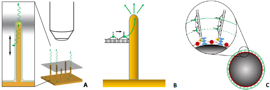
Figure 1. A – lightguiding in a nanowire; B – molecular motor on DNA track attached to a nanowire; C –molecular motors on DNA tracks on a bead.
[1] D. Verardo et al., “Nanowires for Biosensing: Lightguiding of Fluorescence as a Function of Diameter and Wavelength,” Nano Lett., vol. 18, no. 8, pp. 4796–4802, 2018, doi: 10.1021/acs.nanolett.8b01360.
[2] E. H. C. Bromley et al., “The tumbleweed: Towards a synthetic protein motor,” HFSP J., vol. 3, no. 3, pp. 204–212, Jun. 2009, doi: 10.2976/1.3111282.
[3] M. H. Bakalar, A. M. Joffe, E. M. Schmid, S. Son, M. Podolski, and D. A. Fletcher, “Size-Dependent Segregation Controls Macrophage Phagocytosis of Antibody-Opsonized Targets,” Cell, vol. 174, no. 1, pp. 131-142.e13, 2018, doi: 10.1016/j.cell.2018.05.059.
16:05 - 16:10
Prestin is a voltage-sensitive motor protein specifically expressed in the outer hair cells and providing auditory signal amplification in the mammalian cochlea due to the voltage-dependent conformational changes [1]. Its extremely fast voltage-dependent conformational dynamics and a significant mechanical force produced by its molecule, as well as a native expression in the nerve cells of higher animals make it a promising candidate for a voltage sensing domain in the genetically encoded voltage indicators (GEVIs).
In this contribution, we aimed to prove prestin’s ability to function as the GEVIs sensitive core. To that end, we have designed several genetic constructs based on Meriones unguiculatus prestin [1] and FusionRed fluorescent protein [2]. Since little is known about the molecular mechanism of prestin electromotility and therefore there are no ready-to-use principles for prestin-based indicators designing, we have suggested 3 diverse topologies of the indicator molecules. They include “insertion-into-cpFP” (circular permuted Fluorescent Protein) that we implemented earlier for Ciona Intestinalis voltage sensitive domain-based GEVIs [3,4], “insertion-into-FP” that has been widely used in fluorescent indicators development, and “cpFP insertion-into-prestin” that could become a perspective approach applied to the large transmembrane proteins.
Our whole-cell voltage-clamp experiments with live HEK293T cells revealed that the prestin-based GEVI (at least built on “insertion-into-cpFP” topology) demonstrates fast (submillisecond) fluorescent response to the membrane potential changes. Apart from the proof-of-principle of prestin applicability in GEVIs, we have shown that the cpFusionRed insertions into prestin intracellular/external loops often leads to the altered fusion trafficking and/or aggregation [5].
The authors gratefully acknowledge funding RFBR according to the research projects № 18-34-20087 and 19-34-90140, Electrophysiology experiments were supported by RFBR grant 19-015-00022. Experiments were partially carried out using the equipment provided by the IBCh core facility (CKP IBCh, supported by Russian Ministry of Education and Science, grant RFMEFI62117X0018).
Zheng J, Shen W, He D, et al. Prestin is the motor protein of cochlear outer hair cells. Nature 405 (2000), 149–155.
Shemiakina II, Ermakova, GV, Cranfill, PJ, et al. A monomeric red fluorescent protein with low cytotoxicity. Nat Commun 3 (2012), 1204.
Kost LA, Nikitin ES, Ivanova VO, et al. Insertion of the voltage-sensitive domain into circularly permuted red fluorescent protein as a design for genetically encoded voltage sensor. PLOS ONE. 12(9) (2017),
Kost, LA, Ivanova VO, Balaban PM, et al. Red Fluorescent Genetically Encoded Voltage Indicators with Millisecond Responsiveness. Sensors. 19 (13) (2019.), 2982.
16:10 - 16:15
Planar photonic waveguides enable ultrathin sample illumination in fluorescence microscopy over exceedingly large fields of view. Their fabrication is typically based on hard coatings requiring sputter deposition and ion beam lithography, which makes volume production slow and cumbersome. Additionally, they are typically fabricated on top of an opaque Silicon wafer substrate, which restricts their use to upright microscopes. Here we present a photonic waveguide chip based on a standard 170 µm glass cover slip coated with a micrometer-thin layer of EpoCore, a negative photoresist polymer of high refractive index. This enables efficient widefield excitation for fluorescence microscopy and nanoscopy for inverted microscopes using fluorescence detection through the transparent substrate. Channel waveguides with varying widths provide uniform near-field illumination by evanescent waves for fields-of-view up to the millimeter scale. We demonstrate multi-color excitation at 568 nm and 647 nm wavelength resulting in high contrast immunofluorescence and super-resolution imaging.
A beloved imaging modality in optical microscopy is total-internal reflection fluorescence (TIRF), where the illumination beam is reflected at the glass-sample boundary, and the sample is only illuminated by its evanescent tail. This method limits the user to high numerical aperture oil objective lenses, and the illumination is limited to a small field of view. Photonic chips provide ultrathin sample illumination by TIRF, while decoupling excitation and detection paths [1]. Thereby, one can probe a sample with a low-magnification objective and still obtain crisp images from TIRF. Till now, these chips have been based on substrates of silicon, but a disadvantage of this is that the chips are opaque, only allowing for imaging in the upright configuration on a very few select microscopes. Additionally, the waveguide cores typically consist of Ta2O5 or Si3N4, and are fabricated using deposition methods, and typically structured using ion beam lithography, making for a slow fabrication process. To deal with these shortcomings, we designed planar waveguide chips based on standard issue #1.5 coverslips made from borosilicate glass as substrate with a photoresist polymer layer deposited on top of the substrate as waveguide core. EpoCore2 was chosen as the core material as it exhibits a high refractive index (n=1.59 at red light) and flexibility in fabrication, and has already been shown to be useful in biosensorics [2][3].
The fabrication process is described as follows: First the coverslips are carved halfway through using laser ablation to allow for breaking them off into individual chips later. Thereafter they are cleaned in a chemical bath, after which the photoresist is spin coated on top to create a layer on the sub-micrometer scale. The photoresist is patterned into waveguides using UV-lithography and developed by a baking process on a heat plate. In the end, undeveloped photoresist is removed in a chemical bath. The coverslips are then gently cleaved into several chips at the laser ablated positions, creating end facets for coupling into the channel waveguides. No additional polishing is needed.
The waveguides are coupled at the end-facet using a long-working distance objective on a nano-positioner stage. A schematic is shown in Figure 1.
Figure 1: Schematic of the polymer photonic waveguide chip with end facet coupling through a long-working distance objective lens.
Cells can be incubated directly on top of the waveguide and are illuminated by the evanescent field of the electromagnetic waves propagating through the waveguide. Common cellular targets, such as actin and tubulin can be illuminated homogeneously and detected using low magnification air objectives (actin shown in Figure 2). Super-resolution radial fluctuation (SRRF) imaging is achieved by recording short stacks of 200 widefield images [4]. Using Fourier-Ring Correlation provided by NanoJ-SQUIRREL, we calculate mean resolutions of 80 nm over a field of view of 140 x 220 um2, using a 60x 1.20 NA water objective for detection.
Figure 2: (a) A 500 um wide waveguide chip illuminated by waveguide-TIRF, detected with a 20x 0.7 NA air objective. 200 frames were used to reconstruct a super-resolution image using SRRF. (b) Inset from (a).
In conclusion, we show that waveguide chips for inverted microscopes can be produced efficiently and in large volumes by using 170 µm thick cover slides and a commercially available photoresist polymer. Since the substrate material is transparent and its characteristics are well known to microscope users, it allows for easy implementation to inverted microscopes. The resulting chips permit large field of view illumination of biological samples based on total-internal reflection within the waveguide, which then becomes compatible with standard microscope objective lenses.
[1] R Diekmann et al, NATURE PHOTONICS 11 (2017), p. 322-328.
[2] S Hessler et al, SENSORS 19 (2019).
[3] N T Benéitez et al, IEEE Journal of Selected Topics in Quantum Electronics 22 (2016), p. 319-326.
[4] N Gustafsson et al, Nature Communications 7 (2018), p. 12471.
[5] R F Laine et al, Journal of Physics D: Applied Physics, 52 (2019), p. 163001
16:15 - 16:20
With the advances in fluorescence microscopy, conventional methods for intercellular interaction identification are being recognised as unreliable. To address this issue, we suggest applying a combination of novel fluorescence microscopy techniques towards a new approach meant for identification of intact synapses in brain tissue. Finally, we applied this approach in a Mus Musculus model.
Currently, fluorescence signal co-localisation is used to infer biomolecular interactions. However, with the rise of super-resolution microscopy and pixel-based co-localisation, the validity of these methods in precise intercellular interactions is debated [1]. Conventionally, the co-localisation of intracellular pre-synaptic and post- synaptic proteins (e.g. PSD95 and synaptophysin) has been used to identify synapses. This approach is biased as it does not provide evidence of a physical synaptic connection, only an approximation of the two signals coming from near locations. Novel intercellular techniques like eGRASP [2,3] require genetic intervention into the host (e.g. viral infection via adeno-associated viruses) which limit the application of the techniques to specific animal models. Therefore, we have developed a novel approach to effectively detect synaptic contacts which relies on Fluorescence Lifetime Imaging Microscopy (FLIM) and Förster Resonance Energy Transfer (FRET), and immunofluorescence.
In our method, pre-synaptic and post-synaptic cellular adhesion molecules (CAM) are labelled with fluorescent antibodies where the fluorophores are selected to form an efficient FRET pair (Fig. 1). By imaging stained Mus Musculus brain sections with FLIM, only the donor fluorescence lifetime is acquired. A decrease in the lifetime of the donor fluorophore suggests that the fluorophores are in a very close proximity (approx. 5-10 nm). After acquiring a FLIM image, the signal is separated (by lifetime) into quenched and non-quenched donor fluorescence images. Ultimately, the image containing only the quenched donor fluorescence provides accurate localisation and proof of an intact synapse.
Neurexin-1-a and LRRTM-2 were selected as specific synaptic targets for our FRET sensor as these CAMs are only expressed in neuron cells and were shown to be interacting between hippocampal neurons. Moreover, as the selected dyes were compatible with Stimulated Emission Depletion (STED) microscopy, a depletion laser was applied which increased the resolution (and thus the localisation accuracy) 10-fold.
In conclusion, our developed FLIM-FRET synapse sensor can effectively identify intact synapses. With the help of STED, the accuracy of the method is increased markedly. Furthermore, the applicability of the principle is vast: it can be applied in various species, the method is suitable for both live and fixed brain tissue samples, and it does not require genetic interference.
Figure 1. Schematic representation of the FRET based approach of the synapse identification.
[1] J. S. Aaron, A. B. Taylor and T. L. Chew, Journal of Cell Science, 131(3)
[2] J. H. Choi, et al, Science, 360(6387), 430-435
[3] E. H. Feinberg, et al, Neuron, 57(3), 353-363
16:20 - 16:25
Introduction
Tumor cells produce increased levels of sialic acid (SA), a cell surface monosaccharide that can contribute to cellular recognition and cancer invasiveness. Since there is great interest in developing improved tools for detection of SA on cancer cells, we are developing SA-targeted fluorescent molecularly imprinted polymers, SA-MIPs for diagnosis of aggressive cancer. However, when nanoparticles are injected in vivo, it can lead to induction of a host inflammatory response due to non-specific recognition and uptake by macrophages. Therefore, to pave the way for future in vivo studies, we have investigated the effect of SA-MIPs on the immune system by investigating in vitro cultured RAW 264.7 macrophages.
Methods
Analysis of the role of macrophage binding and uptake of SA-MIPs was performed with flow cytometry and fluorescence microscopy. 3D digital holographic cytometry (DHC) and MTS assay was performed for viability measurements of macrophages with or without SA-MIPs treatments. In order to measure the activation of the macrophages after SA-MIPs treatment a TNF-α ELISA was used.
Result and discussion
We have analyzed the macrophage uptake of the SA-MIPs by using fluorescence microscopy and flow cytometry on viable RAW 264.7 macrophages at different timepoints in vitro. Our results showed that macrophages bind and ingest SA-MIPs over time. The cell viability with and without SA-MIPs was analyzed by DHC. The viability was increasing over time but was not affected by the addition of the SA-MIPs. The 3D DHC method was also used to image the uptake of SA-MIPs over time by macrophages. Future studies include SA-MIP degradation and macrophage activation. Analysis with TNF-α ELISA showed an increased TNF-α release from the SA-MIP treated macrophages. This effect needs to be taken into consideration when planning for future in vivo applications.
Conclusion
We have demonstrated that the SA-MIPs influence macrophages, as the latter start to phagocytose the SA-MIPs and thereby secrete TNF-α, due to activation, without any toxic effect. This suggest that the SA-MIPs must be modified in order to target the cancer cells without activating the immune system and become phagocytosed.