To save this page as a PDF, click this button and choose the PDF destination.

Poster Session - Life Sciences

14:45 - 15:15 Wednesday, 25th November, 2020

Meeting Room 1

Track Life Sciences

Presentation type Poster Presentation


14:45 - 14:50

774 Morphology of photoautotrophic PHA producing prokaryotes

Katerina Mrazova1, Kamila Hrubanova1, Jana Nebesarova2, Ines Fritz3, Christina Daffert3, Stanislav Obruca4, Zuzana Sedrlova4, Vladislav Krzyzanek1
1The Czech Academy of Sciences, Institute of Scientific Instruments, Brno, Czech Republic. 2The Czech Academy of Sciences, Biology Centre, Ceske Budejovice, Czech Republic. 3University of Natural Resources and Life Sciences Vienna, Institute of Environmental Biotechnology, Tulln, Austria. 4Brno University of Technology, Faculty of Chemistry, Brno, Czech Republic

Abstract Text

Microbial cells produce a variety of storage materials, which serve as an energy source, carbon source, etc. Polyhydroxyalkanoates (PHA), polyesters of hydroxyalkanoic acids, are biodegradable plastics accumulated as granules in cells of many prokaryotic microorganisms. Primarily PHA are used in microbial cells as storage material, however, recent studies revealed, that cells containing PHA are also more resistant to stress environment such as osmotic imbalances, UV irradiance or temperature changes. Production of polyhydroxyalkanoates is induced in excess of carbon source and also limitation of another element essential for cellular growth. Besides microbial cell storage material, PHA are also considered as a promising bioplastic material, due to its biodegradability and mechanical properties similar to petrochemical plastics, such as polypropylene (PP). [1–3] 

Currently, Cupriavidus necator or recombinant Escherichia coli are considered as one of the biggest PHA producers. Unfortunately, the production of PHA using heterotrophic microorganisms requires large amounts of carbon substrates, such as glucose or fructose, which makes the production quite expensive compared to PP. Carbon substrates could make up to 50% of all production costs. One of the possibilities, how to reduce these expenses, could be the use of waste material (eg. frying oils) as a carbon source or production of PHA by photoautotrophic microorganisms. [1,3,4]  

Promising strain for PHA production is Synechosystis sp., unicellular cyanobacteria. Cyanobacteria are prokaryotic microorganisms commonly occurring in freshwater and also saltwater. Unlike heterotrophic bacteria Cupriavidus necator, Synechosystis salina contains pigments like phycocyanin, carotenoids and chlorophylls and performs photosynthesis. Therefore, these bacteria do not require organic carbon substrate and are able to convert CO2 into various valuable metabolites including PHA. However, compared to heterotrophic organisms, cyanobacteria are not able to produce that high amounts of PHA in their cells [3,4]. This study is focused on deeper morphological study of cyanobacterial cells of strains Synechocystis sp. PCC 6803 and Synechocystis salina CCALA 192, mainly on the content of intracellular PHA granules. 

Cyanobacterial cells were fixed using high-pressure freezing method. After fixation samples were processed using either freeze-fracture method for observation in cryo-SEM or freeze substitution followed by embedding in epoxy-resin for observation in TEM. Freeze fracturing method enables the imaging of intracellular content of cells. PHA granules at temperatures of −130 °C remain elastic and can be observed in cryo-SEM sticking out of fractured cells. Comparison of Synechocystis sp. PCC 6803 and Synechocystis salina CCALA 192 (Figure 1) showed, that strain Synechocystis sp. PCC 6803 contains less PHA granules, but produces higher amount of extracellular substances, forming mass surrounding each cell. This observation was confirmed also by TEM images (Figure 2). After postfixation with OsO4, embedding in Epon and staining ultrathin sections with lead citrate and uranyl acetate images shows higher content of PHA granules in Synechocystis salina CCALA 192 strain, but extracellular matrix was observed mostly in Synechocystis sp. PCC 6803 samples. These findings prove, that cyanobacteria are perspective microorganisms for PHA production and should be further investigated. 


Uncaptioned visual

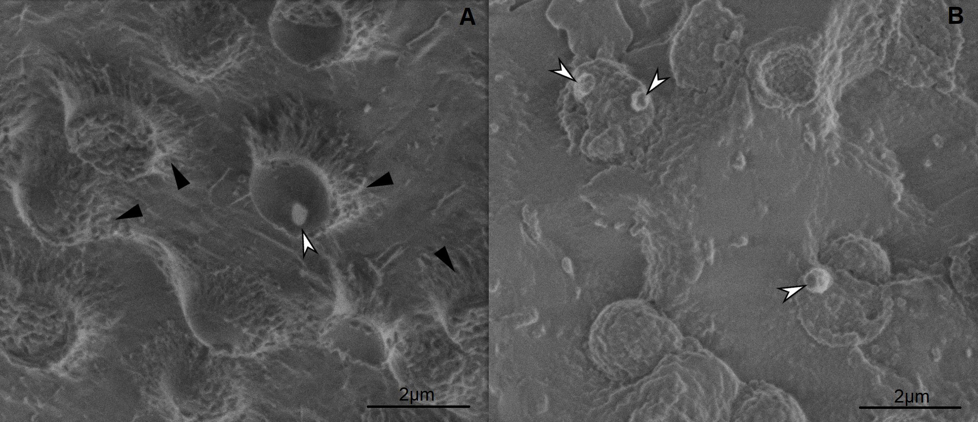
Figure 1: cryo-SEM images of A) Synechocystis sp. PCC 6803, B) Synechocystis salina CCALA 192. Freeze fracturing followed with freeze etching revealed inner structure of the cyanobacterial cells, showing PHA granules (white arrows) and extracellular mass (black arrows).

Uncaptioned visual

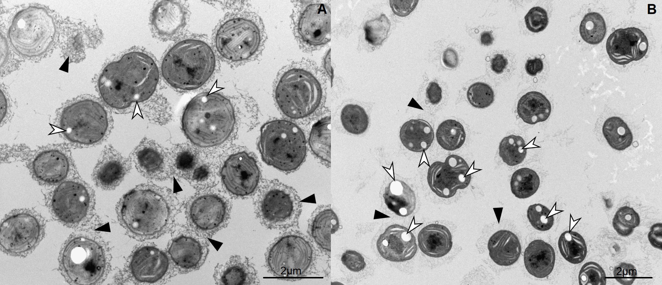
Figure 2: TEM images of A) Synechocystis sp. PCC 6803, B) Synechocystis salina CCALA 192. Staining of ultrathin sections revealed white intracellular PHA granules (white arrows) and dark extracellular mass (black arrows), which is denser in Synechocystis sp. PCC 6803 strain.  

References

[1]   S. Obruca et al, Involvement of polyhydroxyalkanoates in stress resistance of microbial cells: Biotechnological consequences and applications. Biotechnology Advances 36 (2018) p. 856-870.

[2]     P. Sedlacek et al, PHA granules help bacterial cells to preserve cell integrity when exposed to sudden osmotic imbalances. New Biotechnology 49 (2019) p. 129–136. 

[3]     C. Troschl et al, Cyanobacterial PHA Production—Review of Recent Advances and a Summary of Three Years’ Working Experience Running a Pilot Plant. Bioengineering 4 (2017) p. 26. 

[4]     C. Troschl et al, Pilot-scale production of poly-β-hydroxybutyrate with the cyanobacterium Synechocytis sp. CCALA192 in a non-sterile tubular photobioreactor. Algal Research 34 (2018) p. 116-125.

[5]    The research was supported by the Czech Science Foundation (project GF19-29651L), the Technology Agency of the Czech Republic (project TN01000008); the infrastructure by the Czech Academy of Sciences (project RVO:68081731).


14:50 - 14:55

1380 Image based analysis of reproductive toxicity caused by Organochlorine mixtures

Miss Ishita Virmani, ELIŠKA SYCHROVÁ, JIŘÍ NOVÁK, Iva Sovadinova
RECETOX, Faculty of Science, Masaryk University, Kamenice 753/5, 625 00, Brno, Czech Republic

Abstract Text

The effects of an individual chemical have been studied well by the researchers and thus information regarding their toxicity profiles are widely available in the literature. Even, most of the regulations in the European Union (EU) are also based on the testing of single chemicals, thus overlooking the outcomes of mixture interactions occurring during environmental exposure. This is the case of organochlorines that are common environmental contaminants, and we regularly get exposed to the mixtures. Their exposure is highly discussed reason for male sub/infertility which has now become a global issue. Therefore, there is a need to develop a framework for assessing the mixture toxicity, consisting of multiple steps. There are studies confirming an association of individual organochlorines or their mixtures with adverse effects on male reproductive system, however image- and mechanistic-based studies dealing with organochlorine mixtures are scarce [1,2]. Screening toxic effects of a huge number of chemical mixtures in vitro is a central attraction nowadays. Therefore, there is a need to develop high-content analysis (HCA) approaches to gain the information regarding the mechanisms and adverse outcome pathways (AOP) involved from the molecular initiating event (MIE).  


To study the effects of organochlorine mixture on male reproductive system, we developed a semi-automatic and versatile battery of the tests using different in vitro models. Firstly, we studied endocrine-disrupting potential of an environmentally relevant organochlorine mixture using reporter gene assays assessing the interactions with nuclear receptors.  Secondly, we focused on direct effects of mixture on testicular cells, namely on Leydig (TM3) and Sertoli (TM4 cells with different functions in male reproductive health. We performed cell viability assays using 3-(4,5-dimethylthiazol-2-yl)-2,5-diphenyl tetrazolium bromide (MTT) and a combination of three indicator dyes – (alamarBlue, CFDA-AM, and neutral red). To study the inhibitory effects of the mixture on cell-cell communication, we performed the improved multiparametric scrape loading-dye transfer assay (SLDT) [3] for a simultaneous high-throughput analysis of gap junctional intercellular communication (GJIC), cell density and viability. Imaging was performed on two microscopy platforms (Tissue Gnostics TissueFax system and Biotek Cytation 5) as depicted in figure 1 and 2. The data obtained from these two microscopy platforms were analyzed using Image J (Macro) and Gen 5, respectively. To check if exposure to this mixture was leading to the extensive formation of lipid droplets in the testicular cells, we stained the cells with BODIPY and Hoechst for the quantification of lipid droplets and the number of nuclei respectively.


Regarding endocrine-disrupting potential of this organochlorine mixture, strong antiandrogenic and weak estrogenic effects were observed in a dose-dependent manner. The mixture also directly affected testicular cells. In addition to dose and time dependent cytotoxicity in both Leydig TM3 and Sertoli TM4 cells, the organochlorine mixture also rapidly deregulated testicular gap junctional intercellular communication (GJIC) in vitro in both TM3 and TM4 at non-cytotoxic concentrations.  Our set-up allowed quick and convenient multiparametric GJIC assay (different microscopy and image analyzing platforms). The analysis of communicating and not-communicating cells was carried out with open-platform Image J using in-house developed macro.  Additionally, Gen 5 provided us the automatic platform to count dead and all cells for assessing cell density and viability. Using Biotek Cytation 5 and Gen5 pipeline, we were able to evaluate also number of lipid droplets in an automated manner. To conclude, we developed and optimized a versatile battery of imaged-based assays for high-content analysis of in vitro reproductive toxicity it can be highlighted from our results that organochlorine and their mixtures could be a potential etiological agent contributing to reproductive dysfunction in males through an impairment of the testicular GJIC.  


Uncaptioned visual


Figure 1Schema representing the set-up for monitoring level of cell communication in vitro using microcopy platform (TissueFax(a) capturing images (b). This set-up can be applied for various multiwell plates (c), ready for high-throughput application.  Image-based analysis of cell communication is fast and objective and uses in-house developed macro in ImageJ (c and d).  

Uncaptioned visual

Figure 2: Schema representing the set-up for monitoring cell density and viability using microcopy platform (Cytation 5, c). Images showing the dead cells (a) and cell density (e) after treatment with 10% of ethanol. Images depicting the automatic cellular analysis performed using Gen 5 (b and f) (d) Results obtained from the analysis of image (e) showing number of cells per FOV (b).   


References

(1)  Anas MK et al , 73 (2005), p.414.

(2) Enangue et al , 90 (2014) , p.118.  

(3) Dydowiczova et al, 10 (2020) , p.1/ 


14:55 - 15:00

964 Microscope stage for live measurements with magnetic force – design, develop and research.

Daniel Dziob1, Jakub Ramian2, Jan Ramian3, Bartosz Lisowski1
1Jagiellonian University Medical College, Kraków, Poland. 2AGH University of Science and Technology, Kraków, Poland. 3Medical University of Silesia, Katowice, Poland

Abstract Text

Summary


Design and validation of a microscope stages which allows for live measurements with a constant magnetic force acting on a cell or tissue will be presented.  


Introduction


Interest in if and how magnetic field impacts living organisms has a long and well documented history. Scientific literature is full of articles trying to answer how magnetic field influences organisms at macro and micro-level. However, significant amount of research was done for static (constant) magnetic fields, whereas magnetic force can act on an object with nonzero magnetic susceptibility only when a gradient of magnetic fields occurs. Force value obviously depends on this gradient. Here we present a set of originally designed microscopic stages, which allow for obtaining homogenous magnetic forces for microparticle manipulation inside living cells or in buffer. 


Methods


All stages are based on the idea of Halbach array of magnets. We 3d printed and verified several stages, which differ by: (1) the number of magnets, (2) their spatial arrangement, (3) strength and (4) materials from which a given stage was printed. Distribution of magnetic field was verified by measurements of magnetic field components by precise magnetometer (Smart Magnetic Sensor ASONIK SMS-102). Stages were tested for the effects of long-term exposition on temperature, humidity and CO2 concertation, required for observation of living cells. Several experiments were conducted with the use of the stages.


Results and discussion


Obtained results show that by design of the microscope stage with Halbach array of magnets one could adjust the magnetic field gradient and the magnetic force acting on magnetic microparticles placed in a small petri dish, in the buffer or inside the cells. The obtained range of forces varies depending on the architecture of the stage, but it is easy to obtain biologically reasonable forces of the order of piconewtons. Measurements of stages behavior under microscope allowed for the choice of optimal materials for 3d printing as well as the Halbach array architecture of magnets mounted on a stage.


Conclusion


Designing and printing of a functional microscopic stage with Halbach array of magnets, which is a source for constant gradient magnetic field, is nondemanding and accessible method for investigating the impact of biologically significant magnetic forces on living cells. 


References

[1] Wenjun Xu, Jinru Sun, Yangjing Le, Jingliang Chen, Xiaoyun Lu & Xueling Yao  (2019) Effect of pulsed millisecond current magnetic field on the proliferation of C6 rat glioma cells, Electromagnetic Biology and Medicine, 38:3, 185-197.
[2] Barnes, F., Greenenbaum, B. (2016). Some effects of weak magnetic fields on biological systems: RF fields can change radical concentrations and cancer cell growth rates. IEEE Power Electron. Mag. 3:60–68.
[3] Xiaofei Tian, Dongmei Wang, Meng Zha, Xingxing Yang, Xinmiao Ji, Lei Zhang & Xin Zhang (2018): Magnetic field direction differentially impacts the growth of different cell types, Electromagnetic Biology and Medicine.
[4] Michele Destefanis, Marta Viano, Christian Leo, Gianpiero Gervino, Antonio Ponzetto & Francesca Silvagno (2015) Extremely low frequency electromagnetic fields affect proliferation and mitochondrial activity of human cancer cell lines, International Journal of Radiation Biology, 91:12, 964-972.


15:00 - 15:05

1069 DeepACSON: automated segmentation of 3D electron microscopy images of white matter

Ali Abdollahzadeh1, Ilya Belevich2, Eija Jokitalo2, Alejandra Sierra1, Jussi Tohka1
1University of Eastern Finland, Kuopio, Finland. 2University of Helsinki, Helsinki, Finland

Abstract Text

Three-dimensional electron microscopy (3D-EM) is the method-of-choice to access brain ultrastructures and quantify its morphology. Manually or semi-automatically tracing ultrastructures in their entirety are prohibitively time-consuming due to the size of 3D-EM datasets; thousands of images with nanometer resolution yield hundreds of gigabytes to terabytes of image datasets [1]. Deep convolutional neural networks (DCNNs) can automatically trace ultrastructures in such datasets, but the design of the current methods relies on distinctive high-contrast cellular membranes and very high-resolution EM images [2][3]. Acquiring such datasets needs either several months of imaging or limited small tissue volumes.


We developed DeepACSON to segment low-resolution 3D-EM datasets of big volumes of white matter within a reasonable time [4]. DeepACSON performs DCNN-based semantic-segmentation and shape decomposition-based instance-segmentation: a top-down design to account for severe membrane discontinuities inherited with low-resolution imaging. We developed a cylindrical shape decomposition algorithm to automatically decompose under-segmented myelinated axons, via maximizing straightness and cross-sectional homogeneity functions [5]. We trained DeepACSON using human-annotation-free training sets, using segmented high-resolution 3D-EM images of simultaneously acquired volumes from the same tissue by our earlier developed automated ACSON pipeline [6].


With DeepACSON, we segmented ten low-resolution 3D-EM datasets of the corpus callosum and cingulum of rats after sham-operation or traumatic brain injury (TBI) into myelin, about 288,000 myelinated axons, millions of mitochondria, and 2,600 cell nuclei with excellent evaluation scores. DeepACSON quantified the morphology and density of myelinated axons, the spatial distribution of mitochondria, and the density of cells. The analyses indicated that the diameter of myelinated axons in the healthy brain varies substantially along an axon. The cross-sections of myelinated axons were elliptic rather than circular. Myelinated axons were mostly straight than tortuous. Our findings showed significant changes in the axonal diameter, tortuosity, and the density of myelinated axons in TBI rats, five months after the injury. The morphology parameters, such as axonal diameter and eccentricity, were consistent with our 3D measurements in high-resolution 3D-EM datasets.


DeepACSON is a novel technique which improves the state-of-the-art automated segmentation techniques, by dealing with the limitation of low-resolution 3D-EM datasets, noise, and human-annotation-free training sets. DeepACSON offers new ways to unravel the true morphology of the brain and capture ultrastructural alterations in disease [7].

References

[1] Z. Zheng et al, Cell 174 (2018), p. 730

[2] M. Januszewski et al, Nature Methods 15 (2018), p. 605

[3] Y. Meirovitch et al, CVPR (2019)

[4] A. Abdollahzadeh et al, bioRxiv: doi.org/10.1101/828541 (2019)

[5] A. Abdollahzadeh, A. Sierra, and J. Tohka, arXiv:1911.00571v2 [cs.CV] (2019)

[6] A. Abdollahzadeh et al, Scientic Reports 9 (2019), p. 6084

[7] This work was supported by the Academy of Finland (grant 316258 to J.T. and grant 323385 to A.S.), and Biocenter Finland and University of Helsinki (I.B., E.J., and SBEM imaging).